MRI of the mammary glands
Breast MRI is the most accurate method for breast diagnosis. Unlike mammography, it does not have radiation exposure. However - just for prevention, for everyone - it is not suitable because:
- in St. Petersburg there are only a few specialists who can describe MRI of the mammary glands
- High-quality MRI of the mammary glands cannot be done on any MRI machine.
- The research is expensive, the state does not pay for it, and patients look for something cheaper.
Due to the fact that it is difficult to provide all these components, overdiagnosis during preventive MRI of the mammary glands is observed in all countries of the world.
NB! At the First Medical University named after. I.P. Pavlova at st. Lev Tolstoy 17 installed a mammograph with tomosynthesis function - CESM (3rd floor).
CESM is similar to MRI and is effective even in dense breasts.
Anyone can get tested on weekdays from 9:00 to 14:00 without an appointment. Contact the paid services department on the first floor. Cost from 2 (mammography without tomosynthesis) to 5 (CESM-tomosynthesis) tr.
If a tumor is detected, a consultation with an employee of the University Breast Center is free on the day of treatment.
MRI diagnostics
Preventive MRI has shown better effectiveness only in combination with mammography in patients at risk for breast cancer caused by gene mutations (like Angelina Jolie). SEE ABOUT THIS .
MRI is also more justified in cases of dense breast tissue (as a rule, in the absence of childbirth and lactation), in case of small breasts without ptosis - when it is technically impossible to place them in a mammography machine.
The specialists of our Breast Center perform all types of plastic surgery on the mammary gland, including complications (ruptured implants, their displacement, etc.). SEE ABOUT THIS .
MR mammography
The diagnostic and treatment center offers services for performing MRI of the mammary glands (MR mammography) with and without contrast. The study is carried out on a Philips Achieva 1.5T tomograph - this is one of the best tomographs from Philips.
MRI breast diagnostics is a modern research method, which, in comparison with radiography and mammography, is safer for the patient and more informative. During diagnostics, the tissue structures of the mammary glands are studied in detail, which allows the doctor to identify even minor pathological processes and neoplasms.
Currently, breast cancer accounts for 20.9% of all malignant tumors in women and is the leading cause of death from malignant tumors in women aged 40–69 years. Numerous studies have proven that the earlier a tumor is detected, the longer the life expectancy of patients. In addition, early diagnosis of cancer allows organ-preserving operations to be performed without mutilating interventions. For early diagnosis of breast cancer, it is necessary to regularly undergo breast MRI.
In addition to MRI, at the Kutuzovsky Children's Center you can do elastography of the mammary glands as prescribed by a mammologist.
MRI and plastic surgery
In patients after the following plastic and reconstructive surgeries, MRI is used for diagnosis:
- breast augmentation with implants - for implant rupture, the presence of implant-associated lymphoma, and oncological pathology of the mammary gland
- mammoreduction - for early diagnosis of cancer and its difference from granulomas,
- contour plastic surgery with lipofilling or gel - for early diagnosis of cancer and its difference from granulomas,
- breast restoration after removal for cancer - to check for the return of the oncological process in the surgical area.
In all other cases, it is recommended to perform a mammogram . SEE ABOUT THIS .
Contraindications for MRI
The MRI method of the mammary glands is considered safe, but there are still contraindications.
MRI examinations are prohibited:
- When there are metal objects, implants, clips, bracelets, prosthetic limbs and joints in the patient’s body;
- if you have a pacemaker;
- if existing tattoos use paint mixed with metal, there may be a burn or a burning sensation;
- if, due to illness, the patient cannot remain completely still during the study, suffers from a mental disorder or claustrophobia;
- if the patient is at an early stage of pregnancy or is breastfeeding, the study is carried out after consultation with a doctor and if there is evidence of a threat to life and health;
- if you are allergic to the contrast agent, this is very rare, but the consequences are very serious;
- the presence of kidney and genitourinary system diseases;
- if the patient’s weight exceeds more than 150 kg.
Breast MRI
According to statistics, patients themselves find a cancerous tumor measuring 2 cm in size with their hands; a doctor, during a professional examination, finds a tumor in a patient with a size of 1.5 cm in size. The purpose of MRI is to find a tumor before it can be detected with your hands.
There are cases when breast cancer is not visible on mammography. This doesn't happen often. Some patients can afford to do a more expensive test instead of a mammogram - then they can have an MRI.
The rhythm of preventive MRI studies is as follows:
- 35 - 40 years - first MRI. All subsequent MRIs will be compared with this MRI.
- up to 45 - 50 years - once every 2 years
- 50 - 60 years - annually
- after 60 years - once every 2 years.
If a patient takes hormonal contraceptives (especially triphasic ones) for more than 5 years, or hormone replacement therapy, she has a higher risk of breast cancer. She has been prescribed annual MRI since age 40.
If you do not have implants and have not had plastic surgery on your breasts, if you are not at risk for breast cancer, it is enough to perform a mammogram rather than an MRI. SEE ABOUT THIS .
What does a breast MRI show?
An experienced breast MRI specialist will always describe:
- tumor or tumors indicating its (their) size and location in the mammary gland: in which quadrant or on the border of which quadrants
- will indicate the distance from the tumor to the nipple in cm and/or mm, the distance from the tumor to the skin and muscle fascia - the doctor’s decision on the possibility of preserving the skin over the tumor depends on this
- will describe the condition of the skin above the tumor and in the area of the areola (whether there is swelling or not) - the doctor’s decision on the possibility of preserving the skin above the tumor or the need to remove the nipple and areola depends on this
- will describe the condition of the lymph nodes, but not only the axillary ones, but will also indicate the condition of the parasternal ones - which are hidden behind the cartilage of the ribs; will always compare them with the lymph nodes of the opposite side - as a sample of normal lymph nodes in a given patient - the doctor’s decision on whether you can perform a sentinel node biopsy or whether preliminary treatment is necessary depends on this
- will describe the condition of the bone tissue in the scanning area for metastases
- will provide information on the volume of breast tissue - for selecting an implant for one-stage reconstruction
- will provide information on the volume of the installed implant (if the passport for the implant is lost)
The accuracy of MRI is much higher if the images are evaluated “in dynamics” - new images are compared with previous ones. Therefore, when researching, always take the images on disk for your archive.
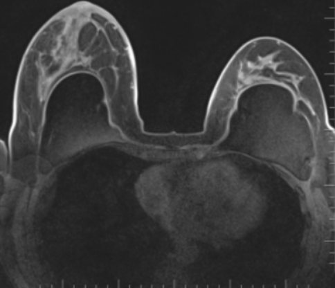
Hypervascularization
Malignant breast tumors on MRI specifically accumulate contrast due to their hypervascularization (increased vascular growth).
Breast diseases
The paired organ has a complex structure. Each breast is formed by cone-shaped lobes with the tip at the nipple. Glandular tissue includes many alveoli. Milk ducts depart from each, merging towards the nipples into larger sinuses. Between the lobes there are lobes of connective and adipose tissue.
The glands are well supplied with blood. The structure of the breast contains lymphatic vessels. The body of each organ is protected by a connective tissue sheath, connected by numerous ligaments to the skin. The glands of the breast are hormone-dependent and perform the function of lactation (this is important during breastfeeding).
As a result of exposure to external and internal unfavorable factors, benign and malignant pathological processes can occur in the organ. The most common:
- cysts - formed as a result of obstruction of the ducts;
- galactocele - a cavity formation filled with milk that occurs during lactation;
- oleogranuloma - necrosis of adipose tissue;
- mastitis is an inflammatory disease;
- fibrocystic mastopathy - a change in the ratio between the epithelial and connective tissue components of the gland;
- cancer - a group of oncological diseases;
- intraductal papillomas - formations on the walls of the ducts;
- atheroma - damage to the sebaceous gland of the skin in the chest area;
- Lipoma is a benign formation consisting of fat;
- fibroadenoma is a structure formed from connective and epithelial tissue.
Magnetic resonance imaging of breasts with implants
MRI of the mammary glands helps in detecting the listed pathologies (except for intraductal papillomas). The method is successfully used for the purpose of differential diagnosis of benign and malignant tumors, but final conclusions about the nature of the identified abnormalities are established based on the results of the biopsy.
MRI for breast cancer
When describing, an experienced specialist will use the Bi-Rads system and avoid evasive terms and phrases with double interpretation. Also, he will always first describe the above points - the main thing, and then mention the less important - blood vessels and heart. Whereas a specialist “inexperienced in the mammary gland” will always first describe in detail what he knows better - the blood vessels and the heart. A conclusion will also be drawn up - an experienced one will first describe oncology, and then about blood vessels and so on.
An MRI of the mammary glands for cancer must be done before a biopsy. After it (due to injury), swelling, reaction of the lymph nodes, hematoma may appear: all this can falsely aggravate the signs of the disease.
THE algorithm for proper screening for breast cancer HERE .
Sometimes, when prescribing treatment for cancer patients before surgery (chemotherapy or hormone therapy, targeted treatment), an MRI of the mammary glands can also be performed to monitor its effectiveness.
The specialists of our Breast Center perform all types of operations on the mammary gland: for cancer and benign tumors, we can combine these operations with breast plastic surgery. SEE ABOUT THIS .
Preparing for a breast MRI with contrast
Before ordering an MRI, the doctor explains what information the MRI scan will add and how this may influence treatment decisions.
To avoid discomfort due to the prone position, it is recommended that you eat before the examination at least 2 hours before the examination.
Before performing an MRI of the mammary glands with contrast, it is necessary to fill out a questionnaire so that the doctor who will describe the tomograms is oriented in the situation as a whole, understands the purpose of the study, and pays special attention to the identified areas of interest - in cases where the patient comes for additional examination from another clinics.
Next, the woman is asked to sign an informed consent form for the study.
Before scanning, you must remove all metal objects (watches, earrings, chains, piercings, hair clips, etc.) and check for tattoos. The duration of an MRI exam is approximately 30 minutes, but may last longer.
Since MRI scanners produce loud noise, ear protection (thick earmuffs) is mandatory.
For those who have difficulty staying in confined spaces, we have good news - you don't have to lie inside a narrow pipe. It will contain your legs and torso, and your head will be located outside the device. For peace of mind, you can take medicine to help you relax. During the examination, you will lie in the machine, being alone in the office, but you will be able to communicate with the MR laboratory assistant, who will see and hear you from the adjacent control room.
In a separate room, you will be asked to take off your clothes and will be asked to put on disposable underwear and a robe, as well as protective headphones so as not to hear the loud sounds of the device. A catheter will be inserted into a vein in your arm to inject a contrast agent.
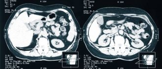
MRI of the abdomen with contrast
MRI of the abdominal cavity with contrast increases its information content. It is indicated if there were any suspicions on the ultrasound.
Specialists of the University Breast Center regularly improve their professional level, attend domestic and foreign conferences to keep abreast of all modern trends in oncology and plastic surgery.
Author: Chizh Igor Aleksandrovich head, kmn, oncologist of the highest qualification category, surgeon of the highest qualification category, plastic surgeon
Start your treatment right now: sign up for a consultation by phone or through the form on the website