- What is breast MRI?
- In what areas is breast MRI used?
- How should you prepare for research?
- What does the diagnostic equipment look like?
- What is the basis for the research?
- How is the research conducted?
- What should you expect during and after the procedure?
- Who reviews the research results and where can they be obtained?
- Benefits and risks of breast MRI
- Limitations of breast MRI
What is breast MRI
An ideal method for examining patients with dense breasts, as well as women using breast implants. To date, the method is considered the most accurate. It helps to check the condition of the gland tissues and lymph nodes. Detects tumors in 96% of cases. This is the highest indicator among all mammographic diagnostic methods. Without contrast enhancement, breast density is checked, cystic formations, hematomas, injuries, and mastopathy are diagnosed. If you suspect cancer, you should do an examination with contrast.
Contraindications to the procedure
Best materials of the month
- Coronaviruses: SARS-CoV-2 (COVID-19)
- Antibiotics for the prevention and treatment of COVID-19: how effective are they?
- The most common "office" diseases
- Does vodka kill coronavirus?
- How to stay alive on our roads?
If a person suffers from claustrophobia or simply has an increased feeling of anxiety, you should inform your doctor in advance. In such cases, the patient may be offered a sedative.
You should also inform your doctor about possible pregnancy. MRI is only possible for pregnant women if the benefits of the procedure outweigh the possible risks. This rule exists despite the fact that during the entire existence of this procedure there has been no recorded case of a negative effect of a tomograph on the body of a pregnant woman or fetus.
It is also important to tell the doctor about all existing chronic diseases and previous operations. Particular attention in this matter is paid to the kidneys. Patients with renal failure should have a blood test done and evaluate the functional state of the kidneys, since the contrast agent is eliminated from the body with their help.
What to take with you
During the examination you must have with you:
- Identification.
- Referral from a mammologist or oncologist.
- Results of previous examinations, biopsy, histology. This will help make magnetic resonance imaging more targeted.
The most convenient way to sign up for an MRI of the mammary glands in St. Petersburg is through the “single center for making appointments for MRI and CT.” Leading diagnostic centers in St. Petersburg cooperate with the service. On the website you can see their addresses, prices for services, information about discounts and promotions. Find out what models of tomographs they use. When registering through the service, patients receive a gift of up to 1000 rubles for diagnostics.
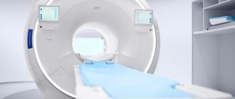
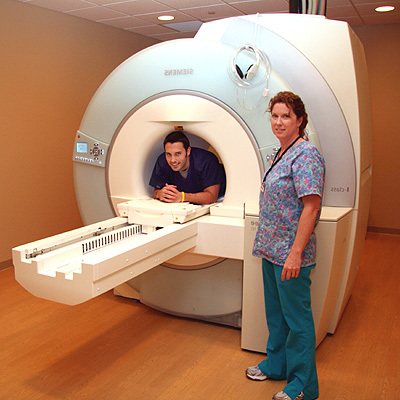
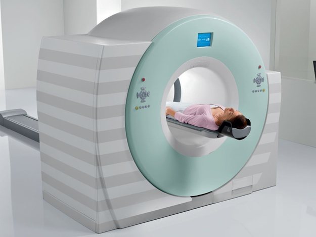
What does the diagnostic equipment look like?
A standard MRI machine is a large cylindrical tube surrounded by a magnet. The patient sits on a movable examination table that slides inside a magnet.
Some CT scanners (called short-tunnel systems) are designed so that the magnet does not completely surround the patient's table. Some devices are open on the sides. Such tomographs are especially suitable for examining obese patients and people who suffer from a fear of closed spaces. Modern open MRI scanners make it possible to obtain very high-quality images for various examinations. However, if an open type machine uses an old magnet, the image quality may be reduced. Some studies cannot be performed on an open tomograph. For more information, consult a specialist.
The computer working system that processes the images is located in the room next to the scanner.
Up
Introduction of contrast
Contrast testing is necessary if cancer is suspected. It allows you to establish not only the presence of tumors, but also their size, structure, nature and location. Shows the degree of spread of the tumor process. Through a pre-installed catheter, the patient is injected with a gadolinium-based substance: Magnevist, Gadovist, Omniscan or Dotarem. Based on the rate of accumulation, excretion and concentration in tissues, the diagnostician assesses the condition of the mammary glands.
At the location of the cancerous tumor, an area with abnormal vascular growths appears. They are characterized by increased permeability. Gadolinium is absorbed and released at a high rate. Elimination time can take up to 2 minutes after entering the body.
Contrast is contraindicated in:
- Liver, heart and kidney failure. Pathologies of the excretory system make it difficult to remove the drug from the body. May lead to intoxication. To determine pathologies, you need to take a biochemical blood test for creatinine content.
- Decompensated diabetes mellitus. A rapid blood or urine glucose test is performed.
- Bronchial asthma in acute condition.
- Allergies to iodine. Before administering the drug, a provocative allergy test is required.
- Pregnancy and breastfeeding. The medicine easily crosses the placental barrier. Lactation is a relative contraindication. After the study, it must be interrupted until gadolinium is completely eliminated from the body.
Read also
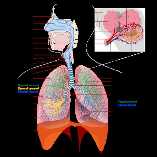
MRI of the lungs and bronchi
The lungs and bronchi are the respiratory organs; the bronchi deliver air to the lungs.
where gas exchange occurs Read more
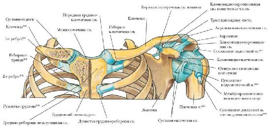
MRI of the sternoclavicular region
The sternoclavicular joint is one of the main joints of the upper limb girdle, attaching the human arm to the chest
Read more

MRI of the heart
The heart is a hollow muscular organ.
ensuring blood circulation in the body Read more
Types of MRI
- MRI of the brain
- MRI of the entire spine
- Abdominal MRI
- MRI of the knee joint
- Whole body MRI
- Other types
It is important to know
MRI for children To obtain the best quality images, it is necessary that the child remains absolutely still during the procedure
More details
What is MRI? MRI is a safe, most informative and painless diagnostic method.
More details
Best MRI Exam There are a few key factors to consider
More details
About the site
- About the site
- Authors of articles
- Add address
- Feedback
- Search
- Privacy Policy
- Terms of use
- Site Map
Progress of the procedure
It is advisable to do the examination from the 4th to the 13th day of the menstrual cycle, unless otherwise instructed by the doctor. The patient undresses to the waist. You must make sure that there are no objects with metal fittings left on your body.
If enhancement with a contrast agent is to be performed, a catheter is inserted into the vein. The drug will begin to flow through it in the 10-15th minute.
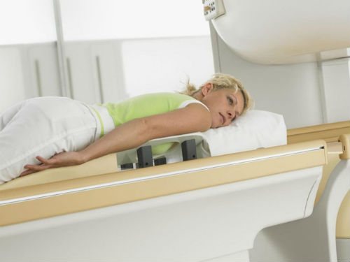
The woman lies down on her stomach on a special couch with holes for the mammary glands. It is secured with wide straps. This will make it easier to remain still. The procedure does not require chest compression. Takes about 30 minutes. The transcript is prepared the next day.
How is the procedure done?
MRI of the mammary glands is performed both in outpatient and inpatient settings. The patient is placed on a special platform, lying on her stomach face down. Inside the platform there are holes for the mammary glands. Thus, MRI images are clear, since there is no displacement or compression of the mammary glands. Electronic sensors are built into the treatment table. It is important to relax and not move. Before you turn on the device, you should make sure that you are in a comfortable position, as tension in the chest or arm muscles can lead to interference in the image.
For screening MRI examination of the mammary glands, as well as checking the integrity of implants, no contrast is required. In other cases, especially for differential diagnosis of various forms of mastopathy and cancer, the introduction of contrast is mandatory.
Alternative
Other methods may be prescribed:
- Mammography. It is carried out using a special device - a mammograph. Refers to radiographic methods that produce small ionizing radiation. Pictures are taken in two projections. Detects cancer, dense nodes, metastases. In women under 40 years of age it is ineffective due to the high density of the gland. Often requires additional techniques. Does not visualize lymph nodes.
- Ultrasound of the mammary glands. Scans the gland in different projections, revealing the structure of the organ and its anatomical features. This method makes it easy to detect cysts and other benign formations. Allows you to monitor their dynamics. Shows the condition of blood vessels. Can be done many times.
- Tomosynthesis. With it, several images are taken in a certain sequence with a fixed interval between slices. This produces from 11 to 90 3D photographs of varying depths. This helps to achieve maximum information about the condition of the breast and nearby organs. Poorly recognizes low-contrast anomalies.
- Laser CT. Newly developed method. Laser radiation in the infrared spectrum helps to identify areas with abnormal accumulation of blood vessels. This may signal an oncological process. Requires expensive equipment and additional training of employees. This means that its cost is very high. Determines only the condition of blood vessels.
What to choose
Patients often ask what is better: breast MRI or mammography? A cheaper method of primary diagnosis of cancer is a breast x-ray. Using mammography, the diagnostician will identify tumors and specify their size and location. But this method cannot clarify the nature of the tumor. To find out whether a malignant or benign breast formation is detected during an examination, you will have to perform a biopsy or do an MRI with contrast. Mammography requires strong compression of the breast during the procedure. This is traumatic for the mammary gland. The patient feels discomfort and pain. A small radiation exposure does not allow this type of diagnosis to be prescribed to pregnant women. Therefore, ultrasound is better suited as a screening method.
Ultrasound diagnostics is the most affordable and harmless option. It is even indicated for women while they are expecting a child. It does not carry radiation exposure, is non-invasive and has no contraindications. It should be carried out in the first phase of the menstrual cycle. If the patient is concerned about pain, changes in the appearance of the gland, or enlarged axillary lymph nodes, the examination is carried out without reference to the cycle.
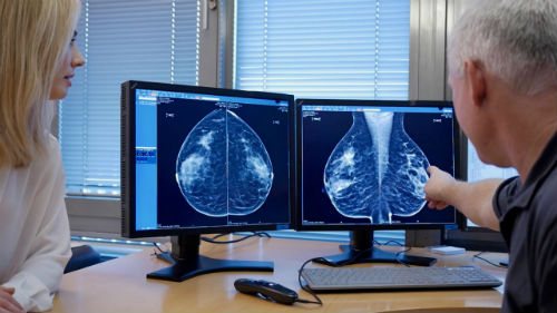
Breast MRI
According to statistics, patients themselves find a cancerous tumor measuring 2 cm in size with their hands; a doctor, during a professional examination, finds a tumor in a patient with a size of 1.5 cm in size. The purpose of MRI is to find a tumor before it can be detected with your hands.
There are cases when breast cancer is not visible on mammography. This doesn't happen often. Some patients can afford to do a more expensive test instead of a mammogram - then they can have an MRI.
The rhythm of preventive MRI studies is as follows:
- 35 - 40 years - first MRI. All subsequent MRIs will be compared with this MRI.
- up to 45 - 50 years - once every 2 years
- 50 - 60 years - annually
- after 60 years - once every 2 years.
If a patient takes hormonal contraceptives (especially triphasic ones) for more than 5 years, or hormone replacement therapy, she has a higher risk of breast cancer. She has been prescribed annual MRI since age 40.
If you do not have implants and have not had plastic surgery on your breasts, if you are not at risk for breast cancer, it is enough to perform a mammogram rather than an MRI. SEE ABOUT THIS .