Clinical Hospital No. 71 – Gynecology – Breast biopsy
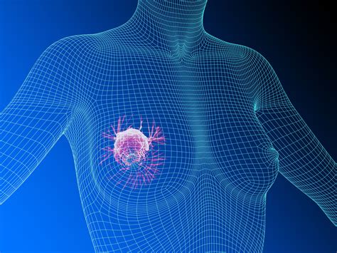
A breast biopsy is a research method in which cells are removed from the breast for examination. A breast biopsy is necessary to determine whether there is cancer in the breast or not. This analysis makes it possible to find dangerous cells.
Based on the results of breast biopsy tests, it is possible to identify:
- the tumor is benign or malignant,
- degree of spread of the disease,
- stage of development
- structure of education.
Carrying out a biopsy in the early stages of the disease makes it possible to detect inflammatory or hormonal processes occurring in the mammary gland. Based on the test results, the doctor selects the correct further treatment regimen. This manipulation is carried out on special equipment using various medical needles.
Indications for breast biopsy
A breast biopsy is prescribed by a mammologist after examining the patient in certain cases. The main indications are:
- palpation of formations in the chest,
- strange changes in the nipple area,
- presence of ulcers on the chest,
- detection of spots on the image in the chest area,
- detection of suspicious areas on the mammary gland on ultrasound diagnostics.
The sources of these problems are established based on the results of a biopsy in order to exclude or confirm the development of a tumor in the mammary gland.
Who is prescribed
A biopsy is a medical procedure in which samples of cells are removed from the breast for study. Often the purpose of such an analysis is to determine the nature of the tumor. A biopsy is performed when other research methods have failed.
It is recommended to prescribe a biopsy procedure only in certain cases. Before it is carried out, a mammogram or ultrasound is already performed for diagnostic purposes. The presence of lumps in the mammary gland, their number, size, and location are determined. Further use of a biopsy may reveal a cyst, fibroadenoma, or oncological tissue degeneration.
Indications for prescribing a biopsy are:
- change in the shape, position of the nipples, formation of crusts on them, retractions, change in color;
- compaction detection;
- discharge of fluid from the nipples, especially with blood and pus;
- change in the shape and size of the breast;
- some diseases - mastitis, cysts, mastopathy, fibroadenoma;
- the appearance of redness on the skin and other skin disorders.
If a multifocal tumor is detected, a breast biopsy will be performed from two lumps.
The psychological side of the procedure is important. The doctor must tell the woman about the purpose of the study and the need for it. Lack of information creates fear and depression, which only worsen the disease. According to medical statistics, in 80% of women the biopsy gives a negative result, that is, oncology is not confirmed.
Contraindications
There are a number of contraindications for which such manipulation is prohibited. These include:
- pregnancy,
- allergic reactions to drugs used for anesthesia,
- the presence in the patient’s body of a device that coordinates the work of the heart,
- state of fever,
- lactation period,
- critical days,
- the size of the formation is up to 5 mm.
When taking medications to thin blood, there is a risk of bleeding. All medications should be stopped 24 hours before the biopsy. Otherwise, this procedure is prohibited. Contraindications also include:
- sharp pain in the spine, neck and shoulders,
- poor blood clotting,
- the tumor is confirmed and is benign,
- infectious diseases.
Possible complications
Severe complications that require emergency medical attention rarely develop after a biopsy. Most often, patients are concerned about moderate pain, inflammatory manifestations (redness of the skin, local increase in temperature), and minor bleeding. If you follow all the doctor’s recommendations, these conditions will go away on their own within a few days. With the development of infectious complications, the symptoms are more pronounced - body temperature rises to 38°C or more, pus may be released from the wound, and severe pain is noted. Some patients experience allergic reactions to anesthetics used during trephine biopsy.
The likelihood of developing undesirable reactions will be lower if the diagnostic procedure is carried out by an experienced doctor who has modern equipment and high-quality consumables at his disposal.
Book a consultation 24 hours a day
+7+7+78
Preparing for a breast biopsy
To successfully perform a breast biopsy, you should follow the doctor’s strict instructions:
- exclude the intake of alcoholic beverages, as well as medications that affect blood clotting processes,
- Pregnant women should not have a biopsy
- If you have allergic reactions to medications, it is advisable to inform your doctor.
You should come to the biopsy with a loved one who will help you get home if you feel unwell.
Also, on the eve of performing a breast biopsy, a woman should undergo additional examinations that will determine the volume of the tumor. This includes ultrasound, mammography, and x-rays.
Benefits and risks of the study
Advantages:
- The procedure is less damaging to tissue than a surgical biopsy, leaves little or no scar, and is completed in less than an hour.
- Ultrasound does not involve the use of ionizing radiation.
- An ultrasound-guided breast biopsy provides a tissue sample that can be used to determine whether the changes are benign or malignant.
- Compared to stereotactic breast biopsy, the ultrasound method allows the examination to be performed faster and without the need for ionizing radiation.
- Ultrasound allows you to track the movement of the biopsy needle in the breast tissue.
- Ultrasound-guided breast biopsy makes it possible to examine tumors in the axillary region or near the wall of the chest cavity, which are not always achievable using stereotactic methods.
- Ultrasound-guided breast biopsy is a less expensive examination method than stereotactic biopsy.
- Recovery after the procedure takes less time, and patients can quickly return to their normal lives.
Risks:
- Since a vacuum biopsy removes a slightly larger tissue sample than using other needles, there is a risk of bleeding and the formation of a hematoma, that is, an accumulation of blood at the biopsy site. The risk, however, is less than 1%.
- Some patients experience significant discomfort, which can be managed with painkillers.
- Any procedure that involves violating the integrity of the skin carries a risk of developing infection. The chance of developing an infection that requires antibiotic therapy is less than 1 in 1000 biopsies.
- Performing a biopsy of a mass located deep in the breast carries some risk of the needle penetrating the chest wall, which allows air to accumulate around the lung, causing it to collapse. This is an extremely rare complication.
Up
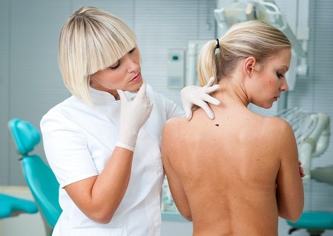
How is a breast biopsy performed?
A breast biopsy is performed in a medical institution by a mammologist. Hospitalization is not required for this procedure.
The essence of the procedure is as follows: the woman is in a lying position and should not move. Anesthesia is performed, after which a needle is inserted and tissue is collected for examination. The procedure does not require stitches. The duration of the procedure is approximately 1 hour. After the biopsy, any physical activity should be avoided.
How is cell research performed?
The sample obtained during collection is cut into thin plates. If the resulting tissue is contaminated with blood or other biomaterials, it is initially washed in saline solution. In order to prevent changes in cell structure under the influence of the external environment, the tissue is stabilized with a 10% formaldehyde solution.
To study atypical cells, tissue is examined under a binocular loupe or microscope. Under magnification, a specialist has the opportunity to see the structure of the tissue, the presence of compactions and necrosis, the presence of psammoma and vitreous bodies. Next comes the study of the neoplasm cell itself - its shape, size. A characteristic feature of tumor cells is their large size and the presence of two or more nuclei. By comparing all the data obtained, the histologist can make a conclusion about the nature of the tumor. Based on this conclusion, the attending physician establishes a final diagnosis and determines further therapy.
Histology results can be of several types:
- incomplete - as a result of the biopsy, too little tissue was obtained or the histologist cannot issue a conclusion due to the ambiguity of the results;
- normal - no abnormalities were found during histological examination of tissues;
- benign – the presence of a neoplasm is confirmed, but its benign nature is established;
- non-cancerous – atypical cells are not a tumor. Most often in this case we are talking about a cyst or mastitis;
- malignant – the diagnosis of cancer is confirmed.
Types of biopsy
There are the following types of breast biopsy that are used in practice.
Fine needle biopsy
A fine needle biopsy of the breast is performed in cases where the breast tumor is easily palpable. The procedure is done in a sitting position. The doctor inserts a thin needle into the gland, which then removes tissue for analysis. During such a manipulation, the patient may feel pain.
Stereotactic biopsy
During a stereotactic breast biopsy, several pieces of tissue are taken from different areas of the tumor. The procedure is performed lying down in the operating room. Using a special apparatus, pictures are taken from different angles, and the result is a three-dimensional image. Based on this image, the location where the needle will be inserted is selected.
Incisional biopsy
Involves taking a small area of the tumor. This material is then examined and a conclusion is made about the malignancy or benignity of the tumor. The procedure is performed under anesthesia, so there is no pain.
Excisional biopsy
A surgical operation is performed, during which a small area or the entire tumor is removed for examination. Everything is done under anesthesia.
The decision about which biopsy to do is made only by the doctor based on the results of tests and other studies.
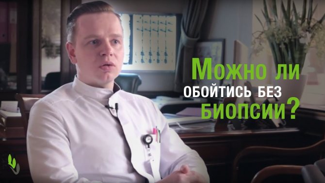
Biopsy methods
The doctor of the Euroonko clinic, V.A. Lisova, talks about the methods of performing a biopsy:
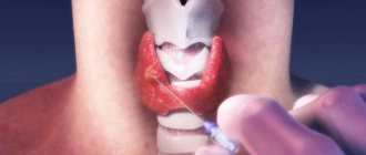
Fine needle aspiration biopsy
This is the simplest and most frequently performed method for assessing a mass in the mammary gland that is not palpable. During this procedure, the patient lies on the table, the doctor, fixing the suspected area of the lesion with one hand, directs a very thin needle connected to a syringe into it with the other, which collects samples of cells or fluid from the tumor. Aspiration biopsy is a quick and easy way to differentiate between a fluid cyst and a tumor.
Needle biopsy
This type of biopsy is chosen if the mass is visible on a mammogram, ultrasound, or felt when examined by a doctor. The procedure uses a slightly larger needle than a fine-needle biopsy. For more precise guidance, the procedure is carried out under the control of mammography, ultrasound or MRI. In a needle biopsy, several tissue samples are taken from different areas for examination.
Stereotactic needle biopsy
This type of biopsy uses X-rays to guide the needle. The patient lies face down on a special padded table with her breasts hanging out of a special hole. The table is raised a few centimeters; the doctor performing the procedure is located at the bottom of the table. The chest is firmly fixed between two plates (cassettes); A mammogram is taken to accurately localize the area of the suspected lesion. An incision no longer than 0.5-0.7 cm is made, and a needle is inserted through it. For a more accurate result, the doctor takes several tissue samples.
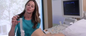
Needle biopsy using ultrasound
This type of biopsy uses ultrasound for imaging. The patient lies on her back, placing her hand behind her head on the same side where the procedure will be performed. This is done so that the soft tissue of the breast stretches and a clearer image is obtained. The doctor localizes the suspected lesion area using ultrasound, makes a small incision and takes several tissue samples.
Needle biopsy using MRI
This type of biopsy uses MRI as an imaging modality, creating multiple images of the breast in many planes, followed by 3-D reconstruction. During the procedure, the patient lies face down on a soft table, and the breasts are placed into special depressions in the table. An incision no longer than 0.5-0.7 cm is made, and a needle is inserted through it. For a more accurate result, the doctor takes several tissue samples.
Surgical biopsy
With a surgical biopsy, it is possible to remove part of the tumor (incisional biopsy, excisional biopsy, punch biopsy) or the entire tumor, followed by histological examination. Surgical biopsy is usually performed in the operating room, under general and local anesthesia. If the formation is not palpable, the surgeon can use a special guide. Before surgery, mammography is used to place the end of a thin guidewire within or through the suspected lesion. During the operation, the surgeon will try to completely remove all tumor masses located along the guidewire. Sometimes, before sending the material for histological examination, the surgeon takes x-rays to check the edges of the tissue. If they contain cancer cells (X-ray positive), then there is probably still cancer in the breast. If the edges of the tissue are X-ray negative, then most likely the entire tumor has been removed.
If all (or almost all) of the diseased tissue is removed during a surgical biopsy, a tiny metal marker may be placed and left in the breast. This is done so that in the future the biopsy area can be easily identified for observation or additional tissue removal.
Biopsy results
The collected biological material is examined in the laboratory over several days. Based on the results of the breast biopsy, the doctor makes a conclusion indicating the size of the formation, location, structure of the affected tissue, and the absence or presence of oncology. If cancer cells are detected in the mammary gland, the type of tumor is indicated.
Based on the results of the study of the biopsy results, there are several options for deciphering these results, which helps the doctor correctly prescribe treatment. Test results may be as follows:
- normal – there are no cancer cells in the breast tissue, their size and shape are within normal limits,
- incomplete – there is not enough biological material to conduct a more detailed study, additional tests are required,
- non-cancerous - minor changes in tissues are detected, however, the development of a tumor is not confirmed. It is possible that a slight inflammatory process is occurring in the mammary gland, cysts are detected, or mastopathy is developing.
If a tumor is detected in the mammary gland, test results may be as follows:
- benign – the presence of a tumor is confirmed, but there are no cancer cells and no tissue proliferation is observed,
- malignant – a cancerous formation has been confirmed, the stage of development of the disease, the location of the tumor, its size and shape have been established.
How to prepare for a biopsy?
No special preparation is required for the procedure. But there are some recommendations that will help you reschedule the study without additional inconvenience:
- Stop taking anticoagulants and aspirin a few days before the procedure, as these drugs can increase bleeding.
- Tell your doctor about possible allergic reactions, pregnancy, or the presence of a pacemaker.
- On the day of the procedure, refrain from using deodorants, cosmetic lotions and other products that are applied to the armpit.
- On the day of the test, wear a bra to keep ice on the puncture site.
After the biopsy, there may be bruising on the chest and slight redness at the puncture site. These effects of the procedure disappear within a few days. If your temperature rises, there is extensive swelling and bleeding, or your breasts change shape, this is a reason to consult a doctor.
Complications after biopsy
Such manipulation, which was performed by experienced doctors, as a rule, does not lead to serious consequences. Dizziness and nausea may be quite natural symptoms after a breast biopsy, but they quickly disappear. In addition, a woman may experience:
- slight swelling, discomfort at the puncture site,
- wound bleeding. To stop bleeding, apply a thick gauze bandage.
More serious complications are extremely rare. After a biopsy, the wound may become infected. At the same time, an inflammatory process begins to develop in the mammary gland, the temperature rises, and there may be discharge from the puncture site.
VACUUM ASPIRATION BIOPSY
If you find a breast lump during a self-exam or your doctor says you have a shadow on a routine mammogram screening, you may need a breast biopsy. Of course, additional tests, such as a repeat mammogram or breast ultrasound, will be performed to fully determine the extent of your condition. However, if any of these tests indicate that the structure or appearance of the breast lump could potentially be cancerous, a biopsy will be ordered to confirm the presence of cancer cells. It can be a scary prospect, but we hope to give you more insight into what you can expect after a breast biopsy.
What is a breast biopsy?
A breast biopsy is performed to obtain a sample of material from the breast to test for the presence of cancer cells. For most women (about four in five), the results are not cancer. There are several different types of biopsies, but the one your doctor recommends may depend on how the lump appears on imaging tests and its size and/or position in the breast.
The tissue sample can be obtained by fine-needle biopsy, trephine biopsy, vacuum aspiration biopsy, or during open surgery. Ultrasound, MRI or X-ray and computer imaging are often used to assist the doctor during sample collection. If the lesion is close to the skin and easily felt, imaging may not be necessary. A surgical biopsy is usually chosen if there is a high enough chance that the tumor is cancer or if the tumor is in an area that cannot be easily reached with a biopsy needle. A small marker may be left inside the breast during a biopsy or surgery so that the doctor can see where the sample was taken if further treatment is needed.
How accurate are biopsy results?
Surgery and needle biopsy have the same high level of accuracy for detecting cancer. At least 98% of cancers are correctly identified by open surgery, and 97-99% of malignancies are identified by core biopsy, depending on the method used.
What are the side effects?
Common side effects include bruising, bleeding, and mild pain. Serious side effects are rare, with fewer than 2 in 100 women having core biopsies experiencing severe bruising, bleeding or infection. Most women do not experience significant pain after the biopsy and usually do not require pain medication. However, if you need pain medications after the biopsy, avoid aspirin as it may make bruising worse.
What happens after the biopsy?
After a core biopsy or vacuum aspiration biopsy (VAB), a tight bandage will be applied and you will be advised to apply pressure to the area for up to 60 minutes to reduce the risk of bleeding and bruising. The dressing should be left on for at least 24 hours. You should wear a firm bra for the next few days and nights after the procedure as this will help the healing process. Don't worry about the procedure affecting your daily activities, but avoid vigorous exercise for the first couple of days. Bruising after a biopsy is normal and will completely disappear within a few days, just like any other bruise.
It's normal to feel anxious about the procedure, as many women say they find any type of biopsy physically or emotionally stressful. However, research has shown that many people prefer VAB to surgical biopsy, and 97% are happy with the way their breasts look after a vacuum biopsy.
When can I expect my results?
After the biopsy, the material taken from you is sent to the pathology laboratory. A pathologist is a doctor who is trained to examine samples from the body under a microscope and look for abnormal or cancerous cells. The pathologist will record their results and send this report to your doctor who performed the biopsy. It is important to remain patient after the biopsy as biopsy results may take 10-15 days to arrive. After receiving your results, your doctor will usually contact you to arrange a follow-up appointment to explain your next steps. If you feel like you need support on this day, take a loved one or relative with you - a friendly face can make a difference.
The biopsy result is part of a triple breast assessment or mammogram. Along with the biopsy, your doctor's clinical examination, as well as ultrasound, mammography, or other imaging, are used to confirm the diagnosis. Sometimes the results of the triple assessment may not be consistent and additional testing may be required to obtain an accurate diagnosis.
Follow-up after the biopsy will depend on the result. Complex cysts require observation, usually for 3-6 months, then a repeat mammogram or biopsy may be prescribed. Other findings, such as atypical ductal or lobular hyperplasia and ductal or lobular carcinoma in situ, are called precancerous lesions: it is not yet cancer, but there is a high risk of developing cancer. Further treatment and follow-up will be required to rule out cancer and eliminate the risk of developing cancer. If the tumor is cancerous, the histological report will provide additional information about how quickly the cancer is growing (grade), the type of cancer, and whether it is sensitive to certain hormones. Your doctor will then explain your treatment options.
Even a result that says there is no cancer and no risk of cancer may make you feel anxious. It is important to talk to healthcare professionals about your feelings and feelings as they will be able to offer support and reassurance. Being aware of your breast cancer risks and, if you are over 50, seeing your doctor regularly for breast exams will help ensure that any future changes are quickly detected and managed.
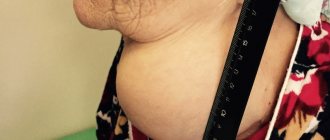
Therapeutic puncture of the breast
If the cystic formation is large and has thickened walls, the patient undergoes a puncture to harden the breast cyst. The procedure is performed by puncturing the wall and removing the contents that are inside the cyst. Then the surgeon introduces a sclerosant based on ethyl alcohol or surgical glue “Sulfacrylate” into the cleaned cavity. As a result, the walls of the cyst stick together and the accumulation of fluid stops. If after puncture the cyst in the mammary gland disappears, this indicates a good result.
The fluid evacuated from the cyst cavity is sent for examination to exclude the malignant nature of the disease. If the cyst reappears, the sclerotherapy procedure is repeated. If, after repeated puncture and sclerosis, fluid continues to accumulate in the cyst cavity, the sector of the mammary gland in which the cystic formation is located is surgically removed.
If the cyst is too small and difficult to palpate, the puncture is performed under ultrasound guidance. In order to reduce the likelihood of undesirable consequences after puncture of a breast cyst, a woman should follow the following recommendations:
- Take blood thinning medications two days before;
- Do not drink alcoholic beverages for 3 days;
- Get a good night's sleep the night before the procedure.
After the puncture, you can place an ice pack in the bra to prevent the formation of hematomas and reduce swelling. For 3–5 days after the puncture, you should avoid vigorous activities, avoid sexual intercourse, and do not wear tight underwear. During the procedure itself, you need to behave calmly and comply with all the specialist’s requirements. You should not make sudden movements, otherwise the doctor may miss and you will have to make a new puncture. There are no consequences in the form of scars after puncture of a breast cyst. If severe pain occurs after the administration of sclerosant, you should inform your doctor. He will prescribe analgesics.