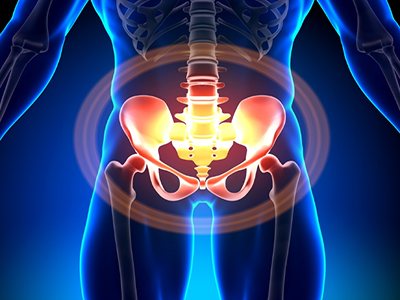
MRI of the sacroiliac joints is a modern non-invasive diagnostic technique aimed at its visualization by using radio waves under conditions of high-intensity magnetic radiation. The procedure is highly informative and harmless, surpassing in these indicators not only ordinary x-rays, but also.
Its competent implementation makes it possible to identify all pathologies and anomalies of the SIJ, as well as determine the condition of the cartilaginous, ligamentous and muscular apparatus of this area. The procedure is effective in identifying diseases of an inflammatory, traumatic and degenerative nature. It allows you to identify neoplasms of benign and malignant etiology and differentiate them.
Depending on the purpose of the study, it can be a survey, or it can be carried out using a contrast agent, which allows you to obtain high-definition images. The price of MRI of the sacroiliac joints depends on the need to use a contrast agent, its type and volume.
MRI of the sacroiliac joints - RUB 5,000.
15-30 minutes
(duration of procedure)
Advantages of MRI of the SIJ at the CELT clinic
The multidisciplinary clinic CELT provides a wide range of diagnostic services. If you need to undergo an MRI of the sacroiliac joints in Moscow, contact our specialists. Their work experience is from 15 years, and they themselves master the technology of carrying out the procedure to perfection.
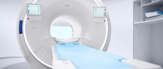
The procedure is carried out on a Philips Achieva high-field magnetic resonance tomograph, which can be called, without exaggeration, one of the best developments of this company. It provides high resolution at maximum scanning speed and maximum comfort for the patient who does not experience discomfort during the scanning process.
The use of MultiTransmit technology allows our specialists to configure RF energy sources taking into account the individual anatomical characteristics of each patient. Using multiple sources increases scanning speed. The ExamCards software module contains all the sequences necessary for highly accurate diagnostics.
The above allows our specialists to achieve high quality diagnostics of a tight joint and identify pathological changes at any stage of development.
You can find out the cost of MRI in the corresponding section of our website by reading the price list or calling us: +7 (495) 788-33-88! Please note: we regularly update our price list, but in order to avoid misunderstandings, we still recommend checking the price with our operators.
How to conduct the study
MRI diagnostics of the sacroiliac joint is usually performed on patients in the supine position. However, given the availability of an open-type MRI machine in our center, the examination can also be completed in the “side” or “half-sitting” position. No special preparation is required. Women are recommended to undergo diagnostics on days without menstruation.
The sequence of actions for a patient during an MRI examination is as follows:
- Immediately before the procedure, all metal objects must be removed.
- Next, you need to take the required position on the table.
- Directly while the tomograph is operating, you must remain in a motionless position for about 10-20 minutes and follow the doctor’s instructions.
- Analyze your well-being after the study - there should be no new complaints.
- Leave the diagnostic room and receive the test results.
If the doctor is dissatisfied with the results of native MRI, during the scanning process the tomograph can be temporarily stopped, a contrast agent can be injected intravenously and the diagnostic process can continue.
MRI of the sacroiliac joints with contrast
The contrast method improves the quality of an MRI image. This is achieved due to the difference in the distribution of the contrast agent between individual tissue types, which increases the image resolution. Thus, the boundaries between pathological and healthy structures are more clearly visible.
Some soft tissue tumors are difficult to distinguish from lymph nodes during a conventional MRI examination, which requires preliminary contrast. In addition, this procedure may be required in case of severe swelling of the soft tissues of the pelvis, when the boundaries of the inflammatory focus are poorly defined, which also requires contrast.
The contrast agent is administered intravenously and is gradually eliminated by the kidneys. Contrast is contraindicated if the patient is allergic to the contrast agent, as well as with end-stage renal failure.
Indications and contraindications for MRI of the sacroiliac joints
| Indications | Contraindications |
|
|
Key benefits of MRI
When examining this area, CT and X-rays are considered insufficiently informative, so an MRI is prescribed. The procedure allows you to better determine changes in the structure of the joint, ligaments, tissues and muscles. The study determines whether the tumor has grown in the bones of the lower spine and pelvis. The procedure is completely safe and can be repeated several times. The patient’s body is exposed to a magnetic field, which is safe and does not cause negative consequences. During the study, the difference in tissue density and changes in their physical and chemical state are determined. As a result, it is possible to determine the occurrence of pathology in the early stages. The use of a contrast agent greatly facilitates diagnosis by improving visualization.
Preparing for MRI of the SIJ
The review procedure does not require the patient to prepare independently. All he needs to do is come to the clinic in a timely manner and provide the diagnostician with a referral from the attending physician, which will indicate a preliminary diagnosis and tasks that need to be solved during the diagnostic process. This approach allows you to improve the quality of the procedure and obtain all the information necessary to make a diagnosis.
If the procedure is performed with the use of a contrast agent, the patient must undergo an allergic reaction and kidney function test in order to exclude contraindications. The study is carried out on an empty stomach and requires refusing to eat no later than six hours before the start.
Diagnosis of the sacroiliac joint: children and patients who cannot remain still for the time required for scanning are sedated with anesthetic drugs. During the procedure, the patient should not have metal objects on them; they must be removed.
Indications for diagnostics
- Suspicion of rheumatism if x-rays are not possible.
- Clarification of information obtained from x-ray examination.
- The presence of symptoms of pathology of the sacroiliac joints and the absence of confirmation of the disease based on the results of radiography and CT.
- Bruise of the lower back, spinal injuries.
If there is suspicion of the formation of ankylosing spondylitis and sacroiliitis
MRI of the sacroiliac joints
is an effective tool for diagnosing sacroiliitis in the initial stages of development. The use of a tomograph can significantly facilitate diagnosis at pre-radiological stages. MRI visualizes swelling of the subchondral bone and changes in the iliosacral joints. Using this method, you can check for exacerbation and remission.
When a patient is diagnosed with osteochondrosis
An informative and accurate MR diagnostic method is used for examination if osteochondrosis is suspected. Using magnetic resonance imaging, the picture of the state of a certain area of study is clarified. The method allows you to effectively diagnose arthritis in the early stages, determine the presence of edema and pathology in the structure of the joints.
When inflammation occurs in the joints of the lower extremities
The study is effective in diagnosing diseases characterized by inflammatory processes in the joints of the lower extremities, especially the ankle. The procedure is prescribed when movement is difficult, to determine the feasibility of the operation.
Methodology for performing MRI of the SIJ
The duration of the procedure is from 15 to 45 minutes; on average it takes about half an hour. It is carried out in the office where the tomograph is located. The patient is asked to lie down on his table, which smoothly moves into a scanning frame that looks like a large ring or tube.
Clicking or cracking noises are heard during scanning. This is normal and should not cause concern to the patient. He needs to remain still during the process, but if necessary, he can contact medical staff through the public address system. This may be necessary in case of a panic attack or intense discomfort.
The procedure itself is painless and does not cause somatic sensations in the patient. Occasionally, some of them note warmth in the pelvic area, which disappears after the end of the study. When using a contrast agent, mild dizziness, nausea, or general weakness may occur.
The diagnostic results are provided to the patient an hour after its completion. The kit includes a written report from the diagnostician, a description of the images taken, and the images themselves: both printed and on digital media.
Video about the service
Magnetic resonance imaging of the sacroiliac joints is a highly effective diagnostic method that allows you to most accurately identify the clinical picture. This study is the only way to detect rheumatoid arthritis and ankylosing spondylitis in the early stages. Compared to X-rays, this diagnosis allows you to obtain a high-definition three-dimensional image, which allows specialists to reliably make a diagnosis. In addition, MRI of the sacroiliac joints is carried out without radiation, which allows diagnostics to be carried out any required number of times. Prices for this diagnostic test in Moscow vary and depend on the equipment used, as well as the loyalty of the pricing policy of each medical center.
Indications
Symptoms indicating the need for MRI diagnostics of the sacroiliac joints:
- A history of congenital structural anomalies, disseminated encephalomyelitis, sclerosis, infectious lesions of the pelvic organs.
- Decreased flexibility of the lower spine.
- Sudden attacks of lameness that occur after prolonged sitting at a desk or improper distribution of load during physical activity.
- Pain in the lower abdomen and sacral region, spreading to the legs.
The following factors are also indications for MRI:
- Injuries of the pelvis and lumbosacral spine.
- Suspicion of tumor development, penetration of metastases, inflammation of bone and soft structures.
- Analysis of pathology dynamics.
- Monitoring the effectiveness of conservative treatment.
- Determining the feasibility of surgery and preparing for surgery.
- Postoperative control.
How much does an MRI of the sacroiliac joints cost in Moscow?
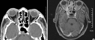
MRI of the sacroiliac joints is an informative method for diagnosing pathological changes in the sacroiliac joints. The essence of the procedure is to sequentially scan a given area of the body using a magnetic field and then obtain a series of images that are converted into three-dimensional three-dimensional images.
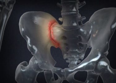
During the examination, the articular surfaces, sacrum, iliac bones and surrounding ligaments and muscles are clearly visualized. Magnetic resonance imaging allows you to quickly and accurately determine degenerative-dystrophic, tumor and inflammatory changes in this area. The ability to visualize inflammatory edema is a unique feature of magnetic resonance imaging. The significance of MRI of the sacroiliac joints in identifying spondyloarthritis lies in diagnosing inflammatory changes in the absence of clear symptoms of sacroiliitis on x-rays.
In some cases, the study is performed using a contrast agent - a special substance based on gadolinium. The contrast is inert, is administered intravenously and is not addictive. MRI of the sacroiliac joints with contrast provides improved visualization of organs and tissues, which allows the doctor to make an accurate diagnosis in the future.
There is no need for special preparation before conducting the study. If a contrast agent is used, the procedure is recommended to be performed on an empty stomach. The doctor will tell you how to properly prepare for an MRI at your consultation.
Symptoms in the presence of which it is recommended to conduct an MRI of the sacroiliac joints: • A history of traumatic injuries to the sacrum or lower back. • Pain in the lower back. • Joint pain that is not associated with injury. • Crunching in the area of the sacroiliac joints. • Sudden attacks of lameness. • Suspicion of rheumatic joint disease. • Confirmation or refutation of the results of other diagnostic research methods.
The diseases that are diagnosed using MRI of the sacrojeal joints include: • Developmental anomalies. • Sacroiliitis. • Bechterew's disease. • Ankylosing spondylitis. • Fractures of the pelvic bones. • Inflammatory processes in joints and surrounding soft tissues. • Benign and malignant tumor neoplasms.
Relative contraindications to MRI of the iliac joints:
· Claustrophobia or fear of closed spaces: during the study, a person is in a device channel about a meter long, motionless. For people suffering from claustrophobia, such conditions may be unacceptable due to restlessness and anxiety. Be sure to warn the clinic staff if you have a fear of confined spaces - sedatives and tranquilizers will help eliminate discomfort.
· Pregnancy: extensive research has not been carried out on the effect of the magnetic field on the developing fetus, due to the ban on experiments with embryos. For three decades, not a single case has been identified that led to harm to the health of the unborn child. In the first trimester of pregnancy, MRI is performed only for health reasons; in the second and third trimesters, when indicated.
· Excess body weight: Coil bore diameter and patient table capacity are limited. Obese people, weighing more than 120-130 kg, are not always physically able to fit into the device. Excess weight can be an obstacle to MRI of the iliac joints.