Why do a CT scan of the heart vessels?
CT scan of the heart and blood vessels is often prescribed when it is necessary to confirm a preliminary diagnosis. Based on the results of the procedure, it will be known which symptoms of the disease are bothering the patient. In this case, you can count on adequate treatment tactics and maximum effect from therapy.
A CT scan is often ordered by a surgeon or cardiologist to evaluate treatment or when planning surgical interventions. Information obtained during a CT scan about the condition of the heart and blood vessels after operations will also be useful.
Preparation
Preparing for a cardiac CT scan
- You must come for the examination on an empty stomach.
- 3 days before the study, avoid taking Cialis, Viagra and Levitra.
- Before the study, if indicated, the attending physician or the Clinic's doctor on duty may prescribe beta blockers - medications that reduce the pulse (heart rate).
IMPORTANT! If there are heart rhythm disturbances (frequent extrasystole or atrial fibrillation), the study is not performed.
General requirements for contrast-enhanced CT:
To perform the test, you must have the result of a blood test with you.
for creatinine, the statute of limitations is no more than 30 days from the date of submission of the biomaterial.
Be sure to tell the radiologist before the examination with contrast:
- Do you have allergies to iodine, medications or food?
- whether you have diabetes, asthma, heart or thyroid disease
How to prepare for cardiac tomography?
In order for a computed tomography scan of the heart vessels to show the most accurate results, it is better to give up alcoholic beverages, as well as products that contain caffeine, about a day before the scan. Before the patient lies down on the tomograph table, you need to make sure that there are no allergic reactions to the contrast agent. To begin the procedure, you must remove all clothing with metal parts, as well as remove jewelry and accessories. In this case, nothing will prevent you from taking clear pictures.
How is the procedure done?
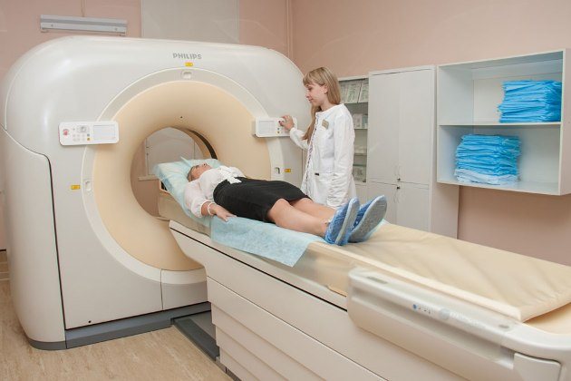
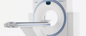
A CT examination of the heart and coronary vessels lasts about 5-15 minutes, depending on whether contrast was used. If a contrast agent was injected, the doctor will take longer to examine the heart and adjacent blood vessels. During the examination, the mobile table of the tomograph slides into the ring of the device, the device produces X-ray radiation and makes a slight noise. The patient will not experience any discomfort, since the method does not require surgical intervention in the body.
CT scan of cardiac vessels with contrast
CT scan of blood vessels and heart with contrast agent is needed in rare cases, usually these include suspected cancer. The contrast accumulates in the affected tissues and makes it possible to assess the presence, size and location of the tumor. The procedure will be useful if it is necessary to find out whether the neoplasms are primary or grow from another organ. If there is no suspicion of a tumor, CT does not require contrast, since the procedure is already very informative.
Indications:
- identified foci of calcification (calcification) of the valvular apparatus of the heart, cardiac muscle (myocardium) or serous membrane of the heart (pericardium) when performing computed tomography of the chest;
- old post-infarction scars, aneurysms of the great vessels or suspected presence of blood clots in the cavities of the heart;
- high calcium index values when assessing coronary calcification (CT “calcium scoring”);
- suspicion of abnormal development of heart vessels or heart valve insufficiency, for example when performing echocardiography;
- chest pain of unknown etiology, atypical pain;
- the appearance of primary or repeated symptoms of angina pectoris;
- deviation from normal indicators of electrocardiography, stress tests, laboratory tests;
- the need to assess the blood supply to the heart muscle (myocardium) after surgical interventions (coronary artery bypass grafting, coronary artery stenting).
[/td]
The high prevalence of cardiovascular diseases and the first place among the causes of mortality in developed countries makes preventive measures and early diagnosis of heart disease . MSCT of the coronary arteries can be performed as a screening during a preventive (dispensary) examination of a person. Atherosclerosis of the coronary arteries is the main cause of the development of coronary heart disease (coronary heart disease) and myocardial infarction. Modern medicine has a wide range of diagnostic capabilities. And with the advent of high-speed multislice computed tomography (MSCT), a completely new, unique opportunity has emerged for non-invasive assessment of the condition of the coronary arteries, which does not require hospitalization of the patient to a hospital for a complex and unsafe surgical procedure (angiography).
MSCT examination takes only a few minutes, does not require additional preparation, and does not cause pain or emotional anxiety. In addition to assessing the condition of the heart vessels ( coronary “tree” ), MSCT of the coronary arteries provides the doctor with additional equally important information, which allows him to assess the condition of the heart as a whole, and develop tactics for further examination and treatment. MSCT allows you to evaluate the morphological structure of an atherosclerotic plaque , study the valve structures of the heart (calcification of the leaflets, anomalies of valve development), identify myocardial lesions (hypertrophy, scars), and assess the condition of the cavities of the heart and pericardium. Valuable information is provided by determining the function of the left ventricular myocardium with identifying areas of impaired contractility.
Types of heart scans
There are several types of tomography of the heart and blood vessels, which are prescribed individually in each case. The choice of research method may depend on what the preliminary diagnosis was, as well as on what exactly needs to be examined during the procedure.
MSCT
This research method involves multi-slice scanning of the organ, which ultimately makes it possible to obtain a three-dimensional model of the heart. Multislice tomography is usually prescribed to identify pathologies of the heart valves, changes in the structure of the pericardium and myocardium, as well as heart chambers. MSCT allows you to detect a lot of deviations from the norm and make the correct diagnosis.
Coronary calcium screening
Screening allows you to assess the presence of excess calcium in atherosclerotic plaques, as well as assess the condition of the blood vessels in general. Using this method, it is possible to identify a number of diseases of the cardiovascular system and prescribe effective treatment.
Vascular angiography

The main difference between this research method is that it requires surgical intervention in the body. The procedure is performed under general anesthesia; to begin, an incision must be made in the upper thigh through which a medical catheter will be inserted. During an angiography, the doctor will carefully move the catheter through the vessels and reach the coronary arteries, after which he will be able to examine their condition. This diagnostic method is prescribed, as a rule, to assess the level of patency of the coronary arteries.
CT coronary angiography: how is it performed?
Contrast enhancement
The procedure itself is performed in a specialized X-ray endovascular surgery room. The study is carried out using local anesthesia. At the discretion of the doctor, the puncture can be made in the femoral, axillary, brachial or radial artery. A catheter is inserted into the vessel and moved to the upper part of the aorta. Through this catheter, a contrast agent is injected, which spreads through the coronary vessels and allows any changes to be clearly seen.
Using a special angiograph apparatus, the results obtained are recorded. Pathological changes in the coronary arteries, tortuosity of the chords, areas of stenosis and a reaction to contraction of the heart muscle can be detected. When taking images, the right and left coronary arteries are visualized. During the examination, the doctor can give the patient additional instructions that are aimed at obtaining images of maximum accuracy.
All manipulations are carried out under the supervision of a doctor; in addition, the presence of a cardiac resuscitator and an anesthesiologist is possible. The patient remains conscious throughout the examination.
On average, diagnosis takes about 40 minutes. If the catheter was inserted through the arm, the patient is usually sent to the ward, and if the femoral artery was used for puncture, then after applying a compression bandage the patient is placed under observation in the intensive care ward. Rest is recommended for the first day after the procedure. Typically, the hospital stay is no more than three days.
At the end of CT coronary angiography, the diagnostician issues a written conclusion on the study, which is required by the attending physician to make a diagnosis and prescribe the correct treatment.
Although coronary angiography using a computed tomograph is considered the most effective and safe diagnostic method, it is not the only one.
Alternative types of research:
- Cardiac MRI – can be used to analyze the condition of the heart and its structures;
- Ultrasound of the heart
- used to diagnose the functional state, allows you to assess the nature of blood flow and the structure of the organ.
Contraindications for examination
Coronary tomography of the heart vessels is not performed in some cases. Contraindications include pregnancy, as well as the patient’s age under 14 years. The procedure is not contraindicated for nursing mothers, but after undergoing a CT scan, the baby will have to be weaned for two days. It is impossible to perform a CT scan with a contrast agent if the patient has diabetes mellitus, an allergy to iodine, or renal failure. For claustrophobia and hyperkinesis, you can undergo a tomography under general anesthesia. If the patient weighs more than 150 kg, you will have to look for another research method.
A procedure with contrast is also impossible if the patient is in a severe state of coma or shock. In this case, the substance can cause significant deterioration in health and become a threat to life.
Indications:
[td]
- high blood cholesterol levels;
- burdened heredity;
- the patient has diabetes;
- high blood pressure (hypertension);
- long-term smoking;
- overweight or obesity;
- insufficient physical activity.
Multislice computed tomography (MSCT) of the coronary arteries (“CT coronary angiography”, “CT cardiac angiography”)
MSCT coronary angiography with contrast enhancement, construction of volumetric (3D) reconstructions and determination of coronary calcification is a non-invasive method for visualizing the arteries of the heart. At the same time, the degree of patency of the artery of the heart (coronary arteries) and their anatomical features are determined with high accuracy, and the overall level of blood supply to the heart muscle (myocardium) is also assessed. Multislice computed tomography (MSCT) of the coronary arteries can serve as an alternative to X-ray angio-coronary angiography.