196
- Introduction.
- Chest X-ray.
- X-ray characteristics of the condition of the heart and its cavities.
- X-ray computed tomography.
- Coronary angiography.
- Aortography.
- Angiocardiography.
- Echocardiography.
- Magnetic resonance imaging.
- Radionuclide studies of the heart.
- Radiocardiography.
- Radiation signs of the most common diseases of the heart and large vessels.
Didactic material, slide show:
- X-ray of the chest in frontal and lateral projections.
- Computed tomogram of the chest.
- Coronary angiography in normal conditions and in cases of atherosclerosis of the coronary arteries.
- Angiocardiography.
- Echocardiogram is normal.
- Radiocardiography is normal and with a ventricular septal defect.
- Chest X-ray for mitral valve insufficiency.
- Echocardiogram for mitral valve insufficiency.
- Chest X-ray for mitral stenosis.
- Echocardiogram for mitral stenosis.
- Abdominal aortic aneurysm (x-ray).
Introduction.
General characteristics of diseases of the cardiovascular system. Cardiovascular diseases remain the leading cause of death in all industrialized countries, including our country. The development of modern technologies for the treatment of cardiovascular pathology is closely related to radiation diagnostics. Every patient with cardiovascular diseases needs radiological diagnostic studies.
Radiation methods for studying the heart:
- Primary methods of radiographic examination of the heart:
radiography in standard projections;
- X-ray television scanning;
- echocardiography (echo-CG);
- Dopplerocardiography (Doppler echo-CG).
- non-invasive:
echo-CG with intravenous contrast;
- angiocardiography;
Cardiology
Cardiology
— a broad integral specialty of medicine associated with the study of the human heart and vascular system: its structure and function, as well as diseases, methods of their diagnosis and treatment. Cardiology also studies the problems of rehabilitation of patients with cardiovascular pathology. The history of cardiology is hardly shorter than the history of medicine - it goes back thousands of years.
Today, the share of cardiac pathology in developed countries is 40-60%. An increase in incidence is increasingly observed in young people. Cardiovascular diseases remain a key global health problem.
The continuous development of cardiology has led to its fragmentation into narrow specializations. Modern cardiology is a broad integral science that combines scientific and practical achievements of a whole group of specialists.
In 2021, a Cardiology Center was opened at the Federal Scientific and Clinical Center of the Federal Medical and Biological Agency of Russia. The purpose of its creation is to unite the efforts of various specialists for the most accurate diagnosis and selection of the most effective treatment tactics for patients with cardiovascular diseases.
The Cardiology Center of the Federal Scientific and Clinical Center of the Federal Medical and Biological Agency of Russia includes:
- 2 cardiology departments with cardiac rehabilitation beds;
- 2 departments of X-ray endovascular diagnostics and treatment;
- department for the treatment of cardiac arrhythmias;
- direction of cardio-oncology.
Key areas of work of the Cardiology Center:
- prevention, diagnosis and treatment of diseases of the cardiovascular system;
- surgical and x-ray endovascular methods for treating diseases of the heart and blood vessels;
- drug and surgical treatment of cardiac rhythm and conduction disorders;
- cardiac rehabilitation;
- cardio-oncology;
- management of patients before and after cardiac surgery;
- control of comorbid conditions (several chronic diseases related to each other).
The Cardiology Center is the base for professional retraining and improvement of the Department of Cardiology of the Institute for Advanced Studies of the Federal Medical and Biological Agency of Russia.
To receive cardiac treatment at the Center's inpatient facility, you must consult a cardiologist. You can sign up for a consultation through the form on the website or by calling the contact center number. If you have current results of instrumental studies (ECG, Echo-CG, MRI, CT, etc.), take them with you to your appointment.
Radiography
Chest X-ray in standard projections: direct, lateral, left and right anterior oblique projections and currently remains one of the most common diagnostic studies. Possibilities:
- Assessment of pulmonary hemodynamics using pulmonary
pattern and roots of the lungs, delineation of venous stagnation in the small circle
blood circulation (which is not available to echo-CG).
- Assessment of the size and configuration of the heart.
- Detection of calcifications of heart valves, pericardium, coronary arteries.
- Exclusion of lesions of other organs (lungs, pleura, esophagus and gastroesophageal junction, chest skeleton), which can be mistaken for heart disease due to the similarity of clinical symptoms.
- Fluoroscopic examination demonstrates a number of functional symptoms.
Complex radiodiagnosis of heart pathology (x-ray + ultrasound) allows in most cases to do without radiography in oblique projections.
Methods of radiation examination of the cardiovascular system
X-ray examination of the heart and large vessels includes:
1) non-invasive methods (fluoroscopy and radiography, ultrasound, CT, MRI) and 2) invasive methods (angiocardiography, ventriculography, coronary angiography, aortography, etc.).
Radionuclide methods make it possible to judge hemodynamics. Consequently, today radiology diagnostics in cardiology is experiencing its maturity.
X-ray examination of the heart and great vessels.
Method value. X-ray examination is part of the general clinical examination of the patient. The goal is to establish the diagnosis and nature of hemodynamic disorders (the choice of treatment method depends on this - conservative, surgical). In connection with the use of URI in combination with cardiac catheterization and angiography, broad prospects have opened up in the study of circulatory disorders.
Research methods
1) Fluoroscopy is the technique with which the study begins. It allows you to get an idea of the morphology and give a functional description of the shadow of the heart as a whole and its individual cavities, as well as large vessels.
2) Radiography objectifies the morphological data obtained during fluoroscopy. Its standard projections:
a) front straight
b) right anterior oblique (45°)
c) left anterior oblique (45°)
d) left side
Signs of oblique projections:
1) Right oblique - triangular shape of the heart, gas bubble of the stomach in front, along the posterior contour on top is the ascending aorta, the left atrium, below - the right atrium; along the anterior contour, the aorta is determined from above, then there is the cone of the pulmonary artery and, below, the arch of the left ventricle.
2) Left oblique - oval in shape, the gastric bladder is behind, between the spine and the heart, the bifurcation of the trachea is clearly visible and all parts of the thoracic aorta are identified. All chambers of the heart open onto the circuit - the atrium is on top, the ventricles are below.
3) Examination of the heart with a contrasted esophagus (the esophagus is normally located vertically and is adjacent to the arch of the left atrium for a considerable length, which allows one to determine its condition). With enlargement of the left atrium, there is a displacement of the esophagus along an arc of large or small radius.
4) Tomography - clarifies the morphological features of the heart and large vessels.
5) X-ray kymography, electrokymography - methods of functional study of myocardial contractility.
6) X-ray cinematography - filming the work of the heart.
7) Catheterization of the cavities of the heart (determining blood oxygen saturation, measuring pressure, determining the minute and stroke volume of the heart).
 Angiocardiography more accurately determines anatomical and hemodynamic disorders in heart defects (especially congenital ones).
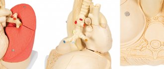
Radiation anatomy of the heart
The heart is an organ of complex shape. On radiographs, fluoroscopy and computed tomograms, only a planar two-dimensional image of it is obtained. In order to get an idea of the heart as a volumetric formation, during fluoroscopy they resort to constant rotation of the patient behind the screen, and during CT, 8-10 slices or more are performed. Their combination makes it possible to reconstruct a three-dimensional image of an object. It is appropriate to note here two newly emerging circumstances that have changed the traditional approach to cardiac x-ray examination.
Firstly, with the development of the ultrasound method, which has excellent capabilities for analyzing the function of the heart, the need for fluoroscopy as a method for studying the activity of the heart has practically disappeared. Secondly, ultra-high-speed computer X-ray and magnetic resonance imaging scanners have now been created, allowing for three-dimensional reconstruction of the heart. Some new models of ultrasound scanners and emission tomography devices have similar, but less “advanced” capabilities. As a result of this, the doctor has a real, and not imaginary, as with fluoroscopy, opportunity to judge the heart as a three-dimensional object of study.
The main projection of cardiac radiography is one - straight anterior, in which the subject is adjacent to the cassette with his chest. In order to avoid projection enlargement of the heart, its imaging is performed at a large distance with a tube-cassette (teleradiography). At the same time, to increase image sharpness, the radiography time is extremely reduced - to a few milliseconds. However, in order to gain an understanding of the x-ray anatomy of the heart and great vessels, a multi-projection analysis of the images of these organs is necessary.
On an X-ray in direct projection, the heart gives a uniform, intense shadow located in the middle, but somewhat asymmetrically: approximately 1/3 of the heart is projected to the right of the midline of the body, and Vi - to the left of this line. The contour of the shadow of the heart sometimes protrudes 2-3 cm to the right from the right contour of the spine, the contour of the apex of the heart on the left does not reach the midclavicular line. In general, the shadow of the heart resembles an obliquely located oval. In persons with a hypersthenic constitution it occupies a more horizontal position, and in asthenics it occupies a more vertical position. Cranially, the image of the heart passes into the shadow of the mediastinum, which at this level is represented mainly by large vessels - the aorta, superior vena cava and pulmonary artery. Between the contours of the vascular bundle and the cardiac oval, so-called cardiovascular angles are formed - notches that create the waist of the heart. Below, the image of the heart merges with the shadow of the abdominal organs. The angles between the contours of the heart and the diaphragm are called cardiodiaphragmatic.
The contours of the heart shadow, normally smooth and clear, have the shape of arcs. Each arc is a representation of the surface of one or another part of the heart facing the contour.
All arches of the heart and blood vessels are distinguished by harmonious roundness. Straightening of the arc or any part of it indicates pathological changes in the wall of the heart or adjacent tissues.
The shape and position of the heart varies in humans. They are determined by the constitutional characteristics of the patient, his position during the study, and the breathing phase. There was a period when they were very keen on measuring the heart on x-rays. Currently, they are usually limited to determining the cardiopulmonary coefficient - the ratio of the diameter of the heart to the diameter of the chest, which normally in adults ranges from 0.4 to 0.5 (more for hypersthenics, less for asthenics). The main method for determining heart parameters is ultrasound. With its help, not only the dimensions of the heart chambers and blood vessels are accurately measured, but also the thickness of their walls. It is also possible to measure the chambers of the heart, and in different phases of the cardiac cycle, using computed tomography synchronized with electrocardiography, digital ventriculography or scintigraphy.
In healthy people, the shadow of the heart on an x-ray is uniform. In pathology, lime deposits may be found in the valves and fibrous rings of the valve openings, the walls of the coronary vessels and aorta, and the pericardium. In recent years, many patients have appeared with implanted valves and cardiac pacemakers. Let us note that all these dense inclusions, both natural and artificial, are clearly revealed by sonography and computed tomography.
Computed tomography is performed with the patient in a horizontal position. The main scan slice is selected so that its plane passes through the center of the mitral valve and the apex of the heart. The tomogram of this layer shows both atria, both ventricles, interatrial and interventricular septa. On the same section, the coronary groove, the place of attachment of the papillary muscle and the descending aorta are differentiated. Subsequent sections are isolated in both the cranial and caudal directions. Turning on the tomograph is synchronized with the ECG recording. In order to obtain a clear image of the cavities of the heart, tomograms are performed after rapid automatic administration of a contrast agent. From the resulting tomograms, two images are selected, taken in the final phases of heart contraction - systolic and diastolic. By comparing them on the display screen, the regional contractile function of the myocardium can be calculated.
MRI has opened up new prospects in the study of heart morphology, especially when performed on the latest models of ultra-high-speed machines. In this case, it is possible to observe heart contractions in real time, take pictures at specified phases of the cardiac cycle and, of course, obtain parameters of cardiac function.
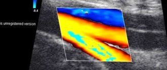
Ultrasound scanning in different planes and at different positions of the sensor allows you to obtain on the display an image of the structures of the heart: ventricles and atria, valves, papillary muscles, chords; in addition, it is possible to identify additional pathological intracardiac formations. As already noted, an important advantage of sonography is the ability to evaluate all parameters of cardiac structures.
Doppler echocardiography allows you to record the direction and speed of blood movement in the cavities of the heart, and identify areas of turbulent turbulence at the site of obstacles to normal blood flow.
Invasive techniques for studying the heart and blood vessels involve artificial contrasting of their cavities. These techniques are used both to study the morphology of the heart and to study central hemodynamics. During angiocardiography, 20-40 ml of X-ray contrast agent is injected using an automatic syringe through a vascular catheter into one of the vena cava or into the right atrium. Already during the administration of the contrast agent, video recording on film or magnetic media begins. Over the entire duration of the study, which lasts 5–7 s, the contrast agent sequentially fills the right chambers of the heart, the pulmonary artery system and pulmonary veins, the left chambers of the heart and the aorta. However, due to the dilution of the contrast agent in the lungs, the image of the left parts of the heart and aorta is unclear, so angiocardiography is used mainly to study the right parts of the heart and pulmonary circulation. With its help, it is possible to identify a pathological communication (shunt) between the chambers of the heart, a vascular anomaly, an acquired or congenital obstacle to the blood flow.
For a detailed analysis of the state of the ventricles of the heart, a contrast agent is injected directly into them. The examination of the left ventricle of the heart (left ventriculography) is performed in the right oblique anterior projection at an angle of 30″. Contrast agent in an amount of 40 ml is infused automatically at a rate of 20 ml/s. During the administration of the contrast agent, a series of film frames is started. Filming is continued some time after the end of the administration of the contrast agent, until it is completely washed out of the ventricular cavity. From a series of frames, two are selected, taken in the end-systolic and end-diastolic phases of heart contraction. By comparing these frames, they determine not only the morphology of the ventricle, but also the contractility of the heart muscle. This method can identify both diffuse dysfunctions of the heart muscle, for example, in cardiosclerosis or myocardiopathy, and local zones of asynergy, which are observed during myocardial infarction.
To study the coronary arteries, a contrast agent is injected directly into the left and right coronary arteries (selective coronary angiography). On photographs taken in various projections, the position of the arteries and their main branches, the shape, contours and lumen of each arterial branch, and the presence of anastomoses between the systems of the left and right coronary arteries are studied. It should be noted that in the vast majority of cases, coronary angiography is performed not so much to diagnose myocardial infarction, but as the first, diagnostic stage of an interventional procedure - coronary angioplasty .
Recently, digital subtraction angiography (DSA)
). DSA based on computer technology allows you to obtain an isolated image of the vascular bed without shadows of bones and surrounding soft tissues. With appropriate financial resources, DSA will eventually completely replace conventional analogue angiography.