Ultrasound screening of the second trimester
At the second planned ultrasound, the specialist evaluates the progress of pregnancy and determines whether fetal development corresponds to normal parameters. The fact is that it is in the second trimester that signs of chromosomal abnormalities (severe developmental abnormalities) may first appear, and the doctor must be very careful not to miss them.
In the second trimester, at 20 weeks, the sex of the baby is usually determined. But sometimes expectant parents have to be patient to wait until the baby in the womb turns as it should.
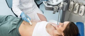
Ultrasound of the pelvic organs
Ultrasound of the pelvic organs in women is performed:
- Transabdominal access (through the anterior abdominal wall). This study is carried out with a full bladder. To fill the bladder, you need to drink at least 500-600 ml of non-carbonated liquid 2 hours before the test. Do not empty your bladder before the examination!
- Transvaginal. No special preparation is required for the study. The study is carried out with an empty bladder.
As an alternative, transrectal (through the rectum) ultrasound is also possible. There is no need to fill your bladder before a transrectal ultrasound, and you should have a bowel movement on the morning of the procedure (either on your own or with a cleansing enema).
Indications for additional ultrasound diagnostics:
- Mother's age is over 35 years;
- Multiple pregnancy;
- The expectant mother has infections or chronic diseases;
- Pathology of the structure of the uterus (or pelvic organs);
- Suspicion of abnormalities in fetal development;
- Threat of miscarriage, signs of frozen pregnancy, fetal hypoxia;
- Abnormalities of placental development;
- Oligohydramnios or polyhydramnios;
- Signs of changes in a child's activity.
Sometimes the doctor prescribes special ultrasound examinations, these are:
- Cervicometry (ultrasound measurement of cervical length). Prescribed from the 16th week if there is a suspicion of a threat of early termination of pregnancy.
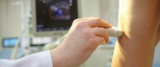
- Doppler. Serves to determine the diameter, length and structure of blood vessels, and calculate the speed of blood flow in the vessels of the fetus and mother's pelvis.
- 3D and 4D ultrasound. Allows you to create a three-dimensional color image of a child and see the baby in motion. This is very important for diagnosing certain fetal conditions. But today, 3D and 4D ultrasounds are increasingly performed simply at the request of parents. And it’s not surprising: the disc with the first video in the baby’s life is a real treasure!
High-precision ultrasound diagnostics of expert level
Highly qualified doctors at the MediArt clinic on the street. Samuila Marshak, 20 conduct high-precision ultrasound diagnostics using a new expert-class ultrasound machine.
Expert level ultrasound
- This is the latest method of instrumental research of the body. It very often becomes decisive for determining the disease and selecting the correct treatment.
What is an expert class ultrasound machine?
This is an ultrasound examination using premium equipment, i.e. high-resolution equipment with many additional functions that improve image quality and, therefore, improve the quality of the examination.
Main criterion
- this is the resolution of modern ultrasound equipment to distinguish the smallest details of structures. This technology is implemented using high-density sensors and high-precision mathematics that are provided in these devices.
Conducting an expert ultrasound allows you to:
- recognize diseases at the earliest stages;
- identify the most accurate diagnosis in a person with a rather vague clinical picture;
- prescribe the correct treatment in a timely manner;
- control the treatment process.
High quality visualization and advanced image processing technologies allow it to be successfully used in a variety of fields.
These are obstetrics, gynecology, abdominal studies and mammology, urology and echocardiography, superficial organs and vascular studies, musculoskeletal studies, as well as transcranial Doppler ultrasound in adults and intraoperative studies.
New methods and technologies used in expert-class devices:
- significantly improves image resolution and realistic reproduction of anatomical features.
- A new imaging technique that dynamically applies transparency to imaged structures to more fully reproduce anatomy and better display internal areas close to solid surfaces.
- New monocrystalline elements, linear and phase scanning sensors provide wider bandwidth, which provides deeper scanning and higher resolution, which is the optimal solution for scanning in specialties such as gynecology, abdominal surgery, cardiology, etc.
- The sensors use a new type of complex piezoelectric material, which significantly improves the acoustic spectrum and reduces acoustic impedance. The sensors provide excellent performance and exceptional image resolution and uniformity when scanning the thyroid, chest, vessels, etc.
- The Smart Face system provides rapid, intelligent optimization of the fetal face image by simply pressing a button. The system can immediately remove interfering parts such as the umbilical cord, placenta, uterus and limbs in volumetric data, and create an optimal image of the fetal face with minimal effort.
- program for automatic measurement of fetal parameters: measurement and calculation of biparental head size (BPD), fronto-occipital head size (OFD), head circumference (HC), abdominal circumference (AC) and thigh length (FL) allows you to reduce the time of the study and get more exact parameters.
- Natural touch elastography improves accuracy in the most challenging clinical environments.
- The unique Smart Track technology allows you to reduce scanning time when examining the vascular system, allows for high-quality examination of blood vessels in real time, and also optimizes the image in color and power Doppler modes.
- use of built-in standard scanning protocols for greater efficiency, and also reduces examination time by up to 50%.
Indications for expert ultrasound in gynecology:
- exclude possible developmental pathologies or diseases in the baby based on the results of instrumental diagnostics and tests
- if you already have children with hereditary diseases
- complications during pregnancy
- after successful in vitro fertilization
- twins in the womb
- for those people who want to have reliable audiovisual information about the results of ultrasound
Expert ultrasound during pregnancy
- Ultrasound using an expert-class device makes it possible to identify even subtle pathologies during pregnancy. Every diagnostic step will be accurately recorded electronically. If you have questions or doubts, you can review the resulting record to make an accurate diagnosis.
- Ultrasound using the device is recommended throughout pregnancy. This research method provides an opportunity to see the correct positioning of the developing fetus and to diagnose impaired blood supply to the unborn child.
- Ultrasound during pregnancy in the third trimester helps to determine the approximate weight and size of the child. An ultrasound is performed both on the recommendation of the attending physician and at the request of the pregnant woman. Ultrasound of the pelvic organs does not pose a risk to the health of the mother and her child.
You can undergo an ultrasound using an expert-class device at the MediArt clinic at the address: St. Samuila Marshak, 20
Ultrasound doctors
- Kurgannikov A. S. Deputy chief physician for medical work (ultrasound and PD), ultrasound diagnostics doctor
- Tyo S. A. Ultrasound diagnostics doctor, obstetrician-gynecologist, Ph.D.
- Blokhina L. A. Ultrasound diagnostics doctor
- Kurgannikova (Zenkina) A. V. Ultrasound diagnostics doctor
- Zhukotskaya (Evstigneeva) M. K. Ultrasound diagnostics doctor, Ph.D.
- Brilliantova N. N. Ultrasound diagnostics doctor
- Voevodin F. S. Ultrasound diagnostics doctor
- Gevorkyan E. G. Ultrasound diagnostics doctor, Ph.D.
- Zhuk N.V. Ultrasound diagnostics doctor, obstetrician-gynecologist
- Kuzmina T. Yu. Ultrasound diagnostics doctor
- Lunkina E. G. Ultrasound diagnostics doctor
- Chuvakhina I. A. Ultrasound diagnostics doctor
Useful information about ultrasound
- S.A. About ultrasound and echography
- Echohysterosalpingography. Tubal patency
- AFC: what does a woman's biological clock show?
- Ultrasound of a newborn
- Who should do an ultrasound?
Reviews
Reviews / Add
- — 4 years ago, our daughter was practically dying, everything was in vain, she was losing half a kilo a day. I took her to every imaginable clinic. When in it...
- — I didn’t want to write a review because... when you praise a specialist, then you can’t find any free time to make an appointment with him, but you also can’t express your deep gratitude...
- — The best ultrasound doctor I have ever seen, and I have a lot of experience. She attended all screenings for as many as three pregnancies. She always accurately determined...
- — I thank Sergei Aleksandrovich Tyo for the ultrasound. Professional, confident, calm. There was a controversial diagnosis, but Sergei Alexandrovich dispelled doubts...
Ultrasound of the Eastern Administrative District: Izmailovo, Golyanovo, Preobrazhenskoye, Novogireevo
Ultrasonography
In the process of diagnostics, ultrasound plays the role of an effective solution, and at the same time, an affordable, safe and acceptable method of examination for every person. All these factors, combined with each other, contribute to ultrasound gaining great popularity. It is worth emphasizing that there is no need to use chemicals to carry out this procedure, irradiate or use harmful methods that are unsafe for health. It is ultrasound examination that is most often the most preferable. Research at the Izmailovo Medical Center using this method makes it possible to see a complete picture of the condition of all human organs of any age category, including a newborn baby, and in the presence of any pathologies. Within the walls of this clinic, specialists conduct a comprehensive examination using modern ultrasound machines, which results in high-quality and high-resolution images. Examinations are carried out exclusively with the help of modern ultrasound machines, which tend to have the best performance and technical capabilities. Carrying out the examination is the responsibility of a diagnostician who has the highest qualifications and the required work experience. If an ultrasound is performed at the Izmailovo Medical Center, you can check the entire body as comfortably and quickly as possible and undergo a comprehensive examination. Our visitors get the chance to have a painless check of all internal organs in just one go. That is, just one procedure, and you already know what state the main systems and internal organs of your body are in. The examination does not take much time, and it is almost painless. After receiving the results, a qualified diagnostician will give comprehensive answers to all the patient’s questions.
What does expert ultrasound mean?
Carrying out an expert examination using an ultrasound machine at the Izmailovo Medical Center implies the presence of the latest type of equipment, which has wide capabilities and works using unique technologies. This is what contributes to obtaining high-quality images and high resolution. Expert ultrasound acts as an achievement of modern diagnostic technology, facilitating a complete examination of the entire body in a short time, while at the same time also obtaining a clear picture. The objectivity of the results obtained depends on the quality and functionality of the equipment used for the examination. Accordingly, our medical center focuses its attention on this aspect.
Types of ultrasound
The Izmailovo Clinic provides the opportunity for everyone to undergo any type of examination using ultrasound machines. You can resort to a comprehensive examination, or you can examine a specific organ or area. Determining the required type of examination is directly influenced by clinical indications. In the center you can check the work using ultrasound:
- Hearts
- Spleens
- Abdominal organs
- Pelvic organs
- Mammary glands
- Liver
- Kidney
- Prostate
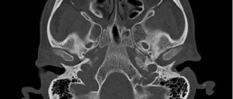
- Sinuses
- Bladder
- Joints
- Vessels
- Conditions of the uterus
Thanks to the use of modern ultrasound scanners, an area or organ is quickly checked for the presence of a certain pathology in it. How safe is it to do an ultrasound? Ultrasound today acts as the safest procedure that cannot cause harm to the body. Therefore, you can be examined in this way many times without fear for your health.
At the EEG Center of the North-Eastern Administrative District, specialists are assigned to each district who will help you prescribe the most optimal examination for you:
At the Ultrasound Center of the Eastern Administrative District, specialists are assigned to each district who will help prescribe the most optimal treatment for you:
| Get an ultrasound of Izmailovo | Sign up |
| Get an ultrasound of Golyanovo | Sign up |
| Get an ultrasound of Preobrazhenskoye | Sign up |
| Get an ultrasound of Novogireevo | Sign up |