Ultrasound examination of the hip joint is a mandatory component of newborn screening. Screening (dispensary examination) is a set of measures aimed at identifying/preventing diseases. It must be carried out at the age of one or two months. The main purpose of ultrasound is to confirm or refute the diagnosis of hip dysplasia. What do you need to know about the disease, how exactly is the study carried out and is ultrasound safe for the baby’s body?
Peculiarities of pediatric diagnostics
The hip joint is a ball-and-socket joint in the human body. It is formed by the lunate surface of the pelvic bone and the articular surface of the head of the femur. The main properties are circular rotation, flexion, extension, abduction, adduction of the hip. The functionality of the musculoskeletal system largely depends on intrauterine development. Initially, the baby is born with “soft” bones in order to safely pass through the birth canal and be born. But already in the first months of life, the bone skeleton becomes stronger, which makes it possible to track norms and pathologies.
Content:
- Peculiarities of pediatric diagnostics
- How does an ultrasound machine work?
- Indications/contraindications for
- What are the advantages of the method
- Preparation and conduct of the study
- Norms and pathologies
Ultrasound of the hip joint is a mandatory procedure as part of newborn screening. It is taken every 1 or 1.5 months to confirm/refute dysplasia. Diagnostics helps to identify changes in the structure, position of the glenoid cavity, the degree and features of the formation of the femoral head, flexibility of the ligaments, etc.
In the treatment of dysplasia, the age of the child and timely diagnosis are fundamentally important. In the period from the first to the sixth month, therapy will consist of massage, soft swaddling or no swaddling, wearing the baby in a sling and other comfortable manipulations. After six months, to get rid of dysplasia, the child will have to wear hard plaster spacers. They limit mobility, and with prolonged wear they trigger the process of muscle atrophy. This disrupts the natural development of the baby and can affect psychological health.
Do not ignore preventive examinations and provide your child with qualified medical care. Timely diagnosis will speed up the treatment process without affecting the baby’s quality of life.
Hip dysplasia

Dysplasia is a congenital defect of the hip joint. The pathology is caused by improper development of the mobile bone joint and leads to dislocation/subluxation of the femoral head.
According to statistics, the disease is diagnosed in 2-3% of newborns worldwide. In 80% of cases, dysplasia occurs in girls. Most often, problems arise with the left hip joint (60%), less often with the right (20%) or with both at the same time (20%).
Dysplasia is diagnosed as a change in the shape/size/structure of the joint. The load is distributed unevenly, which determines the slowdown/acceleration of bone growth. Manifestations, final shape and general condition of the bones are determined individually. In newborn babies, the hip joint is an immature biomechanical structure. His ligaments are too elastic, and the glenoid cavity is thick and vertical. The overall functionality of the body depends on the nature of the development of the mobile bone connection. This is why early diagnosis is so important for overall musculoskeletal health.
There are several stages of dysplasia - preluxation, subluxation, dislocation. With preluxation, the joint cannot be held within the boundaries of the glenoid cavity. Subluxation is characterized by partial displacement of the femoral head. When a dislocation occurs, the head of the femur is completely displaced. Ultrasound examination helps to identify all stages of dysplasia. If diagnosis and treatment are ignored, the child begins to limp, feel severe pain, and may have impaired growth/development of the body, which leads to serious problems in the future.
Treatment
If a pediatric orthopedist confirms the diagnosis of hip dislocation (as well as subluxation or pre-luxation), treatment begins immediately. If therapeutic measures are insufficient, as the child grows, a transition from mild dysplasia to subluxation, and from subluxation to dislocation, is observed. It must be remembered that the treatment of congenital hip dislocation is long-term (usually from one month to one year) and complex. Parents will have to be patient: therapy for hip dysplasia is long-term, continuous, and initially difficult for the child to accept.
In the first month after birth, the baby is swaddled widely . The principle of wide swaddling comes down to the following: a regular flannel diaper is folded into a rectangular spacer 15-17 cm wide and placed between the baby’s legs, which are moved to the sides at 60-80°, bent at the hip and knee joints. The edges of the folded diaper should reach the knees. If you are not swaddling your baby, you can lay the diaper over the diaper and rompers and use ties in the form of rompers to secure it on the baby’s shoulders. The baby quickly gets used to wide swaddling, tolerates it well and, when swaddling, independently holds his legs in the abducted position.
It is also necessary to carry out therapeutic exercises - spreading the hips every time you change the diaper or change the child's clothes. Swimming on your stomach is beneficial.
If wide swaddling and gymnastics are not enough, the orthopedist will prescribe one of the orthopedic aids :
- Pavlik stirrups are the most gentle on the hip joint and the most comfortable aid for the child and parents. Prescribed to children from the third week to 9 months.
- Freyka pillow - plastic pants that support the legs in the “frog” position. Assigned to children from 1 month to 9 months, with benefits changing as the child grows.
- spacer splints (splint with femoral splints, splint for walking, splint with popliteal splints).
Treatment is aimed at fixing the hip joints in a functionally advantageous position - flexion and abduction. The most optimal device from 1 month to 6-8 months is considered to be Pavlik stirrups or an abductor splint with popliteal splints. From 6-8 months, an abduction splint with femoral splints is prescribed, and if the orthopedic doctor allows the child to walk, an abduction splint for walking is prescribed.
How does an ultrasound machine work?
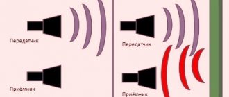
Ultrasound is powerful sound waves. The human hearing organs can perceive frequencies from 16 to 20 kHz, so ultrasonic vibrations (from 20 kHz) are beyond our acoustic perception. However, some groups of animals (whales, dolphins, bats and others) communicate using ultrasound. Each wave is characterized by an oscillation period, frequency and length. They all depend on the elasticity/density of the medium through which this wave propagates. Any environment, including the tissues of the human body, prevents the propagation of sound vibrations. This is called acoustic impedance. The speed and density of sound waves depend on the magnitude of acoustic resistance.
As soon as a sound wave reaches the boundary of two media with different acoustic resistance (for example, soft and hard tissue), one part of it propagates in the new medium and is absorbed by it, and the other is reflected. The intensity of reflection depends on the magnitude of the acoustic impedance. The higher the indicator, the brighter and lighter the signal that the ultrasonic equipment receives. Additionally, the technique records the distance to the separation boundary, the travel time of the wave, the speed of movement, the difference in densities, and so on.
Human skin reflects 99.99% of sound vibrations, making ultrasound examination impossible. That is why the scanned area is lubricated with a special aqueous jelly, which acts as a transition medium.
The reflected sound wave enters an amplifier and special reconstruction systems. They process the information received and transform it into sections of the body part being studied. The picture is painted in black and white, which uses no less than 64 different gradients of the black and white scale. The maximum intensity of sound vibrations is recorded in white, and the minimum - in black.
There are three operating modes of ultrasound - A, B, M. A-mode provides a one-dimensional image, B-mode provides a two-dimensional image of anatomical structures in real time. M-mode is a one-dimensional image with a time coordinate. It is used to diagnose the functionality of the heart.
To make the image as informative and accurate as possible, contrast agents (echo contrast) are used. The contrast agent contains free gas microbubbles (less than 5 microns in diameter). They improve visualization of blood flow or individual organs, increase contrast between tissues and increase diagnostic accuracy.
Indications/contraindications for
Indications for examination of the hip joint in newborns (in addition to screening):
- birth of a baby at less than 37 completed weeks;
- increased tone of the lower extremities;
- heterogeneity of symmetry/depth of skin folds on the baby’s body (particular attention should be paid to the area of the hips and buttocks);
- unequal length of limbs;
- disembryogenesis (disorders of embryonic development that arise due to genetic or teratogenic factors);
- excessive clicking or crunching in joints;
- limited functionality of the hip (for example, it is impossible to fully spread the hips during a massage);
- neurological abnormalities;
- the birth of twins/triplets (one fetus gets unlimited access to the resources of the mother’s body, and several children have to share nutrients among themselves).

There are no contraindications for ultrasound examination. Ultrasound is sound waves that the human ear simply cannot perceive. They do not affect the performance of our body in any way, which means they are absolutely harmless.
Ultrasound of the hip joints in infants with dysplasia

Features of the structure of the hip joints in children
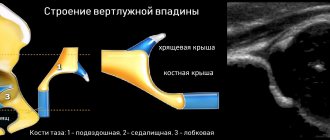
The hip joint consists of the head of the femur and the acetabulum. The acetabulum is formed by the ilium, ischium and pubis. In children, the three bones are connected by a Y-shaped cartilage, the center of which coincides with the center of the acetabulum.
The inside of the acetabulum is lined with cartilage. The articular labrum, a fibrocartilaginous formation, is attached to the acetabulum and transverse ligament.
It increases the depth of the socket by 30%, increasing the contact area of the head with the acetabulum, which promotes joint stability.
The most suitable age for ultrasound of the hip joints is 4-6 weeks. At an earlier age, due to the immaturity of the joint, overdiagnosis of dysplasia is possible.
The femoral head, cartilaginous at birth, provides a window for examining the acetabulum. The head begins to ossify between 2 and 8 months.
What are the advantages of the method
Since 1989, ultrasound has been used in pediatrics. It is noteworthy that the first studies were aimed at studying the hip joint. The problem of dysplasia was encountered everywhere, which means it required an effective solution and constant monitoring. Ultrasound examination still does not lose popularity and remains the safest, most informative and accurate diagnostic method. The production and use of ultrasound equipment is strictly regulated by the World Health Organization, so parents can be confident in their quality and functionality.
The main advantage of ultrasound is safety. Unlike X-rays or computed tomography, after which radiation doses accumulate in the body, ultrasound does not affect a person in any way. It is this type of diagnosis that is suitable for the most vulnerable categories of patients - newborns, pregnant/lactating women and the elderly.
The second advantage is maximum patient comfort. During the examination, the baby does not need to be fastened with soft belts or forcibly held in your arms for the device to record the state of the body. On the contrary, activity within normal limits will help the doctor better study the hip joint and will not darken the mood of the little patient. Additionally, a specialist can track the dynamics of changes, record inflammatory processes or defects in muscles, ligaments, tendons, joint capsule and cartilage.
How the research works
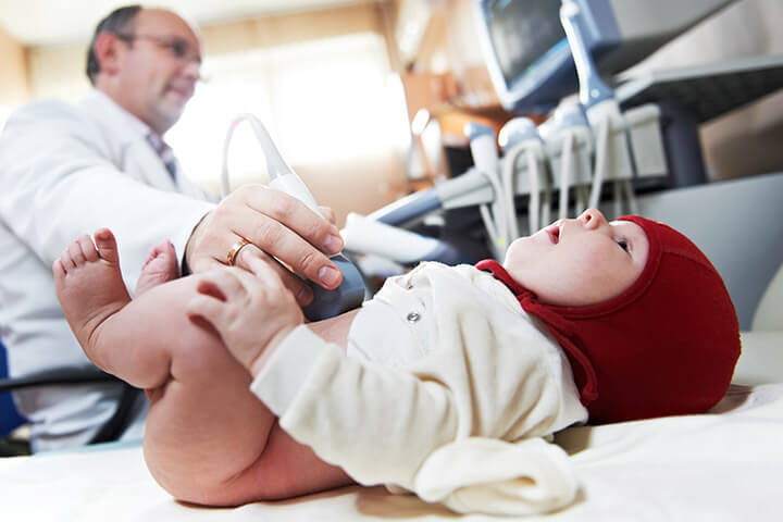
During the examination, the child lies on his side with a slight flexion at the hip joint of 20-30 degrees. After examining one joint, the child is turned over to the other side and the steps are repeated. There is no need to prepare specially for the procedure. The procedure usually lasts no more than 20 minutes on average.
To make a correct diagnosis, the complex of data obtained during the examination (examination by a pediatric orthopedist, analysis of the clinical picture and ultrasound) must be concentrated in the hands of a pediatric orthopedist for the correct interpretation of the studies, making an accurate diagnosis and prescribing effective treatment. If indicated, radiography of the hip joint is recommended.
Preparation and conduct of the study
No specific preparation is required. The main thing is that the baby is well-fed and calm, since excessive activity, crying or fear simply will not allow the doctor to diagnose. Parents should think through the child’s outfit in advance - the lower part of the costume should be easily and quickly removed so as not to waste time.
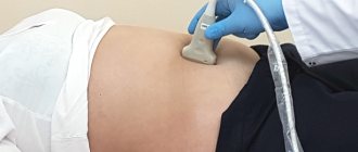
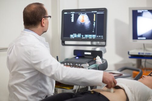
The baby is placed on a couch, previously covered with a diaper, lubricated with a special gel and the ultrasound machine sensor is placed against it. The doctor carefully moves the sensor from side to side, examining the joint. The device simultaneously emits sound waves, records their characteristics and displays an image of the internal cavity on the computer. The doctor and parents can watch what is happening in real time through the computer screen. The ultrasound specialist scans both hip joints with adjacent soft tissue and bone areas. Periodically, the doctor turns the baby onto the left or right side/back/stomach, raises or lowers the limbs, and slightly turns them in order to study the condition and functionality of the bone skeleton in more detail.
Best materials of the month
- Coronaviruses: SARS-CoV-2 (COVID-19)
- Antibiotics for the prevention and treatment of COVID-19: how effective are they?
- The most common "office" diseases
- Does vodka kill coronavirus?
- How to stay alive on our roads?
To diagnose the hip joint, a convex sensor is used. Its frequency varies from 1.8 to 7.5 MHz. The size of the device is minimal, which ensures complete adherence to the patient’s skin. A special feature of the sensor is the size of the final image. Its width is several centimeters larger than the sensor itself. Doctors must take this discrepancy into account in order to navigate the anatomical structures and understand their real size. Typically, convex sensors are used to diagnose deep-lying organs (hip joint, organs of the gastrointestinal tract, reproductive or urinary systems).
The examination lasts only a few minutes. Once all the information has been collected, the doctor completes the examination, wipes off the liquid gel, prints the necessary photographs and talks with the parents about the norms and pathologies of the hip joint.
How does hip dysplasia manifest - symptoms and signs
Parents of an infant are able to themselves suspect the presence of hip dysplasia. Typical signs:
- shortening of the thigh;
- restrictions in hip abduction (in the supine position, the child’s legs, bent at the knee and hip joints, are moved apart, if an angle of less than 160° is formed between them, the likelihood of dysplasia is very high);
- “click” symptom (scientifically called the Marx-Ortolani symptom) - when you slowly open your bent legs, you may hear a click on the affected side. At the same time, the affected leg twitches a little.
- asymmetry of the skin inguinal, gluteal and popliteal folds - asymmetry is most pronounced in children older than 2 months.
Asymmetry of skin folds may be absent with bilateral lesions.