What is the abdominal aorta and where is it located?
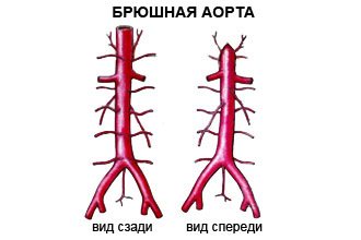
As you know, the largest human artery, the aorta, consists of several sections. Most of them are located within the chest. Only one part (abdominal or abdominal) passes into the abdominal cavity, under the diaphragm. Throughout its entire length, it is located in front of the spine and supplies arterial blood to the entire lower half of the body.
Anatomy of the abdominal aorta
Topographically, this vessel begins at the level of the 12th thoracic vertebra, emerging from the aortic opening of the diaphragm. In the abdominal cavity, the aorta moves anterior to the spinal column, slightly to the left of the midline. Along its entire length, the vessel gives off multiple branches that feed the structures of the abdominal cavity.
The normal dimensions of the abdominal aorta are:
- length – from 13 to 15 cm;
- diameter – 18-20 mm.
The abdominal aorta ends at the level of the 4th or 5th lumbar vertebra, at the point of bifurcation (i.e., bifurcation), where it diverges into the right and left iliac arteries.
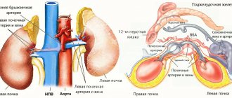
Behind the abdominal aorta is the spine, in front is the root of the mesentery of the small intestine, the pancreas and duodenum. On the right is the inferior vena cava, and on the left are the left adrenal gland and kidney.
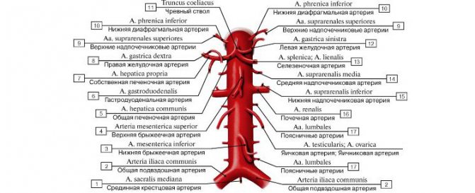
The branches of the abdominal region are divided into parietal (supplying the abdominal wall) and visceral (supplying the internal organs).
The first group includes the following paired arteries:
- lower diaphragmatic;
- lumbar (4 on each side);
- unpaired sacrum.
Visceral branches are paired and unpaired.
Pairs include:
- middle suprarenal;
- renal (renal);
- testicular (in women - ovarian), which supply blood to the genitals.
Unpaired branches:
- the celiac trunk, which gives branches to the liver, stomach, spleen;
- superior and inferior mesenteric, feeding all parts of the intestine.
In the photo you can see the layout of the outgoing branches
:
Structure at the microscopic level
Like the entire aorta, the abdominal section is an elastic type artery, the wall of which consists of three functional membranes:
- Intima is the inner layer that performs a protective, nutritional and regulatory function. The membrane is represented by epithelial cells - endothelial cells, which are most susceptible to pathological influences, including lipid deposition, and this is the cause of atherosclerosis.
- Media is the middle layer that provides mechanical strength and extensibility of the vessel to maintain constant pressure. The shell consists of connective tissue containing elastic and collagen fibers.
- Adventitia is the outer shell that provides a protective function. It is represented by connective tissue cells, but more dense, to create high strength. In addition, it contains nerve fibers and capillaries (the so-called vasa vasorum).
The above layers are not connected very tightly, which is why dissecting aneurysms can form.
Modern approaches to the treatment of abdominal aortic aneurysm
- Abdominal aortic aneurysm is detected annually in approximately 12 to 15 people out of 100,000, and due to the potential for rupture, increases mortality;
- Ultrasound or CT screening is recommended for men over 65 years of age who smoke, and for men over 50 years of age and women over 60 years of age whose parents or siblings have had an aortic aneurysm;
- It is recommended to operate on aneurysms with a diameter greater than 5 cm in women and 5.5 cm in men, or if the aneurysm has increased by 5 mm (or more) in less than 6 months;
- Endovascular surgery for abdominal aortic aneurysm is the treatment of choice in patients over 65 years of age, at high risk due to other diseases, and with previous aortic surgery.
In the hands of experienced surgeons, open aortic aneurysm surgery is safe and long-lasting in most cases, and is well suited for younger patients. During this operation, the aorta is clamped above and below the aneurysm, and the damaged area is replaced with a polyester patch.
Perioperative complications (including cardiac and pulmonary complications, incisional hernias, sexual dysfunction, paraplegia, and death) and recovery time when choosing traditional open surgery may have a poorer outcome in patients who are elderly or at high surgical risk.
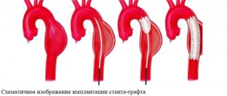
Endovascular treatment of aortic aneurysm using an implantable stent is a safer alternative to open surgery. This procedure produces excellent results in patients with suitable anatomy.
What function and tasks does it perform?
This vessel is very important because it supplies blood enriched with oxygen and nutrients to the entire abdominal cavity and lower limbs. In fact, such an aorta completely ensures the functioning of the digestive and genitourinary systems of the body, therefore pathologies of the vessel can lead to disruptions in the functioning of the corresponding organs.
In addition, this vessel also plays a significant role in maintaining normal blood pressure due to its elastic properties. At the moment of contraction of the heart, a large volume of blood stretches the wall; during relaxation, it returns to its original position. This mechanism prevents too large a gap between systolic and diastolic blood pressure readings.
The blood flow is greatly influenced by the condition of the aortic walls. Normally, a laminar (or linear) flow of blood should be observed. However, if there are any protrusions (or vice versa, pockets, niches), turbulence appears, which causes a turbulent (chaotic) current. It contains a large friction force, which slows down the speed and leads to disruption of hemodynamics and perfusion (blood supply) of tissues.
Treatment and drugs
Here are the general guidelines for treating abdominal aortic aneurysms.
Small aneurysm
If you have a small abdominal aortic aneurysm—about 4 cm in diameter or smaller—and you have no symptoms, your doctor may suggest observation rather than surgery. In general, small aneurysms do not require surgery because the risk of surgery may outweigh the risk of rupture.
If you choose this approach, your doctor will monitor your aneurysm with periodic ultrasounds, usually every 6-12 months; You should report immediately if you experience abdominal tenderness or lower back pain—potential signs of rupture.
Average aneurysm
The average aneurysm measures between 4 and 5.3 cm. It is impossible to say for sure in the case of a medium-sized abdominal aortic aneurysm the risk ratio between rupture and surgery. You will need to discuss the pros and cons and make a decision with your doctor. If you choose surveillance, you will need to have ultrasound scans every 6 to 12 months to monitor your aneurysm.
A large, fast-growing, or “leaky” aneurysm. If you have an aneurysm that is large (greater than 5.6 cm) or rapidly growing (growing more than 0.5 cm in six months), then you will likely need surgery. In addition, a “leaking”, thinned or painful aneurysm requires treatment. There are two types of surgery for abdominal aortic aneurysms.
Open abdominal surgery for an abdominal aortic aneurysm involves removing the damaged section of the aorta and replacing it with a synthetic tube (prosthesis) that replaces the damaged section through an open approach to the abdominal cavity. With this type of intervention, you will likely need a month or more to recover.
Endovascular surgery is a less invasive procedure and can be used to repair an aneurysm in some cases. The doctor uses a synthetic implant, which, with the help of a guide (catheter), is passed through an artery in the thigh into the aorta. The implant—a fabric tube covered with a metal support mesh—is placed at the site of the aneurysm and secured with small hooks. The tissue implant strengthens the weakened part of the aorta and prevents rupture of the aneurysm.
Recovery time for people who undergo endovascular surgery is shorter than for people who have open abdominal surgery. However, more frequent examinations will be required in the future because an endovascularly placed implant may leak. Follow-up ultrasounds should be performed every six months for the first year, and then once a year thereafter. Long-term survival rates are similar for both endovascular surgery and open surgery.
Treatment options for your aneurysm will depend on many factors, including the location of the aneurysm, your age, kidney function, and other conditions that may increase your risk of endovascular or open surgery.
The most common pathological conditions and their complications
Cardiovascular pathologies are among the three leading causes of death. The group of disorders includes diseases of the aorta, including its abdominal section.
The following diseases of the abdominal aorta are distinguished:
- Obliterating atherosclerosis is the most common disease that occurs due to disorders of lipid metabolism. It is characterized by the deposition of protein-fat complexes in the inner lining (intima) of the artery and the proliferation of connective tissue. Because of this, the elasticity of the vessel decreases, plaques form, which narrow the lumen and impede the movement of blood. Also, against the background of such a pathology, thromboembolic complications (most often mesenteric artery infarction) and renovascular hypertension may occur. For treatment, drug therapy (anti-cholesterol drugs) and diet are used.
- Aneurysm - this diagnosis is made if a local increase in the diameter of the vessel is detected by more than 2 times. Most often occurs due to hypertension. At the same time, blood flow worsens and blood clots can form. Characterized by pain and throbbing in the abdominal area. Treatment of pathology is planned or emergency surgery.
- A dissecting aneurysm is characterized by a rupture of the intima, due to which blood flows between the layers of the wall, causing their further dissection and the formation of pathological cavities. It is considered the most dangerous form, since there is a very high probability of a complete breakthrough and death of the patient.
- Arteriovenous aneurysm - usually appears as a result of trauma, which causes an abnormal connection to form between the artery and vein, and blood discharge from the aorta occurs. This leads to significant overload of the right ventricle. As a result, heart failure and venous congestion develop.
- Aortitis is an inflammatory disease of the artery wall due to a bacterial or viral infection, autoimmune aggression. This is a common cause of aneurysms and thromboembolism.
- Nonspecific aortoarteritis (Takayasu's disease) is an autoimmune inflammatory disease, as a result of which the vessel wall becomes sclerotic and perfusion of the lower extremities worsens. One of the complications of this pathology is renovascular hypertension. In the initial stages, conservative treatment is used (glucocorticosteroids, symptomatic therapy); in the future, surgery may be required.
- Leriche syndrome is a disease that is characterized by occlusion (narrowing) of the lumen of the distal abdominal aorta and its branches. This leads to ischemia of the relevant organs. Most often it becomes a complication of stenosing pathologies such as atherosclerosis or nonspecific aortoarteritis. Another cause may be congenital defects. Classic symptoms are intermittent claudication, absence of peripheral arterial pulsatility, and erectile dysfunction.
- Mesenteric artery infarction is one of the most dangerous complications, which is characterized by ischemia of the visceral peritoneum and intestines as a result of blockage of the vessel by a thrombus. Pathology is caused by cardiovascular diseases, congenital and acquired defects, and rhythm disturbances. The result is tissue necrosis and peritonitis. Mortality is up to 60%.
Ultrasound of the aorta in the abdominal cavity
Ultrasound of the abdominal aorta: indications, performance, interpretation of results
Ultrasound is used to diagnose the abdominal aorta. The technique makes it possible to find out about the presence of blood flow disturbances, damage or changes in the walls of the vessel. This procedure is performed with Doppler ultrasound.
Ultrasound of the abdominal aorta: what is it?
During the procedure, the ultrasound doctor will examine the area from the last thoracic vertebra to the sacrum. Standard ultrasound does not make it possible to assess the condition of the vessels in full, so Doppler sonography is used to diagnose the aorta. It is based on the possibility of reflecting ultrasound waves from objects in motion.
During Dopplerography, the branches of the aorta and the aorta itself are painted in different colors, which helps the ultrasound specialist make a conclusion about the state of the blood flow.
In what cases is aorta diagnosed?
The main task of this procedure is to detect the aneurysm. It is characterized by thinning of the vessel wall and the appearance of a sac. This leads to improper blood flow in the vessel. The disease is dangerous due to rupture of the vessel wall and heavy bleeding.
Diagnosis of the abdominal aorta is carried out in the following cases:
- The appearance of throbbing pain in the head;
- The patient has arterial hypertension;
- Severe pain in the abdomen on the left side, spreading to the lower back;
- The patient has memory problems;
- Changing the shape of the abdomen;
- Unreasonable nausea and repeated vomiting;
- The presence of blood in the stool;
- Presence of pulsation in the middle part of the abdomen;
- Decrease or increase in pressure;
- Previously suffered a heart attack or stroke;
- Suspicion of an aneurysm in a smoking patient.
How to properly prepare for the procedure?
If you have been prescribed an ultrasound examination of the abdominal aorta, then you should properly prepare for it. Before such an ultrasound, it is imperative to cleanse the intestines and prevent flatulence. In the absence of preparation, the study may be uninformative, since the object being examined is located behind the intestines, and if it is full, it will be almost impossible to see the artery.
You should start preparing for an ultrasound 3 days before the intended examination. During this time, you should adhere to certain dietary rules and also take certain medications. From the diet you need to completely exclude foods that can cause gas: fermented milk products, legumes/peas, bread (black), cabbage.
If necessary, 2 days before the test, the doctor will recommend medications that improve digestion.
1 day before the ultrasound in the evening, you should cleanse the intestines naturally or with an enema.
The last time you can eat is 8 hours before the ultrasound. You can drink for the last time 2 hours before the test.
Preparation is not carried out only if the patient is admitted to the hospital with complaints of acute pain in the abdominal area. In this case, emergency care may be required, and the preparation time may adversely affect the patient's health.
How is ultrasound of the aorta performed?
Do not be afraid, the examination is completely painless and safe, it is carried out in the same way as a simple ultrasound. During the ultrasound, the patient lies on his back. An ultrasound technician moves an ultrasound probe across the abdomen to diagnose the abdominal aorta. The duration of the procedure is about half an hour.
The patient can watch the procedure on a large screen located opposite the couch. If necessary, the ultrasound doctor asks the necessary questions and comments on what is happening on the screen. Immediately after the study, a conclusion is issued. To prescribe treatment or adjust it, you must contact the attending doctor who referred you for an ultrasound scan.
Diseases that can be detected during ultrasound examination of the abdominal aorta: aortic dissection (which is characterized by a rupture of the walls); aneurysm (depletion of the wall of the abdominal artery will be clearly noticeable); occlusion (impaired vascular patency; a break in blood flow can be seen on the image); atherosclerosis (the appearance of cholesterol plaques, an ultrasound specialist diagnoses them by thickening the artery wall); stenosis (the main branch of the aorta narrows); calcification (formation of calcium salts).
How to decipher the research?
Thanks to Doppler ultrasound, it is possible to obtain information about the exact sizes of blood vessels, identify certain pathologies, determine the possibility of a risk of their occurrence, understand the speed of blood flow and study in detail places with obstructed blood flow.
The abdominal aorta should not be more than 3 centimeters in thickness. With a smaller diameter, there is a high probability of atherosclerosis or stenosis. If the diameter is more than 3 centimeters, an aneurysm is suspected. The ultrasound doctor examines the aorta; the diagnosis is made only by the doctor who prescribed the diagnosis.
If the size of the abdominal aorta exceeds 5 centimeters, surgery will be prescribed to remove the cause of the pathology. Otherwise, it may be dangerous to the patient's life.
The procedure is prescribed for patients predisposed to cardiovascular diseases, as well as for the elderly. By identifying pathology at an early stage of its development and prescribing appropriate treatment, the chances of a speedy recovery increase.
Advanced blood flow pathologies are quite difficult to treat and in most cases require surgery.
Ultrasound of the abdominal aorta in Nizhny Novgorod.
In Nizhny Novgorod, diagnostics of the aorta using ultrasound is carried out at the VIP Academy clinic. The examination is performed on the latest ultrasound machine by a certified specialist.
Cost of ultrasound examination Ultrasound of the abdominal aorta.
The price for an ultrasound of the aorta in a medical clinic is the average city price. The availability of this diagnostic method makes it possible to timely identify and prevent life-threatening diseases. Pathologies of the abdominal aorta are characterized by blood loss, as well as damage to vital organs of the body. It was thanks to timely, high-quality ultrasound diagnostics and the work of surgeons that in many cases patients were able to save their lives.
If a doctor prescribes this procedure, it should not be ignored under any circumstances.
You can sign up for an ultrasound of the abdominal aorta at the VIP Academy clinic through the administrators at: +7 (831) 200-47-38.