Obstetric ultrasound is one of the types of prenatal (prenatal) diagnostics. Together with laboratory diagnostics, ultrasound examination is part of the mandatory routine screening of pregnant women, which is carried out at certain times in order to assess specific parameters and structures of the fetus.
Ultrasound examination is the main method that provides information to the obstetrician-gynecologist and determines the screening result. With the help of ultrasound examination during pregnancy, the doctor can assess the normal anatomy of the fetus, the size of organs and structure, their relative position, determine markers of chromosomal pathologies or abnormalities of the fetal organs, as well as the structure of the placenta, the volume of amniotic fluid and the structure of the umbilical cord. The option of constructing a 3D image of the fetus significantly expands the diagnostic capabilities of the ultrasonography method, while maintaining its complete safety and reliability.
What do you need to know about ultrasound during pregnancy, how often should a pregnant woman be tested, and what information can be obtained using the power of 3D imaging?
How is 3D ultrasound scanning performed?
Content:
- How is 3D ultrasound scanning performed?
- Features and benefits of 3D scanning
- Limitations of the 3D scanning method
- When can you have a 3D ultrasound scan?
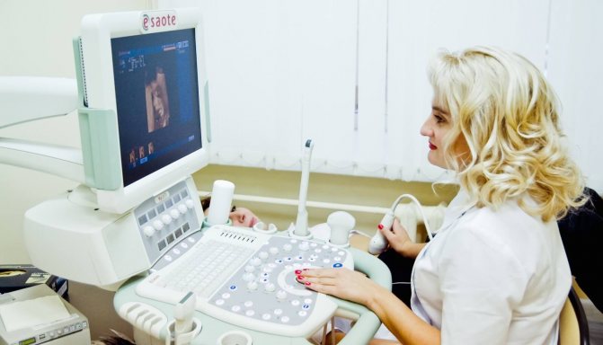
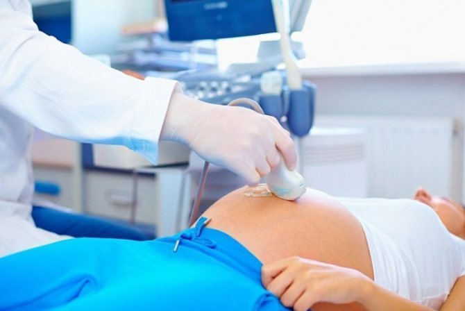
The study does not require special preparation. 3D ultrasonography is performed in a classical way, transabdominal. A special hypoallergenic gel is applied to the skin of the abdomen for better contact with the sensor during the procedure. At the end of the procedure, the gel on the skin is wiped off with a napkin. It should be noted that during this procedure it is advisable for the patient to lie still, as movements degrade the quality of the image. The duration of the procedure depends on the tasks set and on average is about 30-40 minutes.
Why is it important to do a 3D ultrasound, what does it show?
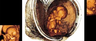
Three-dimensional ultrasound provides not only a three-dimensional image of the fetus, but also allows us to identify various abnormalities in the development of the genital and internal organs, the central nervous system, the structure of the facial bones, chromosomal pathologies and anomalies of the facial structure. Also, 3D ultrasound helps to confirm or refute other external malformations: anomalies of the limbs, anterior abdominal wall and others.
Of course, you shouldn’t do a 3D ultrasound in pursuit of a photo. Only if there is evidence for the expectant mother, a three-dimensional examination is done, but no more than twice during pregnancy .
Features and benefits of 3D scanning
Today, 2-dimensional (standard), 3-dimensional (volumetric) and 4-dimensional ultrasound examinations are used for diagnostic purposes. For a routine routine examination, 2-dimensional ultrasound is used, which provides an image that is quite informative for specialists. 3D ultrasound is used as a complement to conventional two-dimensional imaging studies. 3D ultrasound can be prescribed for medical reasons or performed at the patient’s personal request.
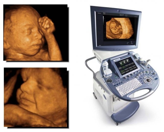
Compared to a conventional device, a 3D scanner has a larger surface area, it is equipped with special sensors and has a specially built-in computer module that produces a three-dimensional reconstruction and displays a three-dimensional image on the monitor. Three-dimensional reconstruction helps in analyzing the ultrasound picture during examination, improves the quality of diagnosis of surface defects, malformations of the face, spinal column, and limbs.
Thanks to the high quality of the picture and three-dimensional image, the specialist has the opportunity to examine the smallest details and better assess the development of the fetus. Instead of a standard two-dimensional picture, we get a three-dimensional image of the fetus. The possibility of diagnosing fetal development anomalies (facial defects, skeletal malformations) increases, since the image makes it possible to see even minor changes and pathologies at the initial stage. It is possible to see not individual parts, but a three-dimensional picture of the entire fetus as a whole and evaluate the proportionality of development.
The main advantage of diagnostics is that the intensity/power of the ultrasound wave is constant for both 2D and 3D images.
The only difference is the time frame of the study. To obtain a three-dimensional image you will have to spend about 45-50 minutes. Parents are given a three-dimensional color image in their hands, on which you can see the appearance, gender and body structure of the baby.
What is 3D ultrasound
Three-dimensional ultrasound, like all other methods of ultrasound diagnostics, is carried out using a sensor that generates ultrasonic waves and receives reflected echo signals, modified into electrical impulses. As a result, all data received in the form of a picture is displayed on the monitor. However, 3D ultrasound uses a larger sensor, the body of which has a built-in two-dimensional sensor that is in constant motion. Ultrasound waves during three-dimensional ultrasound are not directed from top to bottom, as with conventional ultrasound, but at different angles. This allows future parents to see all parts of the baby’s body, as well as get a three-dimensional image of it.
The first “three-dimensional” device appeared in 1989 in Austria. Then the image quality was very low, and it took more than half an hour to wait for it. Therefore, the method did not arouse much enthusiasm. The first scanner, with the possibility of three-dimensional reconstruction in real time, appeared only in 1996.
Wikipedia
https://ru.wikipedia.org/wiki
When is a 3D ultrasound performed?
3D ultrasound is not recommended to be done before the 20th week of pregnancy, since before this period the size of the embryo is too small, and the fetus itself does not at all look like a cute baby.
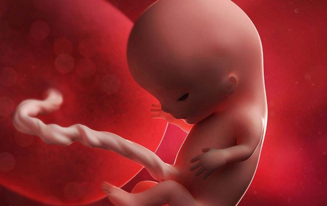
The size of an embryo at the 11th week of pregnancy is approximately the size of a large plum, and it does not look very proportional
It is also not recommended to perform 3D ultrasound in late pregnancy, after the 34th week. Now the size of the embryo is already too large, the fetus hardly moves, there is little amniotic fluid, and the baby occupies almost all the space, so it is simply impossible to see it in a three-dimensional image.
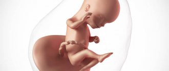
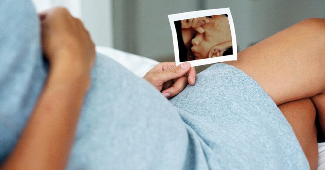
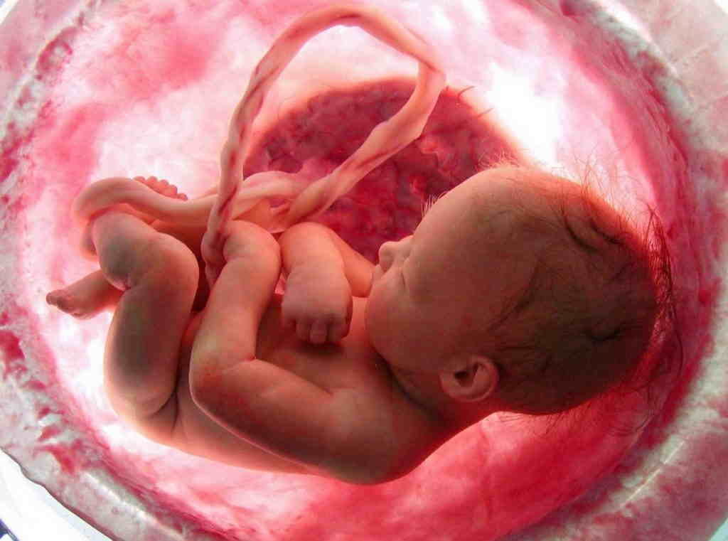
The optimal period for conducting a three-dimensional ultrasound examination is from the 24th to the 28th week of pregnancy. The fetus at this stage is already fully formed, and you can examine the face, all parts of the body and genitals in detail. And future parents will receive the first photo of their baby.
With the help of 3D ultrasound, it is easier to determine the gender of the child, see the features of the skeleton, limbs and face
Differences between 3D ultrasound and other types of ultrasound
The main difference between three-dimensional and two-dimensional ultrasound is the image that we obtain during the study. With a two-dimensional ultrasound, the picture turns out to be flat, two-dimensional, black and white, which is quite difficult for a non-professional to understand.
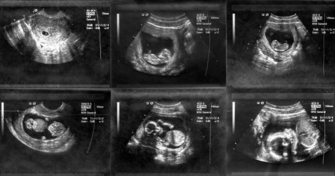
With a two-dimensional ultrasound, the image is black and white, and it is quite difficult for a person not involved in medicine to understand it
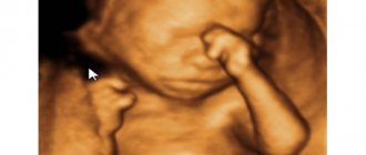
With three-dimensional ultrasound, the image is three-dimensional, three-dimensional, but static. Now we can get to know the baby: see his facial features, facial expressions and even how he yawns or smiles.
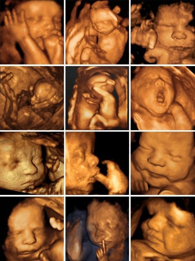
The first photo of the baby is an image obtained with a 3D ultrasound
When it is possible to record an ultrasound study in real time, that is, the picture is no longer static, but moving, then this is already a four-dimensional study. With such an ultrasound, future parents can add a short video to their video library with the baby in the lead role.
Limitations of the 3D scanning method
During pregnancy, 3D ultrasound is used as an additional research method, i.e. it complements, but does not exclude, the standard two-dimensional ultrasound screening carried out in prenatal diagnosis along with a biochemical blood test. We should not forget that in some cases it is impossible to perform a 3D ultrasound.
There are a number of limitations for three-dimensional research related to the timing of pregnancy, the position of the fetus in the womb, etc. It is best to conduct 3D studies between 26 and 30 weeks of pregnancy. Until the 26th week, the fetus still has too little subcutaneous fat, so the skull bones will be visible through the skin. After 30 weeks, the baby's head moves lower into the pelvis and the doctor will no longer be able to get an image of his face.
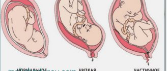
Whether it will be possible to take a photo of the baby's face depends to a large extent on how the fetus is positioned. When the baby lies facing the surface of the abdomen and is surrounded by a sufficient amount of amniotic fluid, it is most likely possible to obtain a fairly clear image of his face. If the child has his back to the sensor, the image will record only his back. A 3D “photo” may not work if there is not enough amniotic fluid around the baby.
Is there any point in trying to get a 3D image of the fetus? This issue is multifaceted and may vary for each party. For example, a two-dimensional image can be quite informative for a doctor. It allows you to evaluate not only the appearance, but also the condition of the internal organs, their compliance with development standards.
As for parents, it is much more interesting for them to observe the child’s face and appearance in the most convenient and understandable format. Often, 3D ultrasound is used as an addition to a conventional two-dimensional imaging study to satisfy the interest of parents. You should be reasonable about the frequency of ultrasound examinations performed and understand why and for what indications a 3D ultrasound will be performed.
Indications
Ultrasound in the second trimester is performed on all women to confirm or refute the results of the initial ultrasound examination. The study is required in the following cases:
- Older pregnant woman (over 35 years old).
- The woman took medications that are contraindicated for pregnant women or a tumor was discovered after the 14th week of pregnancy.
- The child's father and mother are close relatives.
- The pregnant woman has a history of a serious acute illness of a bacterial or viral nature.
- The mother is a carrier of genetically transmitted diseases.
- One of the future parents has hereditary chromosomal diseases.
- This woman had previously experienced miscarriages, fetal death before birth, or spontaneous preterm birth.
- The family already has one or more children with developmental disabilities.
In most cases, the second ultrasound during pregnancy, which can be performed in 3D mode, is a planned study. This repeated examination of the fetus allows us to exclude developmental anomalies that may manifest themselves in the second trimester.
For the sake of image clarity in 3D format, the power of ultrasound and the duration of its exposure are sometimes increased
Causes and consequences of deviations from the norm
During a three-dimensional ultrasound examination, standard measurements are not taken and are not compared with standard ones. It is prescribed in cases where the results of two-dimensional ultrasound require clarification . And then, using 3D ultrasound, specific areas of interest are examined.
The biggest danger in the second trimester of pregnancy is intrauterine growth restriction.
In case of any deviations, the woman expecting a child will be prescribed additional tests and appropriate therapy.
Doctors strongly discourage women expecting a child from making a diagnosis on their own based on the results of a 3D ultrasound.
How to prepare for a 3D ultrasound
A woman expecting a child does not need to do any special preparation before conducting a three-dimensional ultrasound examination. At the 20th week of pregnancy, the uterus and fetus already occupy most of the abdominal cavity and are clearly visible, therefore, whether the bladder is full or empty will not affect the course of the study in any way.
Experts advise that two or three days before a 3D ultrasound, a woman in an interesting position should refrain from eating gas-forming foods: legumes, cabbage, black bread, fresh sweet fruits, etc., to avoid difficulty with acoustic access.