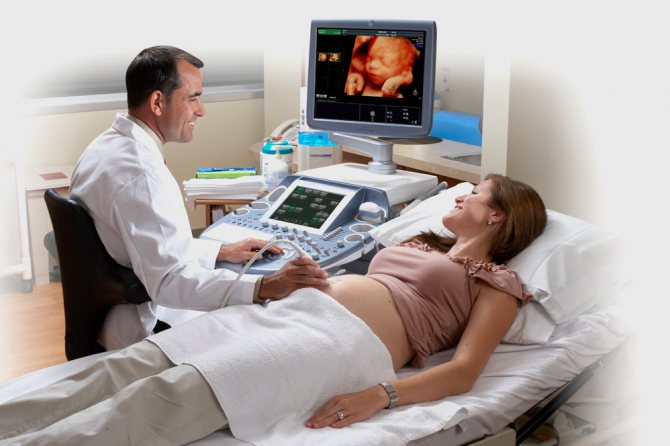
Ultrasound during pregnancy - a planned event that is included in the mandatory program during pregnancy. Fetal ultrasound is a kind of acquaintance with the baby. Ultrasound is performed 2-3 times throughout pregnancy: at 10-12, 20-24 and 32-37 weeks. In some cases, a fourth ultrasound is also prescribed, before birth, at approximately 34-36 weeks. Basically, it is aimed at determining the possibility of natural childbirth or by caesarean section.
Currently, there is no research method in obstetrics that could be comparable in information content to ultrasound. From an additional method, ultrasound examination becomes one of the main ones in obstetric practice. Doctors consider the main advantages of ultrasound to be highly informative, painless and safe.
In what cases is ultrasound used?
- When diagnosing pregnancy;
- When determining the age of the fetus;
- When diagnosing congenital anomalies;
- When determining the position of the fetus;
- When diagnosing multiple pregnancy.
Ultrasound in the mode used in obstetrics is safe for the baby and mother. This has been confirmed by clinical studies and many years of experience in its use for diagnosis during pregnancy. However, it is obvious that this diagnostic method should be used only as prescribed by a doctor, and not when desired.
You should try to perform ultrasound during pregnancy in strictly regulated periods - that is, when the diagnostic value of the method is especially high. An individual research schedule is decided upon in consultation with a gynecologist.
3D ultrasound during pregnancy
Nowadays, one of the most informative and accurate diagnostic methods for studying the condition of the fetus during pregnancy is ultrasound. The essence of this method is to use the phenomenon of uneven reflection of acoustic waves from tissues with different densities. The higher the frequency of the emitted ultrasonic waves, the deeper you can “look” into the developing body of the fetus and the better the quality of the resulting image will be.
A regular two-dimensional ultrasound is included in the list of mandatory diagnostic procedures that every pregnant woman must undergo. There is an alternative research method that is more accurate, informative and of high quality - 3D ultrasound.
Why is 3D ultrasound performed?
3D ultrasound is prescribed to clarify diagnoses identified in previous studies. For example, if a 2D ultrasound showed markers of a chromosomal disorder (for example, the thickness of the fetal nuchal translucency is not normal), then a 3D ultrasound may be prescribed to confirm or refute Down syndrome or other chromosomal disorders.
When is 3D ultrasound prescribed?
If we talk about prescribing a 3D ultrasound, at what time it is better to do it, then this usually happens at the 24th week or later. It is possible to carry it out earlier, it depends on what results the 2D screening showed, as well as blood tests and other diagnostic studies. If genetic pathologies, heart defects or other developmental anomalies are suspected, 3D ultrasound can be prescribed at any time, starting from the 8th week of pregnancy.
How does ultrasound work?
Ultrasound is elastic sound waves, inaudible to the human ear, whose frequency exceeds 20 kHz. They contain, for example, the noise of the wind and sea. Some of our smaller brothers - bats, dolphins and fish - communicate using ultrasound. In a word, such waves are not something alien and introduced from outside into the nature around us.
Almost all devices used for ultrasound during pregnancy operate in a pulsed mode, which significantly reduces the total exposure time. Plus, only half of the ultrasonic energy reaches the object under study due to the effect of absorption and reflection of supersonic waves by the underlying tissues.
The ultrasound procedure itself, which fits into the following scheme, does not raise any special safety questions. An ultrasonic emitter (it is also a scanner for receiving signals reflected from body tissues), in which sound with a frequency of 20 kHz to 1 GHz is concentrated into a narrow beam and directed to the desired location. The doctor moves the emitter over the skin at the observation site, which, in the role of a scanner, receives signals and transmits them to the control panel. And it converts ultrasonic waves into an image on the monitor.
That's it, the session is over, be it a regular two-dimensional ultrasound or a 3D ultrasound. But the “cinema” will be different. And the future of “actors” raises many more questions among specialists when using 3D.
3D/4D ultrasound: myths, reality and possibilities.
- 29.02.2016
- 5570
Nowadays, 3D/4D volumetric ultrasound has become very widespread, especially in the field of obstetrics. On the facades of private, and often budgetary, medical institutions, on the Internet, in the media, you can often find advertisements for such a service with beautiful pictures and promises of unforgettable experiences recorded on CD or other media. What is behind all this?
Let's first understand the terms again. Routine ultrasound examination is performed in 2D mode (D, dimension - measurement; synonymous with B-mode), when two-dimensional “flat” gray-scale ultrasound slices are obtained, which are updated at a speed of more than 20 frames per second, which is perceived by the human eye as real-time video . Additionally, so-called Doppler modes (color and spectral) are used, which make it possible to obtain qualitative and quantitative information about blood flow in the vessels under study.
In 3D mode, a third spatial dimension appears - depth. A 3D examination captures multiple 2D slices from which the software very quickly reconstructs a single, still, 3D volumetric image, which can then be presented in a variety of ways. In 4D mode, volumetric images are updated very quickly, the volumetric image is perceived as a movie in real time.
Time is considered as another dimension (3+1), but the essence of the mode is similar to 3D. To ensure rapid updating of the volumetric image over time, the system captures 2D images with a lower density; accordingly, the quality (“density”) of volumetric information in 4D mode is lower than in 3D mode. Specialists are often forced to use only the 4D mode if the object under study (most often the fetus in the uterus) is actively moving. 3D/4D is always an additional investment of time.
To obtain 3D/4D images, special volumetric sensors are used; as a rule, they are larger and heavier than sensors for routine studies. During the examination, the patient may feel a slight vibration from such sensors, because There are moving components inside when the sensor is operating in volumetric mode. Progress does not stand still; the latest models of volumetric sensors (especially the so-called “matrix”) are almost no different in appearance and dimensions from conventional sensors.
An ultrasound machine looks like a regular one; to obtain a three-dimensional image, special software and specialized sensors are required. In part, the class of the device can be judged by the size of the monitor (for expert ones from 19 inches or more) and the presence of a touch panel (“touch screen”) on the control panel of the device.
We are accustomed to the fact that 3D/4D ultrasound is an ultrasound that is usually used to obtain a photograph or video of the fetal face, legs, arms, genitals, etc. This is the so-called surface volumetric reconstruction mode (photorealistic, surface, etc.). At short stages of pregnancy (up to approximately 13-14 weeks), it is possible to obtain a superficial three-dimensional image of the entire fetus.
The most realistic image of the face can be obtained at 22-30 weeks, when human features are already more formed and there are conditions for obtaining high-quality surface reconstruction. The best modern devices have a variety of versions of this type of three-dimensional image, sometimes even “frighteningly realistic”.
Receiving such an image is an exciting, joyful moment for parents, establishing emotional contact with the unborn child. At the same time, for a competent ultrasound specialist, this type of three-dimensional image allows one to assess possible malformations of the face, limbs, hernias in various areas, etc.
There are often situations when parents will be disappointed: there is little amniotic fluid, the unborn child presses his face tightly against the wall of the uterus or even turns his face down, umbilical cord loops are applied, very poor “visibility” in 2D mode. In such situations, it is impossible to obtain a high-quality three-dimensional image of the face and other parts of the body, unfortunately...
Unfortunately, in many medical institutions, 3D/4D ultrasound is limited to only beautiful three-dimensional images of the face or leg recorded on disk. This is due both to the professional level of the ultrasound diagnostic specialist, and to the level of the ultrasound machine, with the capabilities of processing the resulting volumetric image, which are often additional expensive software components.
For expert examination of various internal organs of the fetus (for developmental defects), more advanced and complex three-dimensional modes and technologies are used, the use of which, of course, requires certain experience and knowledge.
For example, the use of 3D/4D ultrasound allows one to evaluate the anatomical structures of the fetal brain simultaneously on three mutually perpendicular planes (multiplanar reconstruction) or on a series of parallel sections, as with MRI or CT, which is impossible with standard ultrasound.
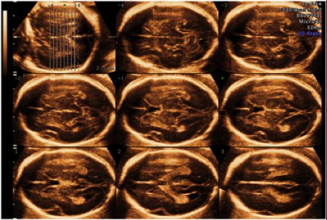
In many foreign clinics, 3D/4D ultrasound of the brain (neurosonography) is a mandatory research method, both in normal conditions and in the presence of a congenital malformation. Before sending a woman for an MRI of the fetus, which is recommended in the presence of a congenital malformation of the brain, it is necessary to conduct a detailed ultrasound examination of the fetal brain in three-dimensional imaging modes.
In the hands of an expert in prenatal (obstetric) ultrasound, 3D/4D neurosonography can replace MRI (for some types of fetal brain malformations), which is not available everywhere today, and is also an expensive research method.
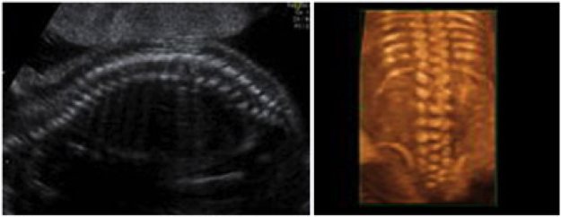
To study the fetal skeleton, the “pseudo-X-ray” volumetric imaging mode is most often used. In particular, when assessing the spine, 3D/4D is used to assess the integrity of the spine and exclude spinal malformations, such as underdevelopment of the vertebral bodies, spina bifida, and spina bifida. Below is an ultrasound image of the spine in a conventional 2D ultrasound (left) and in a 3D ultrasound, pseudo-X-ray imaging mode (right).
The fetal heart is the most difficult organ to study and identify developmental abnormalities. Timely detection of heart defects directly affects the further management of pregnancy and the prognosis of the life of the unborn child. The heart rate in the prenatal period is about 150 beats per second (60-80 in an adult), the heart moves quickly, all its anatomical elements (a set of obligatory two-dimensional slices) and blood flows inside are not easy to evaluate in 2D mode.
The frequency of volumetric frames, even on the most advanced ultrasound machines, does not allow obtaining a 4D image of the heart in real time. To solve this technical problem, STIC (Spatio-Temporal Image Correlation) technology was created, which allows reconstruction of a single high-quality volumetric cardiac cycle. This volumetric data can then be displayed in the form required by a specialist for thorough analysis.
In our country there are few devices equipped with this technology, and even fewer experts who can practically apply the study of the fetal heart using STIC technology. Very painstaking work is required to obtain and process volumetric images of the heart; unfortunately, there are often cases when high-quality capture of volumetric data is impossible.
Below are images of multiplanar (multi-plane) reconstruction of slices and 4D of the heart in combination with color Doppler mapping mode (displaying blood flow) on the left, and on the right, a superficial volume of the chambers of the heart and large vessels in combination with inversion.
These are just a few examples of real diagnostic applications of 3D/4D ultrasound; in fact, the scope of application of volumetric diagnostic ultrasound is much wider. Competent specialists use this type of research with high information content in gynecology, cardiac surgery, and less often when examining the abdominal organs and superficial organs. In addition, a big advantage of 3D/4D is that analysis of the internal organs of the fetus can be carried out after the study, off-line.
The volumetric image is stored in a database on an ultrasound machine, or on an external computer with special software, and, if necessary, is “extracted” from it. This makes it possible, firstly, to clearly explain the malformation, if present, to the woman herself (or both parents), and, secondly, it allows for a joint analysis of the congenital anomaly to obtain a “second” opinion or during a consultation with other specialists to decide question of tactics for further management of the patient. In this case, additional ultrasound and the presence of the patient himself are not necessary.
The ultrasound research method is the safest medical research method for both ordinary patients and pregnant women, and for the fetus itself.
A number of multicenter and multinational studies have been conducted in which children under 8 years of age were examined and underwent a series of ultrasound examinations in the prenatal period. The results of these studies prove that ultrasound does not have a harmful effect on the human fetus: it does not lead to the development of malformations, malignant neoplasms, or retardation in neurological development and growth.
A number of studies have been conducted on the effects of three-dimensional ultrasound technologies on the human fetus: no harmful effects of 3D/4D ultrasound have been identified. A wave of publications against the use of 3D/4D ultrasound, which appeared at one time in the media, was aimed at the non-medical, non-diagnostic use of three-dimensional ultrasound, when ultrasound machines began to be purchased privately, installed almost in hairdressing salons, and many hours (!!) of monitoring the behavior of the fetus were carried out. in the womb in 4D mode.
It is usually used to show a woman her baby in motion. For diagnostic purposes, static 3D modes are mainly used.
What does 3D ultrasound show during pregnancy?
3D ultrasound allows you to obtain data on the condition of the fetus, its position inside the uterus, the presence/absence of any pathologies, and the correspondence of size to gestational age.
3D ultrasound is also used as one of the tools for predicting childbirth.
A 3D scanner allows you to obtain not just a still image, but also process a series of echograms in video format. This makes it possible to see the baby in dynamics, visualize his movements and even facial expressions. You can see the child's reaction to the mother's voice, music or touching the stomach. All this is not done just for fun - such studies allow a more accurate assessment of the activity of the central nervous system of the fetus.