| Name of service | Price, rub |
| Ultrasound of the fetus from 10 to 21 weeks | 1800 |
| Ultrasound detection of pregnancy in the 1st trimester (up to 10 weeks) | 1500 |
| Ultrasound monitoring of a growing follicle (1 study) | 1000 |
| Fetal ultrasound 1st trimester to 12th week (SCREENING) | 2000 |
| Fetal ultrasound 2nd trimester after 12 weeks (SCREENING) | 2500 |
| Fetal ultrasound 3rd trimester after 21 weeks | 2200 |
| Ultrasound determination of fetal sex | 1200 |
| Ultrasound of the fetus up to 21 weeks (multiple pregnancy) | 2200 |
| Ultrasound of the fetus from 21 weeks (multiple pregnancy) | 3500 |
| Vascular Doppler | 1200 |
| Ultrasound monitoring of a growing follicle (folliculometry) | 900 |

Pregnancy management is one of the most important areas of gynecology, and ultrasound diagnostics is one of the main ways to monitor the condition of the fetus and the pregnant woman. Women are screened for the first time at 11–13 weeks. Its main tasks are to clarify the gestational age, assess how well the fetus is formed, measure its heartbeat, and determine the length of the cervix in order to prevent a possible miscarriage (if it turns out to be shorter than it should be). Also, during the first screening, it is possible to identify markers of chromosomal pathologies.
In the second trimester, it is important to continue monitoring the development of the fetus, and ultrasound is included in the list of mandatory diagnostic procedures. To the important question of how many weeks should 2nd trimester screening be done, doctors answer: 18-22 weeks of pregnancy.
Our clinic constantly hosts promotions
Ultrasound at 20 weeks of pregnancy
The second trimester (14-26 weeks) of pregnancy is considered the easiest period when bearing a child. The expectant mother is no longer tormented by toxicosis, the baby in her stomach begins to move slightly, which, of course, pleases her.
During this trimester, monitoring of fetal development continues, and ultrasound is included in the list of mandatory diagnostic procedures. The recommended time for the second screening ultrasound is 20 weeks of pregnancy.
Why is an ultrasound performed at 20 weeks of pregnancy?
Ultrasound at 20 weeks of pregnancy
carried out for the purpose of:
- Assess fetal development parameters and compare them with normative ones.
- Identify/refute the presence of chromosomal or genetic disorders in the fetus. Perhaps the second screening will show defects that were not noticed during the first ultrasound examination.
- Determine the sex of the child. At this time, you can find out who will soon be born with almost 100% probability (although mistakes still happen).
- Assess the length of the cervix and identify possible prerequisites for premature birth (if the length of the cervix is less than normal).
- Determine the amount of amniotic fluid. At 20 weeks, low or polyhydramnios can be diagnosed.
- Assess the condition of the umbilical cord, see the number of its vessels.
- Assess the functioning of the placenta, its maturity, measure its thickness.
- Assess blood flow in the feto-placental and utero-placental complex. This study is called Doplerometry.
- Identify pathologies of the female reproductive system (for example, fibroids, etc.).
Ultrasound to determine the sex of the child (in addition to the main examination)
Currently, for many parents, the topic of determining the gender of their unborn baby is relevant. Some, after the floor is installed, begin to choose a name for the baby, others - arranging the children's room and purchasing the necessary supplies.
Popular questions
Often future parents ask specialists:
- can there be an error in ultrasound when determining the sex of the child;
- the likelihood of an error in the ultrasound procedure when determining the sex of the child?;
- If the task is to determine the sex of the child using ultrasound, then at what time will the result be accurate?
So, is ultrasound wrong when determining the sex of a child? And what is the probability that the diagnostician provided incorrect information?
An ultrasound to determine the sex of a child cannot provide an absolute guarantee of the correct result. The probability that the doctor will name the sex of the baby correctly is ~93%. That is, out of ten embryos, the gender of one is indicated incorrectly. And this error is not always due to insufficient qualifications of the specialist.
Deadline for determining the sex of the child by ultrasound
The sex of the child is determined in the DNA at the time the egg is fertilized. The reproductive system begins to form at 5-6 weeks of pregnancy. In some cases, it is possible to find out the sex of the child when the 11th week of pregnancy occurs, but the most suitable period for this is the period from 20 to 25 weeks.
Reasons why ultrasound is wrong when determining the sex of a child:
- A short period of pregnancy during which the sex of the child is determined (up to 20 weeks of pregnancy).
Many parents want to receive information about the gender of the embryo during the first screening, signing up for sessions of the latest 3D and 4D ultrasound examinations. But even with the use of modern technologies, the sex of the child cannot always be determined with reliable accuracy at 11, 12 or 13 weeks (the time when the initial ultrasound is performed).However, we should not forget that the first screening has completely different goals, namely to identify the nature of the pregnancy and exclude its ectopic location. And also see how the fetus develops. At this stage, even 3D or 4D ultrasound can still be wrong because during this period many female embryos have slight swelling of the genital organs, which is not a pathology, and they are mistaken for male. So, the most optimal period for determining the sex of the child is 20-24 weeks of pregnancy.
- Peculiarities in the behavior of the unborn baby in the second trimester
Even if the pregnancy has reached the 25th week, determining the sex of the child using ultrasound does not always have a reliable result. An important role here is played by the fact that the fetus is already quite large, so it is often curled up in the uterus, which prevents a good view of the genitals. - Fetal size in the third trimester
Here the situation is in many ways similar to the second trimester, but the fetus has become even larger and takes up even more space. - Quality of ultrasound equipment
Of course, if the equipment is technically outdated, then the screening result to determine the sex of the child cannot be considered reliable. - The experience of the specialist who carries out the procedure.
The qualifications and experience of the doctor also play an important role in determining the sex of the baby using an ultrasound device. So specialists, whose work experience is extensive, can accurately determine the gender of the child.
The gender of the unborn child is important not only to future parents, but also to doctors. After all, knowing the sex of the baby, you can exclude the likelihood of various genetic diseases.
In Novocherkassk, you can sign up for an ultrasound to determine the sex of your unborn baby. The presence of qualified specialists and the latest equipment make it possible to identify the gender of the unborn child with maximum accuracy and exclude many pathological conditions.
What does an ultrasound show?
Ultrasound at 20 weeks of pregnancy
(we’ll look at the normal indicators in the decoding tables below) very informatively and reliably shows how the fetus develops and how comfortable it is in the womb. The correspondence of the fetal size to the established gestational age is assessed.
Important points of the second ultrasound are the examination of the internal organs of the fetus and the identification of their pathologies.
Examination of fetal organs
By the 20th week, the unborn child has already formed all the internal organs. Using ultrasound, the doctor assesses their condition, structure and degree of maturity. He examines the heart, kidneys, liver, brain, stomach. Attention is also paid to assessing the development of the fetal limbs, its spine, facial bones, and so on.
A detailed examination of internal organs using ultrasound allows one to identify heart defects of various types and predict the further course of pregnancy, as well as assess the level of viability of the child after birth.
Basic fetal indicators
There are a number of indicators by which the level of fetal development is assessed:
- BPP is the biparietal size of the fetal head. At 20 weeks of pregnancy, it should be approximately 36 mm (plus/minus a few mm).
- LZP – fronto-occipital size. At week 20, this parameter should normally be 56-68 mm.
- Fetal abdominal circumference. At 20 weeks, this figure should average 144 mm (plus/minus a few mm).
- Fetal head circumference. At the 20th week of pregnancy, the normal OG can be 154-186 mm.
- Length of the femur (normal 26-38 mm).
- Cephalic index, that is, the ratio of BPR to LZR. This index allows you to accurately identify the type, size and structure of the fetal head.
Ultrasound also evaluates the size of the fibula, tibia, radius, forearm bones and other parts of the body.
Placenta
An ultrasound examination necessarily evaluates the condition of the placenta. First of all, you need to determine the degree of its maturity. At 20 weeks and up to the 27th week, as a rule, the degree of maturity is assessed as zero. Then comes the first stage - until the 32nd week. Before the 34th week, the second stage of placental maturation should normally be diagnosed, then the third stage begins. The longer the pregnancy, the older the placenta becomes, the more difficult the process of transferring nutrients to the child is.
Ultrasound helps to see where the placenta is attached to the walls of the uterus. The norm is attachment to the back wall.
An important parameter is the thickness of the placenta. This figure should constantly increase. For example, if in the early stages the placenta may have a thickness of 10-11 mm, then by the end of the third trimester (by the 36th week) its thickness may be 35 mm.
Premature aging of the placenta, its thinning, and detachment become alarming signals, and in such situations a decision is often made about premature delivery.
Umbilical cord
The second ultrasound involves a detailed examination of the umbilical cord, through which the baby receives intrauterine nutrition.
It is necessary to examine her vessels - normally there should be three of them. There is another, more rare, but not pathological option - when the umbilical cord has only two vessels (vein and artery). In the second case, monitoring of pregnancy should be enhanced, since it is likely that in the last weeks of pregnancy the fetus will lack the oxygen that reaches it through the two vessels of the umbilical cord.
At the second ultrasound, they also pay attention to the number of umbilical cord loops and check whether there is any entanglement.
How and when does sex formation occur in the fetus?
From the moment of conception, each embryo has a “program” that will allow the fetus to become a boy or a girl.
The formation of the genital organs begins at 8 weeks of pregnancy. On ultrasound this moment remains unrecognized. The shape and size of the baby do not allow us to determine the presence of genitalia.
By 12-13 weeks, the diagnostician can observe the fetal genitals on an ultrasound scan. They are visualized in the form of a tubercle, which cannot yet be reliably identified as a penis.
Until the 20th week of fetal development, even the most experienced diagnostician can make a mistake and not give the mother an answer about the sex of the unborn child.
Modern high-tech ultrasound machines can give a visual picture of the genitals at 15=16 weeks. The correct interpretation of the observed objects depends primarily on the experience of the doctor performing the diagnosis. On the monitor screen you can calculate the angle between the back and the baby's genital tubercle. If this angle is more than 30 degrees, then the likelihood that a boy will be born increases.
From 21 weeks, the chances of accurately determining gender double. The fetal genitals are already fully formed. Girls have labia, boys have a penis.
Even with the modern level of diagnostic equipment and highly qualified medical personnel, difficulties arise in the gender identification of the fetus even at late stages of planned ultrasound (32-34 weeks).
Ultrasound during pregnancy is indicated for early diagnosis of pathologies; determining the sex of the child is a pleasant bonus.
Useful information on the topic:
- Determining pregnancy by ultrasound
- Ultrasound during pregnancy
- Ultrasound in the first trimester of pregnancy
- Ultrasound during pregnancy in the second trimester
- Ultrasound in the third trimester of pregnancy
- Ultrasound in late pregnancy
- Ultrasound in early pregnancy
When can parents see the gender of the baby?
The most accurate way to examine the sexual characteristics of the unborn child is an ultrasound examination, which pregnant women need to undergo every trimester. When registering with a gynecological consultation, the first examination is carried out at 11–12 weeks of intrauterine development. During this period, it is possible to identify the presence of various developmental anomalies in the fetus and, if the unborn baby does not turn away or cover his limbs, try to discern the first hints of gender.
3D and 4D ultrasound of the fetus
Of course, it is worth considering the fact that the formation of the genitals is not yet complete. However, a qualified specialist will already be able to see the boy’s formed testicles - they are still in the tummy, and will descend into the scrotum only in the seventh month of pregnancy.
It is necessary to take into account the high percentage of errors in such diagnostics, which is why the sex of the child at birth may differ from what the ultrasound showed. The likelihood of an accurate result increases with each subsequent week of gestation.
At the 14th obstetric week, the ultrasound specialist not only examines the external genitalia, but also measures the angle between the skin of the back of the fetus and the genital tubercle: if the value is less than 30°, the baby will be a girl, if more, the baby will be a boy. When an expectant mother doubts whether the doctor said the gender of the child correctly, she can check the instrument readings by undergoing an ultrasound scan at the 20th week of pregnancy - during this period the likelihood of error becomes minimal.
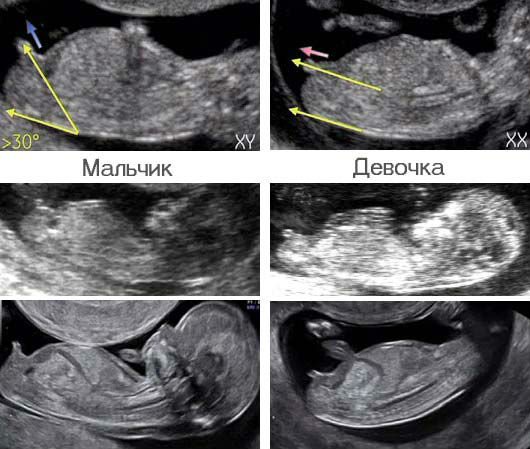
Our readers can see what the difference between babies looks like in the picture
Is it possible to make a mistake when determining gender?
According to the latest results of medical research, a diagnostic method such as ultrasound during pregnancy cannot guarantee absolute accuracy in determining the sex of the unborn child. Thus, it was found that this procedure is only 90–92% accurate. That is, errors occur in rare cases, but there are some explanations for this phenomenon.
The use of old-generation equipment when performing ultrasound increases the likelihood of miscalculation. When the rays emanating from the ultrasound sensor do not penetrate so deeply into the body of the expectant mother, the structures being studied are visualized quite poorly.
But in fairness, it should be noted that medical equipment has been actively improved recently, and equipment with flaws is becoming less and less common in hospitals. If a woman expecting the birth of a baby sees such a device in a sonologist’s office, she has the right to refuse an ultrasound, which will be performed using an outdated model of the device - this way, you can reduce the risk of incorrect interpretation of the diagnosis and determination of the sex of the child.
We should not lose sight of the human factor, which is becoming a more common cause of errors. Often, doctors who do not have sufficient experience in conducting research provide parents with incorrect information related to the gender of the baby. A qualified specialist, in turn, cannot take such “liberty”; in stories from medical practice, there were doctors who were able to identify the gender of a baby even at an extremely early stage - 9-12 weeks.

Before an ultrasound, you should make sure of the doctor’s professionalism: the diagnostic result will depend on his experience