Interesting Facts
| Options | Indications |
| Time from conception | 16 weeks |
| Period by month | 18 weeks |
| What month | 5 |
| Dimensions and weight of the fetus | 170 mm, 150-200 g |
| Uterus dimensions | At the middle of the distance between the womb and the navel or slightly above |
| Pregnant weight | Gain 400-500 g in 1-2 weeks |
Your baby is the size of
Large potatoes
170mm Size
150-200 g Weight
The eighteenth week of pregnancy is a vivid, memorable time when a woman may for the first time feel the movements of the baby inside her. This is an amazing and incomparable feeling. The fetus becomes very active, its movements are more coordinated. What else is going on with baby and mom this week? Let's find out more, but first let's look at the calculations:
Feelings of the expectant mother
At 18 weeks of pregnancy, vaginal discharge becomes more abundant. If they are still clear or light in color and you feel itching or burning, there is nothing to worry about. A curdled discharge with an unpleasant odor is a likely symptom of thrush. Be sure to consult a gynecologist.
Fetal movements
At 18-20 weeks of pregnancy, a first-time mother can already feel the baby’s movements. Now they are light, similar to vibration or fluttering. The doctor will ask you to track the number of movements. Normally this is 4-8 times per hour. If you count more or do not feel any movements at all, inform your gynecologist.
Sex
Intimate relationships benefit a pregnant woman, confirming her own attractiveness. But it should be taken into account that as the tummy grows, partners need to take special care and choose comfortable, safe positions.
Contraindications to sexual intercourse at week 18:
- Placenta previa;
- Increased tone of the uterus;
- Infections in a partner;
- Pregnancy with twins.
How the fetus develops at 18 weeks of pregnancy
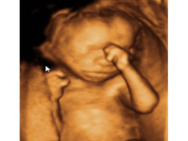
The baby performs many different actions: hiccups, yawns, winces, sucks his thumb, rolls over, pushes off the walls of the bladder with his arms and legs, grabs the umbilical cord and his own legs.
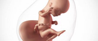
Its body length is about 17 cm, weight is 150-200 grams. The fetal skeleton already consists of 200 bones, by the time of birth their number will increase to 300. For comparison, an adult has 206 bones. As they grow older, they do not disappear, but grow together.
The nervous system is improved:
- more neural connections appear in the brain;
- myelin is formed - the sheath of the nerve, protecting it from damage and accelerating the transmission of nerve impulses;
- The functioning of the senses improves: the baby hears and recognizes sounds, distinguishes the taste of amniotic fluid. It is influenced by the foods that mother ate.
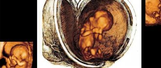
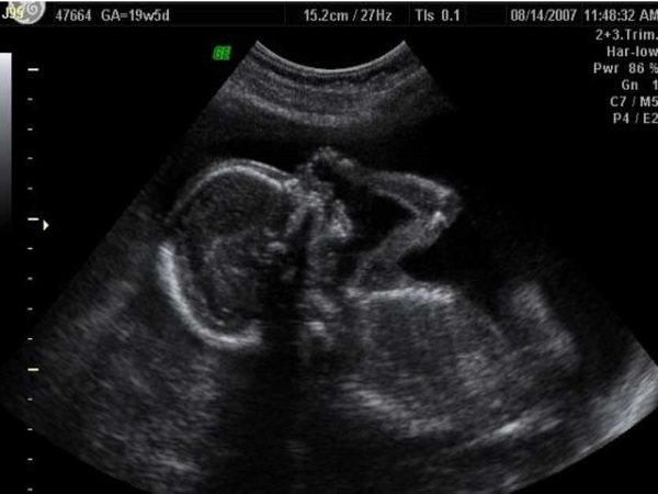
Ultrasound in the fifth month of pregnancy
Ultrasound scanning in expectant mothers can be performed in two ways: transvaginally and transabdominally. Depending on the individual characteristics and indications for the study, the doctor chooses the most suitable method.
Transabdominal scanning is performed with a special sensor using conductive gel. During the manipulation, the woman does not feel discomfort except for the apparatus moving across the abdomen. The procedure lasts from 5 to 30 minutes. A transabdominal study is performed to study the fetus: determine its size, obtain information about development and behavior.
Transvaginal ultrasound is needed when the first method cannot provide the required amount of information. For example, if the embryo is rotated in a certain way and does not allow the specialist to measure the necessary organs and parts. Also, a transvaginal sensor can provide more information about the condition of the cervix than its counterpart.
If the expectant mother has cervical insufficiency or is suspected of premature opening of the internal os, then an examination is performed through the vagina.
Why is ultrasound prescribed: indications
During the entire gestation period, a woman will undergo three main scans. These are called screening studies. The results obtained after the first such diagnosis are considered very important and the most revealing. If these data do not fit into established and generally accepted norms, then the doctor may prescribe a routine ultrasound at 18 weeks of pregnancy. During this period, it is much easier to assess the child’s condition.
Why don’t they want to wait the required 20 weeks for the second screening? The fact is that if it is necessary to terminate a pregnancy, it is much easier to carry out such manipulation at a period of 18-19 weeks. The procedure will entail fewer negative consequences than an abortion at 22 weeks. So, the main indication for a routine ultrasound at 18 weeks is poor results of the first screening.
Ultrasound examination may have other reasons for carrying out. Scans are sometimes performed for emergency reasons. The doctor may prescribe a diagnosis for you if you have the following complaints:
- bleeding from the reproductive organ or beige-brown spotting;
- pain in the lower abdomen;
- discrepancy between the volume and size of the abdomen and the expected period;
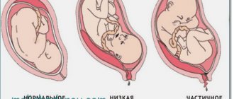
- abnormalities in the location of the placenta identified earlier.
In addition to ultrasound, the woman is prescribed additional tests that can tell about the reasons for this or that deviation.
What can you see during a scan?
An ultrasound at 18 weeks of pregnancy is designed to evaluate the development of the embryo. The specialist takes measurements of body parts: arms and legs, head circumference and tummy. Attention is drawn to the presence of internal vital organs. At this stage, the nasal bone and the thickness of the collar space are no longer indicative, so attention is not focused on them.
During ultrasound manipulations, you can see the condition of the uterus. The doctor determines the location of the placenta and determines its maturity. The umbilical cord must be examined (the number of vessels and its length are studied). It is important to establish the condition of the internal pharynx. In the fifth month of pregnancy it should be tightly closed.
During an ultrasound performed at week 18, developmental defects that were not previously noticed can be detected. If the first screening showed poor results, then the next examination can already give a clear assessment of the developing baby.
Gender
Many couples wonder: will an ultrasound at 18 weeks of pregnancy show the gender of the baby? The answer to this will most likely be positive. An ultrasound scan performed by a competent specialist using a modern device can determine the gender of a child with 100% certainty.
Some women preparing for motherhood specifically go for research to find out the sex of the baby. You shouldn't do that. Increased interest will be satisfied, but frequent scanning is not the best for a normally developing pregnancy.
There are also situations in which it is necessary to find out the sex of the baby in advance. This is necessary for inherited autoimmune diseases or other pathologies.
What do the genitals of an embryo look like at this stage? In girls, the labia can be clearly seen. But from a certain angle they can resemble testicles. Therefore, some experts may predict that you will have a boy, but the birth will be a girl. It is much more difficult to confuse the male and female genders. At this stage, the male embryo has a clearly visible penis.
Note: the testicles of a male fetus at 18 weeks of pregnancy have not yet descended into the scrotum. This will happen closer to childbirth. If the doctor shows you the testicles and promises the birth of a boy, be wary: most likely, the result is incorrect.
What do the resulting numbers mean?

After monitoring, the ultrasound diagnostic specialist will issue you a protocol.
The form indicates all the values and characteristics of the fetus obtained during the ultrasound. It also sets out norms that are considered generally accepted. If your values fall within the standard range, then we can talk about a normal pregnancy. Any deviations in one direction or another must be recorded by a gynecologist. The doctor monitoring your pregnancy can make a specific diagnosis based on the findings.
You should not try to decrypt it yourself. Most likely, you won't succeed. If you find values that do not fit into the established framework, you will only worry once again, which is extremely undesirable. At 18 weeks of pregnancy, the standard values will be as follows:
- the weight of the embryo is about 200 grams, and its length is from 12 to 15 centimeters;
- the fronto-occipital zone is from 4.9 to 5.9 cm, and the BPR (biparietal size) is from 3.7 to 4.7 cm;
- the circumference of the future baby's tummy is 10.4-14.4 cm, and the circumference of the head is 13.1-16.1 cm;
- the length of the femur is about 3 cm, and the length of the tibia is 2.5 cm;
- the forearm is approximately 2 cm, and the humerus is half a centimeter larger;
- the degree of maturity of the placenta is zero, the thickness at this stage has not yet been measured;
- the volume of amniotic fluid ranges from 80 to 220.
Depending on the equipment, normal ultrasound readings may vary slightly.
Possible deviations
If the diagnostics revealed indicators that do not fit into the norms, then you should not immediately panic. Deviations can be natural and pathological. You cannot make a diagnosis based on ultrasound alone. To confirm or refute suspicions, additional studies are carried out: dynamic scanning; Dopplerography; blood test for hormones, antibodies, and so on.
If the size of the embryo does not fit into the established standards, then this may be hereditary. Parents of short stature and thin physique do not give birth to heroes. The baby may be larger than the stated norms. This often happens in obese couples or women with diabetes.
Polyhydramnios and oligohydramnios are also deviations. In the first case, the cause may be chronic diseases of the mother or Rh conflict. If you had an infection during pregnancy, this is also accompanied by polyhydramnios. Low hydramnios is a more dangerous phenomenon. It becomes a consequence of fetal defects, for example, lack of kidneys. Doctors still cannot reliably establish the causes of this pathology.
Deviations from the norm may appear due to congenital anomalies. They are often accompanied by intrauterine growth retardation, growth retardation, and non-compliance with normal values. If the fetus is missing any vital organ, then this is an indication for termination of pregnancy. In other cases, possible drug correction or surgical intervention is carried out.
Tests and ultrasound
The main study at 16-20 weeks is the second pregnancy screening. It includes an ultrasound and analysis of 3 hormones: hCG, estriol and alpha-fetoprotein. Helps determine the risks of developing genetic diseases such as Down syndrome, Patau syndrome, Edwards syndrome, as well as disorders associated with abnormal structure of internal organs. If the ultrasound results are normal, no blood test is usually performed. However, there are cases when you cannot do without it.
Who is particularly recommended for a triple hormone test?
- Pregnant women over 35 years of age, when, due to age-related changes, the risk of the child developing genetic disorders is high.
- Patients with a family history, that is, when someone in the family had similar pathologies, or if the first child was born with chromosomal abnormalities.
- If you have previously had premature birth or miscarriage.
- When a woman suffered from an infectious disease in the first trimester, she took teratogenic drugs without knowing about pregnancy.
- If the results of 1 pregnancy screening were questionable.
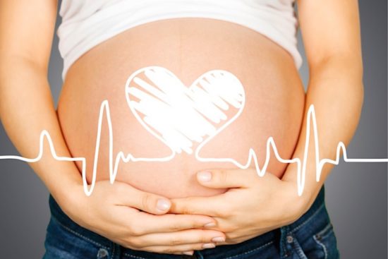
Starting from the 18th week of pregnancy, the gynecologist will listen to the fetal heartbeat at each scheduled visit. This gives the specialist an idea of the baby’s condition and presentation in the uterus.
Developmental norms in the fetus at 17, 18 weeks
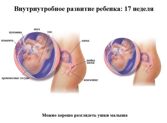
A baby at 17-18 weeks is already quite big. The normal height at 17 weeks is 12 cm, the baby’s weight reaches 100 grams. An ultrasound at 17–18 weeks shows the development of the fetus and the presence/absence of pathologies.
Normal fetometry indicators at week 17 are:
- BPR. Biparietal size is approximately 34 – 42 mm;
- OG. Head circumference is 112 – 136 mm;
- LZ. The fronto-occipital size is 41 – 49 mm;
- coolant. Abdominal circumference ranges from 121 to 149 mm.
As for the size of the long bones of the fetus, they are normally:
- forearm bones – 15 – 18 mm;
- femurs – 20 – 28 mm;
- shin bones – 15 – 21 mm;
- humerus – 15 – 21 mm.
In addition to the size of the fetus at 17 weeks, the specialist also examines the condition of the placenta and the baby’s brain during an ultrasound examination.
By the 18th week, qualitative changes occur in the baby’s body. By the end of the month, his weight will reach 150 grams, and his height will be about 12.5 - 14 cm.
- BPR. Biparietal size is approximately 37 – 47 mm;
- OG. Head circumference is 131 – 161 mm;
- LZ. The fronto-occipital size is 49 – 59 mm;
- coolant. Abdominal circumference ranges from 104 to 144 mm.
As for the size of the long bones of the fetus, they are normally:
- forearm bones – 17 – 23 mm;
- femurs – 23 – 31 mm;
- shin bones – 23 – 31 mm;
- humerus – 15 – 21 mm.

From the 18th week, a specialist can already determine the sex of the unborn baby and show it to you in a photo. At 17–18 weeks of pregnancy, the specialist not only examines the external and internal organs of the child, he also receives information about:
- number of fruits;
- condition, location of the placenta (it should be localized on the back wall of the uterus);
- the cervix (its length should not be shorter than 30 mm, the external and internal pharynx should be closed);
- amount of amniotic fluid;
- condition of the uterus (the specialist must make sure that there is no hypertonicity of the uterus.
- presentation of the fetus (normally it should be cephalic at this stage).
What to discuss with your doctor
- If you are worried that you have a small belly for the 18th week of pregnancy, then know that, firstly, this is subjective and depends on the woman’s general physique. And secondly, to dispel worries, discuss with your doctor whether the rate of fetal development corresponds to obstetric standards. If the baby is developing correctly, there is no point in worrying about waist circumference.
- Tell your doctor if you have frequent back pain and see if you should wear a back brace.
What's happening
During this period, the development of the child’s brain continues, and subcutaneous fat gradually accumulates. By the end of the 18th week of pregnancy, the external and internal genital organs of the fetus will be fully formed, and during an ultrasound you will be able to find out who has settled in your tummy.
The baby’s immune system is also improving: by the 18th week of pregnancy, his body begins to produce interferon and immunoglobulins, designed to protect the baby from infections and viruses.
A pattern unique to him has appeared on your baby’s fingers. The rudiments of teeth have formed, the child is able to hear, but the development of the organs of vision has not been completed, but by the 18th week of pregnancy the baby can already distinguish between light and dark.
Possible complications
You may experience lower back pain. They arise because the center of gravity shifts due to the growing belly. Massage, a warm shower, swimming and exercises that strengthen your back muscles will help you cope with this.
If at 18 weeks of pregnancy you periodically experience tightness in your lower abdomen, do not worry. The muscle ligaments holding the uterus are stretched. Therefore, short-term pain occurs. However, for prolonged spasms, consultation with a gynecologist is required.
If you stand up suddenly, you may feel dizzy. It is caused by low blood pressure or low blood sugar. Therefore, in the morning, do not rush to get out of bed, do it smoothly. You can leave some fruit on the nightstand for a light snack in the evening.
Hormonal changes and increased blood volume cause anemia in many pregnant women. Most often it is associated with iron deficiency. In this case, diet adjustments are not enough; long-term treatment with special iron supplements is necessary.
Recommendations
Continue to eat a healthy diet during your 18th week of pregnancy. Have a full breakfast and for lunch eat a meat dish (beef, rabbit, chicken, turkey): protein is needed for normal fetal development. Fresh fruits, berries, vegetables, biscuits, and unsweetened yogurt without artificial additives are great for a light snack. To prevent constipation, drink a glass of kefir at night.
17 - 18 weeks of pregnancy is a period when your kidneys are working harder, so you have to run to the toilet more often. Empty your bladder on time and avoid stagnation of urine: this will help prevent the development of infection.
If you experience dizziness with a sudden change in body position, do not get up immediately after sleep. First, sit on the edge of the bed for a few minutes, and only then you can slowly get up. And if at 18 weeks of pregnancy you feel tired during the working day, try to retire to a secluded place and rest for 10 - 15 minutes.
Dietary recommendations
Throughout pregnancy, your diet should be balanced and nutritious. What does it mean? Your daily menu should include:
- 4-5 servings of fruits and vegetables;
- 4 servings of dairy or other calcium-rich products;
- 3 or more dishes made from whole grains (porridge, whole grain bread, durum wheat pasta);
- 2-3 – lean protein, that is, meat and a variety of legumes.
You should have fish on your table 2 times a week, but don’t often choose fish that can accumulate mercury, for example, tuna, marlin, swordfish.
Do you need vitamin supplements?
If you eat a varied diet, are not a vegetarian, and have access to vegetables and fruits all year round, then you do not need additional vitamin intake. The exception is folic acid in the early stages of pregnancy and vitamin D, the deficiency of which in our region is associated with the number of sunny days per year.
If you have any chronic diseases, for example, the thyroid gland, you may need additional iodine. The issue of taking medications should be decided together with the doctor monitoring the pregnancy.
Prohibitions and recommendations for expectant mothers in the 18th week of gestation
The advice that pregnant women should follow remains the same. This is emotional peace and a healthy way of life.
Nutritional Features
Many women during this period are faced with an irresistible desire to eat more. If a pregnant woman is not bothered by heartburn, she may gain a few extra pounds. It is important to follow a diet, control the composition of the diet and its calorie content. The nutritional value of food can be gradually increased. Dairy products, various cereals, eggs, lean meats, and vegetables are suitable for this. It is recommended to choose low-calorie and unsweetened fruits (bananas and grapes are not suitable).
From this week you need to gradually limit the amount of salt you consume. This will prevent swelling in the following weeks.
Taking vitamins
To prevent hemorrhoids and varicose veins, pregnant women should take calcium. It is better to use it in combination with vitamin D3. Both microelements will complement each other and help prevent the development of rickets in the baby in the future. Most expectant mothers in the second trimester do not receive the optimal amount of nutrients from their diet. You can consult a gynecologist about taking additional multivitamin complexes.
Sex at 18 weeks pregnant
In the absence of contraindications, intimate relationships are not limited. In addition, sexual contacts will only be useful. As long as the stomach does not cause discomfort, sexual intercourse will be comfortable and can bring the pregnant woman many new sensations. Some women believe that uterine contractions during orgasm are dangerous for the fetus. This opinion is wrong and there is no need to worry about it. Pressure on the abdomen, deep penetration positions and anal intercourse should be avoided.
Reasons for limiting sexual relations during this period may be:
- Multiple pregnancy;
- Multiple miscarriages;
- Leakage of amniotic fluid;
- Placenta previa;
- The appearance of cramping pain in the lower abdomen.
Physical activity
The expectant mother should avoid excessive exercise, but moderate exercise will be beneficial. You can sign up for dancing, yoga, fitness or gymnastics for pregnant women. Water aerobics or swimming are also suitable. If you can’t go to the gym, you can walk outside. The main thing is to lead an active lifestyle. This will help avoid excess weight gain and deep vein thrombosis.
Medical procedures, medication administration
Any medical manipulations during this period should be performed under the supervision of an obstetrician-gynecologist. You should avoid taking any medications, especially antibiotics. If possible, treatment is delayed until the postpartum period. If absolutely necessary, therapy is carried out using the safest methods possible.
Checklist for 18 weeks of pregnancy
- Talk to your baby more often, he hears you and understands your emotional state. Music and your singing also have a beneficial effect on a child's development.
- When you lie down or sit for a long time, change your body position more often so as not to cause blood stagnation in the pelvic organs.
- If you experience leg cramps at night, avoid prolonged vertical exercise. Consult a specialist about the need to take magnesium. Often its deficiency causes involuntary muscle contractions.
Entrust your pregnancy management to the specialists of the Medical Women's Center. We guarantee an attentive and sensitive approach to each of our patients.
Visiting an obstetrician-gynecologist
At the 18th obstetric week of pregnancy, a woman can have a scheduled appointment with a gynecologist. Before the appointment, it is necessary to take urine and blood tests (directions were issued at the previous appointment).
The doctor will conduct an examination on the chair, measure temperature, blood pressure, weight and abdominal circumference. Conduct a survey about your well-being and changes that have occurred since the last meeting. Listen to the fetal heartbeat through a stethoscope.
At the end of the appointment, he will issue directions for the next tests and set a date for the second screening.
If everything is fine with the tests, the gynecologist may advise you to see a dentist, if necessary. Now is the time to check the condition of your teeth and, if necessary, carry out minor treatment.

Visiting the doctor at 18 weeks
Ultrasound and tests
A routine ultrasound and “triple test” (blood test) can be scheduled at 18 weeks of pregnancy. The main purpose of the study is to identify possible abnormalities in fetal development.
During an ultrasound, the doctor examines:
- position of the fetus in the uterus;
- fetal bone structure, correspondence of parameters to gestational age;
- the condition of the internal organs of the unborn child, compliance with the deadline;
- place of attachment of the umbilical cord, its condition;
- volume of amniotic fluid;
- cervical condition;
- main parameters of the fetus: head circumference (HC), length of the femur (HB) and humerus (HB), width of the head from temple to temple (BPR).
At the current stage, if the unborn baby is in the right position, doctors can name his gender. The probability of an error at the current time is low.
The triple test at 18 weeks of pregnancy is the 2nd screening test. In some cases, the doctor may not prescribe it if there is no indication. But it is not necessary to worry prematurely if the gynecologist talks about the need for this study. A triple test at 18 weeks of pregnancy measures the levels of:
- hCG;
- free estriol;
- alpha-fetoprotein.
Based on the results of this analysis, the doctor can make a conclusion about possible congenital diseases in the unborn child. If the results are not very good, additional invasive tests will be suggested.
Uterus and belly
At the 18th week of pregnancy, the uterus continues to increase in size. At the current stage, it has risen by 17-19 cm. The belly is almost under the navel and can no longer be hidden under loose clothing.
At the current stage, you can already see which side of the abdominal cavity the unborn child is on. The abdomen shifts slightly in the right direction and looks uneven.
Pain in the abdomen and other parts of the body
At the 18th week of pregnancy, pain may normally be present, but a woman needs to clearly know what sensations should alert her and force her to urgently consult a gynecologist for medical help.
Stomach ache
Very often, at the 18th week of pregnancy, a woman experiences discomfort in the abdominal area. If the manifestations are not severely painful and are accompanied by itching, then most likely this is caused by stretching of the abdominal muscles. You should talk about this symptom at your next appointment with a gynecologist.
If there are painful sensations in the abdomen and back, accompanied by tension across the entire surface of the abdomen (it seems to be turning to stone), then most often we can talk about the tone of the uterus. If the pain appears once or after a long walk or physical activity, then you need to talk about it at your next appointment. If you experience frequent pain, you should consult a gynecologist in the coming days. You may need to take additional medications to relax the muscles of the uterus.
Severe and/or cramping pain is a reason to immediately seek medical help to identify the causes.
Leg pain
Pain in the legs can be caused by various reasons: from fatigue to varicose veins. The doctor must identify the source of the discomfort. Before your next appointment, it is advisable to:
- rest more often with your legs raised high;
- reduce the amount of salt consumed to a minimum;
- engage in permitted physical activity (swimming, walking).
Back pain
In most cases, discomfort in the back during the current week of pregnancy is caused by physical fatigue and a change in the center of gravity. If they are not accompanied by abdominal pain or other unpleasant symptoms, it is advisable to give your back rest and start wearing a prenatal bandage. At your next appointment, tell your gynecologist about the sensations you are experiencing.
If back pain is localized on the sides, accompanied by discomfort when urinating, uterine tone or other symptoms, you should consult a gynecologist for advice. Be sure to take a urine test before taking it.
Headache
Most often, headaches in the 18th obstetric week of pregnancy are caused by changes in blood pressure. The occurrence of this symptom should be reported to the doctor at your next appointment.
In addition to the indicated types of pain, at the 18th week of pregnancy, a woman may experience manifestations of hemorrhoids, carpal tunnel syndrome, pain in the feet, and thoracic spine. You should definitely tell your doctor about any pain you experience at your next appointment.
Discharge
The amount of vaginal discharge may increase significantly during the 18th week of pregnancy. Normally, their color should be light or milky, the smell should be barely perceptible sour. Any change in color, consistency or smell should be reported to your gynecologist.
If a woman notices red or brown discharge or clots of pus, she should immediately seek medical help.
Bleeding and menstruation
There should be no bleeding or menstruation at week 18. Any appearance of blood or red or brown discharge is a reason to urgently seek medical help.