Ultrasound during pregnancy
Ultrasound during pregnancy is a planned diagnostic procedure that needs to be done at least three times during the entire period of gestation. If any abnormalities in the development of the unborn child are detected, the attending physician may prescribe a larger number of ultrasound scans to assess the dynamics of changes in the condition of the fetus, amniotic fluid, placenta, and so on.
Ultrasound allows you to accurately assess the growth and development of the unborn baby, determine its gender, identify developmental anomalies, genetic disorders, and threats to pregnancy.
A subtype of ultrasound examination is fetal Dopplerometry, thanks to which the doctor can analyze the blood flow in the vessels.
Also, for future parents, ultrasound is an excellent opportunity to see their baby before he is born.
Why do you need to do an ultrasound?
So, the doctor ordered an ultrasound scan for the patient. Not everyone understands why such an examination is done. It must be done in order to:
- recognize the onset of the development of most diseases (including very dangerous ones) even at the earliest stages);
- establish a diagnosis using ultrasound, and do it most accurately (in the presence of certain clinical symptoms);
- to effectively monitor the treatment of many pathologies (including in a hospital setting);
- to prevent the development of dangerous complications as a result of treatment and development of the disease.
Types of ultrasound examinations during pregnancy
Ultrasound in the first trimester
The first ultrasound examination should be carried out at 11-14 weeks of gestation, and ideally before the 6th day of the thirteenth week. It is during this period that gross abnormalities in fetal development can be identified or markers of genetic or chromosomal pathology can be detected.
Ultrasound in the 1st trimester
carried out to:
- confirm pregnancy and determine its type - uterine or ectopic;
- determine the number of embryos;
- assess the size and structure of the embryo;
- identify pathologies of a woman’s internal genital organs that can affect the development of the fetus;
- identify threats to pregnancy (miscarriage, fading of fetal development due to weak heartbeat, and so on).
Although ultrasound during pregnancy is not harmful to the fetus, it is not worth doing such a procedure before 11 weeks, as the results may be erroneous.
Ultrasound in the second trimester
The second ultrasound is performed in the second trimester
, from the 18th to the 21st week. The goals of ultrasound at this stage are as follows:
- Determination of fetal presentation, its position in the uterine cavity.
- Determination of the main parameters of the fetus - weight, head size, hip size, and so on.
- Determination of fetal heart rate.
- Assessment of the condition of the fetal facial bones, its spine, limbs, and so on.
- Determination of the amount of amniotic fluid.
- Assessment of the condition of the placenta (its degree of maturity), cervix and umbilical cord.
- Study of the anatomy of the internal organs of the fetus. First of all, the heart is examined for the presence of defects. The condition of the brain, lungs, liver and kidneys is also assessed.
At the second ultrasound, it is already possible to determine the sex of the child, although this is not included in the mandatory protocol filled out after the study.
Ultrasound in the third trimester
During the period from the 32nd to the 34th week of gestation ( in the third trimester
) it’s time to conduct a third ultrasound, which helps determine:
- fetal size (head, limbs, abdominal circumference and other parameters);
- size, level of development and structure of the internal organs of the fetus;
- accurate presentation of the fetus;
- amount of amniotic fluid (diagnosed as oligohydramnios/polyhydramnios of varying degrees);
- level of oxygen supply to the fetus (ultrasound can reveal signs of hypoxia);
- level of blood flow in the umbilical cord;
- the fact that the fetus is entwined with the umbilical cord.
Indications for unscheduled ultrasound
An unscheduled study can be carried out at any time if there are appropriate indications. Before the 10th week of pregnancy, it may be required in the following cases:
- suspicion of a frozen or ectopic pregnancy;
- the occurrence of sudden bleeding from the vagina;
- pain in the lower abdomen of varying intensity;
- a woman’s history of miscarriages or abortions;
- the likelihood of multiple pregnancy;
- risk of miscarriage;
- pregnancy occurred as a result of artificial insemination (using IVF);
- The woman underwent a procedure to stimulate ovulation.
At any time, an unscheduled ultrasound can be prescribed for:
- multiple pregnancy;
- acute or chronic infectious diseases in the expectant mother;
- disorders of the female endocrine system (diabetes mellitus, etc.);
- pathologies of the structure and diseases of the pelvic organs;
- identifying isthmic-cervical insufficiency;
- threat of early miscarriage or late premature birth;
- suspected developmental defects and chromosomal abnormalities of the fetus;
- identifying placenta previa (ultrasound is performed in the later stages to ensure that the presentation is intact);
- detection of early aging of the placenta;
- deviations from the norm in the volume of amniotic fluid (oligohydramnios, polyhydramnios);
- changes in the baby’s behavior, lack of movements;
- monitoring the condition of the scar if the previous birth was performed by cesarean section;
- The woman is over 40 years old.
In the later stages of pregnancy, additional ultrasound monitoring can be carried out if a transverse or pelvic presentation was detected during the planned examination. There is a possibility that the baby will turn over shortly before birth. It is also necessary to assess the degree of umbilical cord entanglement when it is detected.
When does an ultrasound show pregnancy?
At a gestation period of more than 3 weeks, an ultrasound scan can already show pregnancy. At such early stages of fetal development, the transvaginal method of examination is usually chosen as it can give more accurate results. When using the transabdominal method of examination, an accurate result can be obtained only at the 4-5th week of gestation. At the same time, such early diagnosis of pregnancy should be carried out in a complex: in addition to ultrasound, it is necessary to conduct a blood test to detect the level of hCG (a hormone secreted by the placenta when pregnancy occurs).
What kind of ultrasound is done in the early stages of pregnancy?
As part of the first screening, a planned ultrasound is prescribed no earlier than the 10th week of the gestational period.
| The first ultrasound during pregnancy The first ultrasound during pregnancy shows what happens to the mother and baby at a certain period of development. If the size of the fetus is more than 5 mm, it can be visualized and the first unique photo of the baby will be obtained. |
Timely examination allows you to identify and prevent some congenital pathologies, such as: Down syndrome, pathological development of the neural tube, triplodia (the presence of three chromosome sets), Edwards syndrome and others.
However, in some cases, ultrasound is performed unscheduled:
- a woman suspects pregnancy, a home test shows a positive result, and menstruation occurs. In this case, a study is prescribed at any time to confirm or refute pregnancy and eliminate the risk of ectopic implantation of the embryo;
- after in vitro fertilization, ultrasound in the earliest stages of pregnancy is prescribed in parallel with laboratory tests to compare the dynamics of fetal development. Ultrasound after IVF monitors the condition of the fertilized egg (or several eggs in case multiple pregnancy develops);
- the woman has a history of spontaneous miscarriages. The condition of the cervix is checked, if there is a risk, the treatment is adjusted and special sutures are applied to prevent premature cervical dilatation;
- patients over 35 years old. Hormonal balance may become disrupted with age, leading to spontaneous abortion. The purpose of ultrasound is to identify and prevent possible complications;
- the mother or father of the child has genetic diseases in the family or the baby’s parents are close relatives;
- There were previously frozen pregnancies, there are already children with congenital pathologies.
An ultrasound is especially important if fertilization occurred under the influence of alcohol. Even if the father and mother are absolutely healthy, it is difficult to predict how the fetus will develop. Carrying out screening in the first weeks will allow you to examine in detail how correctly the child’s body is forming.
How many times can an ultrasound be done during pregnancy?
The question of how many times and how often an ultrasound can be done during pregnancy is difficult to answer unambiguously, since everything here depends on the individual health indicators of the woman and her unborn child. There is an average number of scheduled ultrasounds - there are three, one in each trimester. If the results of each subsequent ultrasound show that the fetus is developing normally and there are no threats to it, then there is no point in going through this procedure again.
If the next planned ultrasound reveals any abnormality (for example, the weight and height of the fetus do not correspond to the average normal values, the blood flow between the placenta and the fetus is disrupted, and so on), the doctor observing the pregnant woman may prescribe her certain therapy. After completing a course of treatment (outpatient, in a day hospital or at home), it makes sense to prescribe a repeat ultrasound, which will show the effectiveness of the therapy.
Additional ultrasound can be performed immediately before birth - in the maternity hospital. Such a study can help doctors determine the correct tactics for managing labor or prescribing a cesarean section.
Is ultrasound during pregnancy harmful to the fetus?
The question of whether ultrasound during pregnancy is harmful to the fetus worries most expectant mothers. Studies of the effect of this procedure on the development of the unborn child have shown that two-dimensional ultrasound (which is most often prescribed) does not harm the fetus. In such a study, the ultrasound machine emits rather weak waves that are not capable of disturbing the structure of the embryo, affecting its growth, and so on.
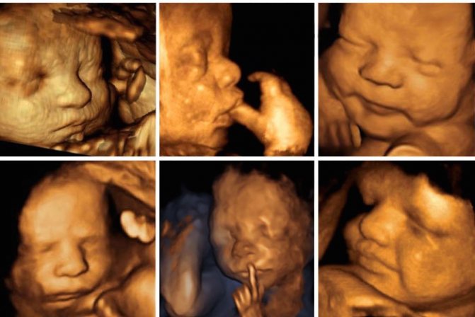
Another question is 3D and 4D ultrasound. Are such studies harmful to the unborn child? To obtain a three-dimensional/four-dimensional image of the fetus on the screen of an ultrasound machine, deeper penetration of sound waves into the structure of the fetal tissue is required. In this case, such waves may have a negative effect on tissues, for example, overheating or destruction. Therefore, 3D and 4D ultrasound are prescribed only in cases where the benefit from them far outweighs the possible harm. Such procedures should under no circumstances be prescribed for “entertainment” purposes, for example, when parents simply want to take a closer look at their baby or get a three-dimensional photo of him as a souvenir.
How not to miss an ectopic and non-developing pregnancy
Often pregnant women at the very beginning are afraid of two conditions - an ectopic or frozen pregnancy. Those who have already experienced this once are especially worried.
In an ectopic pregnancy, the fertilized egg does not reach the uterus and is implanted in other places - most often the fallopian tubes. If you do nothing, the embryo will grow and rupture the tube. In this case, the woman may not be saved. Therefore, an ectopic pregnancy should be terminated without hesitation. Since the embryo cannot be removed separately, the entire tube is cut out. This can be done twice in a lifetime, after which pregnancy occurs only after artificial replantation with the help of IVF. An ectopic pregnancy can be suspected indirectly by the level of β-hCG, but it is definitely refuted only by the presence of a fertilized egg in the uterus on an ultrasound.
But the experiences of the expectant mother do not end there. It is impossible to predict a frozen pregnancy. In some cases, doctors shrug their shoulders, and why this happened remains unknown. Waiting a long time for a non-developing egg to come out on its own is dangerous, as it can begin to decompose and the uterus can become inflamed. Up to 6 weeks, the gynecologist may suggest a less traumatic medical termination, and after that, classical abortive methods. It is possible to definitely identify a frozen one only by ultrasound - it shows that the fertilized egg is smaller than it should be for its term or a heartbeat cannot be heard. Such a diagnosis is never made at random, because the life of the unborn child depends on it, and most often an ultrasound is prescribed again after a few days. If it turns out that the dimensions have not changed, and there is still no heartbeat, then - alas!
What is the procedure like during pregnancy?
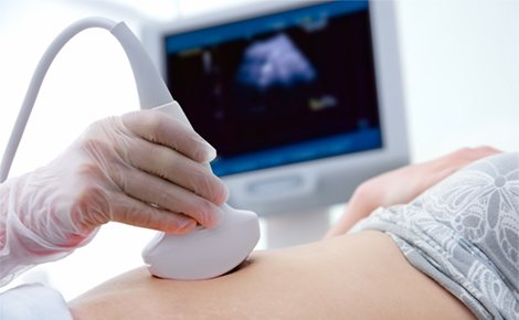
To conduct the study, the woman lies on the couch on her back. Legs bend slightly at the knees. The transvaginal ultrasound method (used in the early stages) involves inserting a special sensor into the vagina with a condom on. For a transabdominal examination, a special gel is applied to the exposed part of the patient’s abdomen. Next, the doctor begins to move the sensor over the patient’s abdomen.
3D and 4D ultrasound
3D and 4D ultrasounds, which can be done during pregnancy if there are certain indications (infrequently, usually 1-2 times during the entire gestation period), demonstrate more accurately both the size of the fetus and the structure of its internal organs. A three-/four-dimensional image of the fetus appears on the monitor screen. On it you can evaluate its structure in detail, see facial expressions, count the fingers and toes, and examine in detail the sex of the child. If any heart defects (or other organs) are detected on a 2D ultrasound, a 3D or 4D ultrasound procedure allows a more detailed assessment of the severity of these defects and gives an accurate prognosis for the further course of pregnancy.
How to prepare for the procedure
The procedure does not require special preparation. If it is performed vaginally, then the woman only needs to empty her bladder immediately before the ultrasound. You should also take a condom with you.
The main requirement for a transabdominal ultrasound is a full bladder. To do this, a woman should drink 1 - 1.5 liters of non-carbonated liquid (and not dairy!) 1 hour before the procedure, and then not empty the bladder. Alternatively, the pregnant woman may not urinate for 3 to 4 hours before the test so that she feels a strong urge to urinate at the time of the test. The procedure is carried out on an empty stomach.
Additionally, the woman must take with her to the hospital a medical card with the results of previous ultrasounds. At the state consultation, you are required to bring a towel or disposable diaper to lay on the couch, and napkins to wipe off the gel from the body.
Ultrasound and pregnancy
Experiments conducted on the harmfulness (harmlessness) of ultrasound examination for the mother and fetus prove that even repeated studies of this kind are harmless for infants. It has also not been proven whether a woman who has had ultrasound scans many times can give birth to a child with genetic abnormalities.
In other words, there is no evidence of the harm of ultrasound examination, just as there is no evidence of its harmlessness. That is why such examination is carried out by women strictly at the time specified by the doctor. This is especially true in cases where 3D research is used for examination.
Recently, information has appeared according to which babies in the womb can hear ultrasound very well, and it is unpleasant for them (in the form of a loud, piercing scream). In addition, they are well aware of the vibration coming from the ultrasonic sensor.
According to some researchers, a small child feels the same as a person who is directly next to the plane at the moment of takeoff. And this, you see, is far from a pleasant feeling. However, a very strong and piercing sound can negatively affect the neuro-emotional state of a small child.
For those who are especially worried about the harm of ultrasound to a child, we can cite this fact: doctors can perform an ultrasound on a pregnant woman even at the 38th week, that is, just before giving birth. And in these cases it will be completely safe.