Modern research method
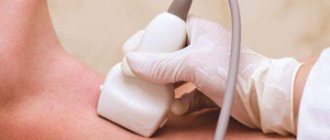
In a medical clinic, you can do rheovasography (RVG) of the vessels of the upper and lower extremities.
Rheovasography (RVG) of the upper and lower extremities is a modern method of functional diagnostics, with the help of which the intensity and volume of blood flow in the arterial vessels of the extremities is determined.
The principle of this research method is to measure the resistance of a skin area when an electric current of minimal strength (absolutely harmless), voltage and a certain frequency is passed through it using special sensors. Depending on the intensity of blood supply to the tissues, their resistance changes. The worse the blood flow, the higher the resistance of the skin and tissues. Changes in the resistance parameter are displayed on a paper tape in the form of a curved line, along which the functional diagnostics doctor determines the nature of blood flow in the area of the body being studied.
Suspicion of diseases
Some symptoms may indicate obvious diseases that can be identified through examination:
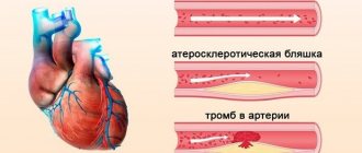
- atherosclerotic lesion due to stenosis or complete occlusion of the supply artery;
- autoimmune pathology with abnormal hemodynamics in the hands;
- diabetes mellitus with complications;
- inflammation of the walls of arteries from the inside;
- varicose veins
In addition, using RVG it is possible to determine the etiology of the disease. Understand what influenced the subsequent development of the pathology, the structure of the canals or reasons caused by lifestyle.
Indications
RVG is necessarily prescribed based on the patient’s subjective complaints about:
- leg cramps or swelling;
- the appearance of spider veins;
- pain and/or weakness when walking, arising and disappearing for no reason;
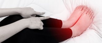
- numbness, chilliness, paleness of the feet or hands;
- pain in the arms with light exertion or at rest,
- change in skin color,
- long-term non-healing ulcers and cracks on the feet or heels.
The procedure can also be prescribed for smokers who complain of leg pain. RVG of the vessels of the lower extremities is used for diagnosis when the following diseases are suspected:
- phlebeurysm;
- atherosclerosis of leg vessels;
- thrombophlebitis;
- Raynaud's syndrome.
Main indications
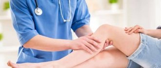
The disease can be identified and a procedure prescribed based on a person’s complaints.
The patient must list the symptoms manifested over a certain period of time. Complaints:
- Swelling of the extremities;
- leg cramps;
- visible venous network;
- sharp pain in the limbs when walking, which disappears as spontaneously as it appears;
- numbness of the feet or hands;
- pale skin color on the extremities.
Preparation for RVG of vessels
Before you begin examining the patient’s upper and lower extremities, the basic requirements preceding the rheovasography procedure must be met:
- Within 15 minutes, a person should receive complete relaxation (preliminary rest);
- 2 hours before RVH, smokers should stop taking nicotine into the body;
- 24 hours before the study, it is necessary to suspend the use of any medications by patients undergoing treatment;
- For RVH of the lower extremities, the legs should be freed from clothing.
Next steps for the patient
With a deciphered rheovasogram of the vessels of the lower extremities, the patient is sent to the doctor who prescribed the examination. Based on the results obtained, a diagnosis is made and treatment tactics are selected. In non-dangerous stages of vascular damage, the doctor prescribes special medications that strengthen the vascular walls, dietary nutrition, rational physical activity, and compression garments. In some cases, it is recommended to change your field of activity if work aggravates the disease.
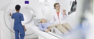
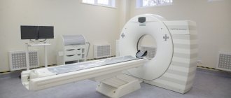
Serious changes in the blood flow system are an indication for additional research. A more detailed analysis of the hemodynamic process includes the following methods:
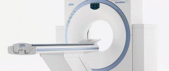
- RK angiography (x-ray contrast). Diagnosis of vascular diseases using x-rays with a contrast agent;
- MR angiography. Magnetic resonance imaging examination;
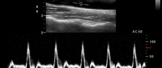
- Ultrasound Doppler. Ultrasound examination using Dopplerography.
Depending on the final results, the patient is prescribed conservative therapy or surgery. If you experience systematic discomfort in the lower or upper extremities, you should consult a phlebologist or vascular surgeon. The sooner the pathology is identified, the less radical the method of eliminating it will be.
Samples
In some cases, doctors use special tests during diagnosis that help give a more accurate picture. The most common:
- Nitroglycerin test. It consists of recording vascular resistance while taking nitroglycerin tablets. The bottom line is that the study is carried out before and after the drug is absorbed. This test allows you to assess the ability of spasmodic vessels to expand. If the drug does not cause any changes in the rheogram, the test is considered negative, and the spasm is considered an organic vascular lesion (this is most often observed with vascular damage by atherosclerosis). This test is contraindicated for pregnant women and children under sixteen years of age.
- Cold test. Most often used to study microcirculation of the fingers. Such a stress test will be very informative in the development of Raynaud's disease, so it is used in special cases. After a single examination, the patient is asked to hold his hands under running cold water for one and a half to two minutes, after which the examination is repeated. The test is considered negative when blood flow and vascular pulsation are restored by the eighth minute after cooling. If nothing has changed within fifteen minutes or the restoration of blood flow occurs slowly, then the test is considered positive.
- Compression test. It is carried out after examining the vessels in a calm state. Special rubberized cuffs are placed on the limbs, which compress the tissues, as a result of which the inflow and outflow of blood is disrupted. Next, sensors are applied, the cuff is removed, and the rate of blood flow restoration is assessed. This method is considered informative if the patient has thrombosis of small and large veins.
- Turns and tilts of the head. When diagnosing pathologies of the blood vessels of the head and neck, the doctor may ask you to turn or tilt your head in one direction or another and fix it in this position for a few seconds. Such a test is necessary to monitor the blood filling of the vascular bed.
Types of rheovasography
To comprehensively assess the characteristics of blood circulation in the extremities, doctors use different methods of applying electrodes:
- Longitudinal - when the electrodes are attached sequentially to one side, front or rear surface.
- Transverse - electrodes are located on different surfaces at the same height, allowing you to measure the resistance of all vessels at the same level (section) of the limb.
- Longitudinal-transverse - a combination of methods.
Operating with different options, the angiologist clarifies the location of the blood clot or changes in the lumen of the vessel that caused circulatory disorders. It also determines whether the disorder is functional or organic. Knowledge is necessary to choose a treatment method.
The patient is asked to remain motionless, except when performing functional tests, which the doctor asks for. This is a recording of a blood flow diagram after flexion-extension of the limbs, muscle tension, and turns. A compression test can be performed, squeezing the limb with a cuff. In some cases, measuring vascular resistance before and after taking a nitroglycerin tablet is indicated.
With the help of rheovasography, the patient can find out the type and degree of circulatory disorders in the extremities. And the doctor receives important information to develop a treatment plan.
To sign up for rheovasography at a medical clinic in Veliky Novgorod, contact the reception desk in person or by phone. A preliminary consultation with a neurologist is necessary.
How it goes
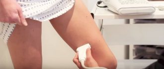
The room where the procedure itself takes place must be quite warm, since the patient is forced to take off his clothes, freeing his limbs for examination.
In order for the fixed electrodes to show accurate data, the person must lie in a motionless position. For convenience, a soft couch is available.
Before fixing the sensors, the surface fatty layer is removed from the skin. They are installed clearly according to the principle of longitudinal or transverse placement. This depends on the limbs being examined.
If the mechanical activity of the vessels of the arms is examined, then the sensors are placed in four places throughout the limb. On the legs these are the feet, legs and thighs.
Rheovasography (RVG) in Belgorod
Rheovasography is one of the ways to study blood circulation in the upper and lower extremities. RVG is one of the most informative, accessible and safe methods for assessing the condition of blood vessels in certain parts of the body. Since the vessels of the legs are subject to the greatest load, rheovasography of the lower extremities is most often performed. In addition, RVG of the upper extremities, head and neck can be performed.
Price for the service
| Rheovasography (RVG) | 400 ₽ |
What is rheovasography?
This research method is used to determine the tone and elasticity of blood vessels. In addition, it allows you to identify areas of blood vessels with partial or complete obstruction of blood. The results of rheovasography allow you to get a general idea of the state of the circulatory system, as well as identify specific diseases.
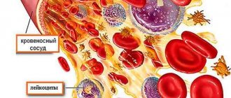
During the study, high-frequency alternating current is applied to a specific area of the patient's body. In this case, the resistance of certain body structures to alternating current is studied. Our body is a heterogeneous conductor. Blood is the best conductor of current, but bones are the worst conductor.
The level of electrical conductivity, depending on the degree of tissue saturation with blood, is recorded and recorded by a rheovasograph. The shape of the rheovasogram, its symmetry, as well as the distribution of peaks characterizing the inflow and outflow of blood give the diagnostician an idea of the condition of the blood vessels.
Why is vascular rheovasography performed?
Most often, the RVG procedure is prescribed for diagnosis:
- circulatory disorders in the extremities caused by diabetes mellitus;
- Raynaud's syndrome - a vasospastic disease characterized by circulatory disorders in peripheral vessels;
- vascular insufficiency.
And also to study the condition of blood vessels in thrombophlebitis, varicose veins, atherosclerosis, obliterating endarteritis and other vascular diseases.
In addition, RVG of the extremities can be performed to determine the causes of the following symptoms: feeling of coldness in the arms and legs, cramps, pain, swelling, numbness and tingling in the extremities, change in skin color.
How is rheovasography performed?
RVG is a non-invasive, safe and painless study. It has no contraindications or restrictions. Before conducting RVG you should:
- one day before, stop taking all medications that may affect blood pressure levels or the condition of blood vessels (a detailed list of medications will be provided to you by your attending physician);
- do not drink alcohol at least three days before the examination and refrain from smoking at least three hours before the procedure;
- try to avoid strong physical and emotional stress on the day of the study.
- 15-30 minutes before the examination, the patient is advised to remain completely at rest to relax the muscles.
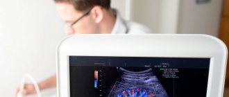
During the procedure, the patient lies down on a couch. The diagnostician attaches special sensors to his body. On average, rheovasography of the extremities takes 15-20 minutes.
Along with the high information content and ease of performing RVG, the price of this procedure is much lower than other methods for diagnosing vascular diseases, such as angiography, venography, Dopplerography and duplex scanning of blood vessels.
Reception is conducted by:
Getman Igor BorisovichCardiologist, functional diagnostics doctor
Key Indicators: What They Measure
The RVG results show a curve. Wave interpretation is based on qualitative and quantitative values. These include: the steepness of descents and ascents, intervals, amplitude, peaks.
Based on the values, control indices are derived, which are:
- rheographic - it controls the strength of blood flow in the arteries;
- arterial walls shows IE;
- IPS – peripheral resistance index;
- numerical value of venous outflow.
The most basic indicator is considered to be RI - rheographic index.
Decoding the results
Obtaining research results does not take much time.
Within 30 minutes, a specialist can decipher the received line and display the indices. RVG decoding is output in Ohms. As mentioned above, RI shows the total volume of filling of blood vessels. With readings of 0.05 Ohm and above, we can say that their condition is normal.
With deviations of one unit, mild deficiency is observed. If RI is sharply reduced, the patient is diagnosed with a blood supply disorder.
Standards for vascular tone data without deviations are calculated from 0.4. Values that do not reach the specified units are already considered violations. The lower the indicator, the lower the tone of the vascular wall.
The outflow index shows the speed of blood movement in the venous canal. RVG should range from 0.2 to 0.5. Anything that exceeds this line indicates serious illness.
The IPI responsible for counteracting vascular channels should be in the range of 0.2-0.45. In case of obvious blood flow pathologies, the index may be higher or lower than the norm.
Additional samples
Violations that were recorded during RVG do not always indicate damage.
The resulting index may have a functional nature of deviations. Often such results are obtained when preliminary preparation is not followed. There are two additional samples. They help determine what is really happening, a temporary spasm or pathology:
- Nitroglycerin. Using the drug, the rheovasography procedure must be completed several times. The first occurs before the drug is used, and the second after its action. The doctor receives data from two studies and conducts an analysis. If comparable results do not differ significantly, then we can conclude that there is a pronounced pathology on the face. In the case where the drug had an effect and the indicators improved, one-time disturbances caused are assumed.
- Compression. The essence of the procedure is to use a cuff that compresses the required area of study. As in the case of the RVG drug, the patient undergoes it twice. Before and after applying the cuff to the limbs. This test helps determine how quickly blood supply is restored when the deep veins are blocked.
The latest rheovasography devices have a large number of positive aspects. They greatly simplify the work of specialists. The system itself calculates the results, which is much faster than routine calculations.
At the same time, the patient does not receive any radiation or other negative effects on his body. The method is more than safe and informative. Due to this, this diagnosis is relevant in medical practice.