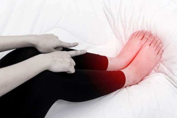
Vascular diseases of the lower extremities are a common problem in the modern population.
Vessels are divided into two groups: arteries and veins.
Through the arteries, blood flows to the tissues and supplies them with oxygen. Through the veins, blood flows from the legs, carrying away metabolic products and carbon dioxide.
Impaired arterial patency occurs in people:
- Smoking
- Having high blood pressure
- Diabetes sufferers
- Overweight
- Suffering from diseases of the liver, thyroid gland, adrenal glands
- Having high cholesterol or sugar in blood tests
- Not involved in physical education
Signs of diseases of the arteries of the lower extremities:
- Muscle pain during exercise
- Tendency to get cold feet
- Muscle and skin atrophy (muscle volume and strength decrease, skin becomes thin and pale)
- Lameness, weakness in legs
- In severe cases, pain in the lower leg muscles at rest, pain in the toes.
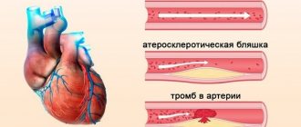
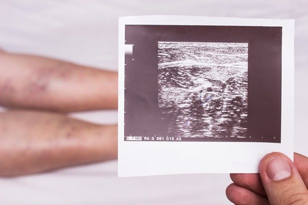
The reason for these changes is the narrowing of the lumen of the arteries by plaque, which gradually grows and further disrupts arterial blood flow.
The danger of narrowing the lumen of the arteries is that in severe cases it can lead to gangrene, amputation of the feet and even the entire leg.
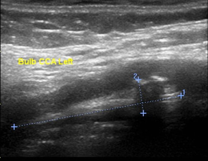
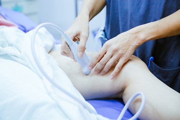
Timely diagnosis will help avoid irreversible changes. Once diagnosed, stenosis (narrowing of the artery) will allow vascular microsurgery to be performed (installation of a stent that widens the lumen of the artery), restoring the patient’s health and normal blood supply to the legs. One of the most informative diagnostic methods is color duplex scanning of the arteries of the lower extremities. The study is carried out on an expert-class ultrasound scanner, specifically designed for the study of blood vessels. Our specialists are highly qualified in performing vascular studies.
Blood flow in the femoral artery is normal
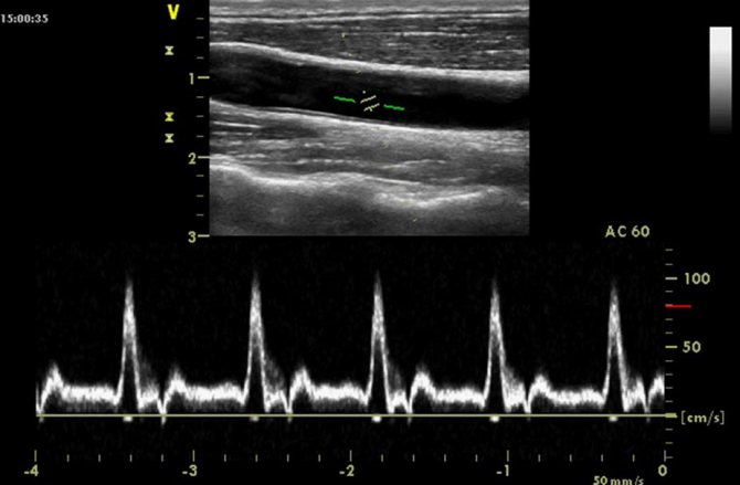
Color Duplex Mapping Mode
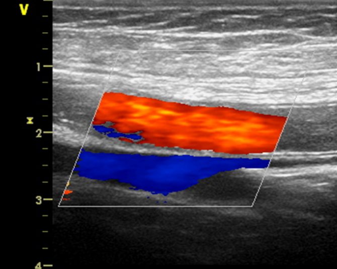
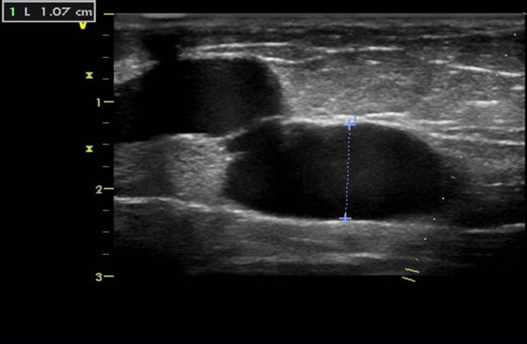
Large plaque in the lumen of the artery, narrowing by 70%
Vein diseases are divided into two groups: disruption of the venous valves, which leads to varicose veins; violation of venous patency - thrombosis.
Varicose veins of the lower extremities occur in every tenth person; vein thrombosis is somewhat less common, but is much more dangerous due to the risk of a blood clot breaking off and embolism into the pulmonary artery, which can cause death.
Causes of varicose veins:
- Heredity
- Low physical activity
- Excess body weight
- Work involving prolonged standing or sitting without moving
- Chronic lung diseases (increased pressure in the chest impairs venous outflow)
- Heart failure
- Frequent bloating
- Late pregnancy or the presence of a tumor in the pelvis compressing the inferior vena cava
Causes of thrombosis:
- Presence of varicose veins
- Heart failure
- Heart rhythm disturbances
- Previous heart attacks
- Chronic liver diseases
- Alcoholism
- Condition after injury associated with long-term limitation of mobility
- Condition after surgery, especially on the leg joints
- Presence of plaster or other structures for leg bone fractures
Signs of diseases of the veins of the lower extremities:
- The appearance of varicose nodes under the skin
- Tendency to swelling of the legs
- Change in color of the lower leg and foot (darkening, appearance of pigment spots)
- In severe cases, trophic ulcers on the skin of the lower leg.
- When varicose veins become inflamed, there is acute pain, redness of the skin and fever.
Color duplex scanning of the veins of the lower extremities is the most informative diagnostic method, allowing to identify both varicose veins and blood clots.
Varicose nodes
Thrombus in the femoral vein
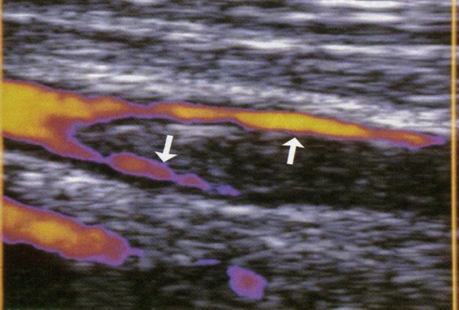
Thrombus in the great saphenous vein against the background of varicose veins

Varicose vein thrombosis

Types of ultrasound: Doppler ultrasound, duplex scanning and color Doppler mapping of the veins of the lower extremities
You can often hear the question, what is an ultrasound examination called? The question arises due to the fact that there is more than one ultrasound technique: triplex angioscanning, duplex ultrasound (USDS or USAS), Doppler ultrasound (Doppler ultrasound or USDG) of the veins of the lower extremities. The complexity of the terminology causes some confusion, but only for the uninitiated person. Regardless of how an ultrasound examination is done, it is always an ultrasound. Only the hardware method for diagnosing the veins of the lower extremities is changing. Let's look at both the terminology presented above and the differences between these research methods:
Doppler ultrasound or Doppler ultrasound of veins
The ultrasound system of veins and arteries of the lower extremities is based on the Doppler effect - the reflection of ultra-high frequency sound waves from red blood cells moving in the blood vessels. Based on this, the specialist receives information about the speed of blood flow, the lumen of blood vessels, the presence and absence of varicose veins, weakness of the vascular walls, etc.
!
Doppler is the name of an Austrian physicist, but now it has become a household name.
You can often hear that an ultrasound examination with Doppler is prescribed. This means that a procedure called Doppler sonography is ahead. At its core, it is a conventional ultrasound examination. To obtain the most informative data, Doppler ultrasound can be performed in duplex or triplex modes. Doppler ultrasound is considered one of the simplest ways to diagnose venous valve pathologies and assess blood flow.
Duplex scanning or ultrasound scanning of veins
What does duplex scanning do? What is the difference with Doppler ultrasound? This ultrasound examination of the veins of the lower extremities is considered the most complex and combines several research techniques.
Expert opinion
Doppler helps to study blood vessels, blood flow and their characteristics. A scanner operating in B-mode visualizes the received information on the monitor in on-line mode as two-dimensional gray scale images of anatomical structures. Ultrasound scanning makes it possible to differentiate pathological conditions of blood vessels that affect the decrease in blood flow, including cholesterol plaques and blood clots.
Vascular surgeon, phlebologist
Osipova Ekaterina Yakovlevna
Ultrasound ultrasound with color mapping / color Doppler mapping / triplex angioscanning
How is ultrasound performed using this technique? Triplex angioscanning allows you to maximally examine the blood flow of not only superficial and deep veins and arteries, but even the smallest connecting capillaries of the lower extremities. This is possible due to the combination of all the methods described above and the visualization of all the vessels of the legs with blood flow in color. This type of study is the most highly accurate in terms of determining not only the patency of blood vessels, but also their stenotic lesions.
Indications for ultrasound examination of the veins of the lower extremities
What parameters are being studied
During the examination, the following parameters are examined:
- Diameter of superficial and deep veins.
- Vessel lumen, areas with impaired blood flow.
- Venous patency.
- Functionality of valves.
- Absence/presence of blood clots.
- Condition of perforating veins.
- The condition of the vascular walls, their thickness, etc.
- Degree of vessel deformation.
Preparation for the procedure
Duplex scanning of the lower extremities does not require any preparation.
It can be performed at any time of the day. If necessary, manipulation can be carried out immediately after consultation with a phlebologist.
Features of ultrasonic testing
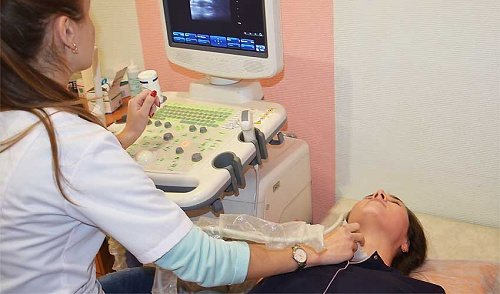
Diagnosis is carried out in the supine position. A special contact gel is applied to the skin of the legs, after which an ultrasonic sensor is installed. The doctor slowly moves it in the direction of the main veins and arteries. Additionally, the patient may be asked to take a deep breath and hold his breath to perform the Valsalva maneuver. A specialist can also conduct an examination of the lower extremities in a standing position - this is necessary to clarify the patency of the perforating veins.
The total diagnostic time is about 30 minutes. The patient immediately receives the scan results.
What can be revealed
The data obtained are necessary to determine vascular pathologies and venous insufficiency. The technique also allows you to assess the degree of blood supply disturbance and determine whether the patient requires surgical intervention.
Using the procedure, the following diseases can be diagnosed:
- Varicose veins
- Deep vein thrombosis.
- Thrombophlebitis.
- Atherosclerosis.
Norms and interpretation of results
Normally, a person should have the following diagnostic indicators:
- Veins – no thickening or widening of the walls, patency – good.
- Arteries – no areas of narrowing.
- Valves - no reverse blood flow.
- The speed of blood flow in the arteries of the leg is about 50 m/s.
- The speed of blood flow in the femoral artery is about 100 m/s.
- There are no blood clots or plaques.
Remember that if your results do not correspond to the data presented, this does not mean that you have pathologies. Only a phlebologist can evaluate the diagnosis, so be sure to make an appointment with him after the scan.
We will be waiting for you at our International Hemostasis Clinic for an ultrasound scan of the veins of the lower extremities. We offer high-precision diagnostics at a price starting from RUB 2,900.
Our medical center is located at the address: Moscow, metro station Kropotkinskaya / Arbatskaya, B. Afanasyevsky lane, building 22. You can make an appointment with us using the online form on the website or by phone.
Ultrasound of the veins of the lower extremities: indications and contraindications
Ultrasound of the leg veins is informative and, most importantly, a safe diagnostic method, as more than 60 years of medical practice has shown. In this regard, there is more than one indication for which an ultrasound of the legs is performed to check the veins and arteries:
- constant feeling of fatigue and heaviness in the limbs;
- pain syndrome;
- increasing or constant swelling;
- constant or sudden redness along the veins, accompanied by a burning sensation;
- manifestation of the vascular pattern – asterisks;
- the formation of trophic ulcers localized in the lower legs and feet;
- paroxysmal contractions of the leg muscles;
- changes in skin color;
- tingling feeling;
- cold feet.
All these signs may be symptoms of vascular disease. If they are detected in oneself, a person should consult a specialist for advice. He will examine the limbs and conduct a survey.
!
If necessary, if pathology is suspected, the doctor will certainly prescribe an ultrasound scan of the veins and vessels of the lower extremities.
To be able to monitor the state of health and the dynamics of diseases, a phlebologist prescribes an ultrasound scan of the veins of the lower extremities for:
- varicose veins;
- thrombophlebitis;
- diabetes mellitus;
- atherosclerosis;
- lymphatic edema;
- chronic venous insufficiency;
- thromboangiitis;
- postthrombotic disease;
- postoperative period, etc.
As mentioned earlier, ultrasound is a safe diagnostic method. But does this mean a complete absence of contraindications? Despite the safety of the method, it is important to note that there are absolute contraindications to its implementation. Therefore, it is impossible to find out the condition of the arteries and veins of the lower extremities using ultrasound if:
- infectious diseases and skin lesions;
- myocardial infarction;
- exacerbation of heart failure and arrhythmia;
- asthmatic attacks;
- cerebrovascular accidents in the acute stage;
- mental disorders during their decompensation;
- burn disease.
Carrying out an ultrasound examination in any of these conditions can provoke complications of the disease. However, it is worth noting that ultrasound of the veins of the lower extremities is not prohibited during pregnancy. As you know, during this period of a woman’s life, safety comes first.
Ultrasound with color circulation
What is CDK
Color Doppler mapping is one of the types of ultrasound examination based on the Doppler effect. When using this method, not only a black and white image of the internal organs is displayed on the monitor screen, but also color inclusions that show the direction and speed of movement of fluids through the ducts.
Red color indicates the movement of liquids towards the sensor. Blue – from the sensor. The intensity of the hue allows you to determine the speed. For this purpose, the device has a special color scale.
Ultrasound with Color Doppler at Dialine
You can get an ultrasound with colorectal dosage in Volgograd at Dialine clinics. We use the latest high-precision equipment, staffed by qualified, experienced specialists. With us you can undergo the examination at a time convenient for you, immediately receive the protocol in your hands and consult a doctor for decoding.
Ultrasound of organs with color circulation
This type of ultrasound examination makes it possible to obtain data on the direction of blood flow, its speed, the patency of arteries and veins, the presence of resistance in the form of blood clots, plaques and vasoconstriction. During the examination, the thickness of the walls of the blood canals can be measured and the risk of an aneurysm determined.
Ultrasound of the thyroid gland with colorectal dosage
It is prescribed for suspected thyroid tumors, as well as for patients at risk: those with a hereditary predisposition, those living in areas with a lack of iodine in water and food, those exposed to radiation.
Ultrasound of the breast with colorectal dosage
It is carried out to diagnose pathological formations in the chest. For maximum information content, the examination is recommended to be carried out from days 4 to 14 of the cycle.
Ultrasound of the abdominal cavity with color circulation
Includes examination of a whole complex of organs: stomach, large and small intestines, spleen, liver, pancreas, kidneys, ureters, adrenal glands.
Ultrasound of vessels with color circulation
Allows you to identify pathologically tortuous vessels, the presence of atherosclerotic plaques and blood clots, narrowing of blood vessels and impaired blood supply to vital organs. Can be used to examine the vessels of the lower and upper extremities, neck and head.
Ultrasound of the scrotum
This method of examination allows you to visualize the organ and determine by the nature of the blood flow whether there are pathological neoplasms.
Ultrasound of the pelvic organs with color circulation
An effective, affordable and highly accurate method for diagnosing a wide range of diseases of the female reproductive system. Used for screening pregnant women.
Ultrasound of the uterus and appendages with color circulation
Allows you to determine the state of blood flow in the uterine and ovarian arteries, endometrium, as well as pathological formations, if any.
Ultrasound of the gallbladder with color circulation
Thanks to visualization of blood flow, it will allow you to distinguish a stone from a polyp.
Ultrasound with color circulation in gynecology - features of gynecological ultrasound
In order to diagnose neoplasms in the ovaries, it is carried out on days 5-7 of the cycle, to diagnose endometriosis, the presence of fibroids and other formations in the uterus on days 22-26 of the cycle.
Ultrasound of soft tissues with color circulation
The skin, subcutaneous fat, tendons, ligaments, and nerve trunks are examined.
Ultrasound of the liver with colorectal dosage
It can be performed either in combination with an examination of the abdominal organs or separately.
Indications for ultrasound with color circulation
The list of indications for such a study is wide:
- pain in the arms, neck and sternum;
- swelling of the legs;
- cramps in the limbs;
- enlargement of the thyroid gland;
- pregnancy against the background of Rh conflict between mother and fetus;
- to assess fetal development;
- gestosis;
- the appearance of atherosclerotic formations.
Preparation for ultrasound with color circulation
Preparation recommendations vary depending on the area of examination. The general recommendation is to refrain from drinking alcohol and heavy meat foods, which can slow down blood flow, for several days before the procedure.
Carrying out the procedure
The procedure is similar to a regular ultrasound. It is performed in a lying or sitting position (depending on which part of the body is being examined).
Where to do an ultrasound with CDK in Volgograd, Volzhsky and Mikhailovka
At Dialine clinics you can undergo the widest range of tests. A complete list of clinic addresses is on the website. Call and specialists will help you make an appointment.
Preparation for ultrasound of the veins of the lower extremities
In order for the result to be reliable, most laboratory and instrumental diagnostic methods require certain preliminary preparation.
How to prepare for an ultrasound examination? There is no advance and complex preparation for venous ultrasound. There is no need, for example, for a diet. But it is important to exclude negative factors that affect the condition of the vessels of the lower extremities. This includes smoking and alcohol – at least 24 hours before. These 2 factors provoke dilation of veins and arteries, which can indirectly affect the result obtained.
Expert opinion
Before giving a referral for this diagnostic procedure, the patient must receive preparation recommendations from the doctor. If the patient is taking medications that have a vasoconstrictor or vasodilator effect, the doctor will most likely stop them a day before the test. This is necessary in order to eliminate diagnostic inaccuracy.
Vascular surgeon, phlebologist
Osipova Ekaterina Yakovlevna
On the day of the study, some time before it, the patient performs hygienic measures. Takes a shower or washes lower limbs. If you wish, for your own convenience, you can choose comfortable clothes that will allow you to free the part of the body being examined to the maximum and without temporary delays.
How is ultrasound diagnostics of leg veins performed?
How is ultrasound examination of the veins of the lower extremities performed?
If an ultrasound scan of the veins of the lower extremities is prescribed, the question may arise as to how it is performed? Most likely, each of us has undergone ultrasound diagnostics at least once, and even in general terms, we have an idea of how this process is organized.
When you need to check the condition of the blood vessels in your legs using ultrasound, it will take from 20 minutes to an hour of your time. The first thing the doctor will ask the patient to do is to remove clothing covering the lower limbs. This is necessary for unhindered access to the area requiring research. After which the patient takes one of the positions required to begin the diagnosis - lying on his back and stomach, as well as a standing position. The doctor will tell the patient the order of positions.
When the patient has taken the required position, the specialist applies a contact medium – a universal gel – to the skin of the legs. It is on top of this product that the doctor will advance the ultrasound sensor.
In some cases, accuracy will require the application of special cuffs that allow blood pressure to be measured in the diagnostic area. Testing may also be required. What are they and how are they made? These are actions with the help of which an ultrasound diagnostic specialist will be able to establish the absence or presence of vascular damage.
!
Some of the most common ultrasound examinations of leg veins are: Valsalva stress and the Hackenbruch-Sicard test (cough test).
For varicose veins on the legs, these tests allow for effective analysis and identification of the disease even at its earliest stages. Valsalva stress is a specific breathing technique, and the Hackenbruch-Sicard test is a series of cough movements.
If the specialist considers that these manipulations are not enough for a complete diagnosis, then additional studies may be required.
Interpretation of ultrasound results of leg veins
Information obtained using the CDC
This method can now be considered the most used in the world. After all, information obtained with the help of modern technology helps to understand the degree of damage to the endocrine gland, as well as the entire system as a whole. If you use DK in color, the doctor can examine not only the thyroid gland, but also the fluid around it. No other equipment has this capability. The specialist will check the condition of the patient’s blood vessels.
Color Doppler mapping makes it possible to analyze information “live”. On the display, the endocrinologist sees red and blue colors, which transmit data about blood movement. It should be noted that colors are not the type of vessels and arteries. To do this, the doctor uses a special list that describes in detail the problem and type of disease.
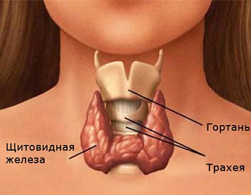
Basic data received by the equipment:
- Blood flow speed.
- Color information about the vascular system.
- The structure and type of tissue of a certain vessel.
- The rhythm of the thyroid gland during operation.
- Presence or absence of tumors.