Duplex ultrasound scanning of the brachiocephalic arteries, or abbreviated ultrasound BCA, is a modern ultrasound method for diagnosing the vessels of the head and neck, including the carotid and vertebral vessels, which supply blood to the brain, and the subclavian arteries.
First of all, the person who is scheduled for this study may have a question: what are the brachiocephalic arteries and where are they located.
Brachiocephalic vessels are the largest arteries and veins that are responsible for blood flow to the tissues of the head, brain and upper extremities. They are also called main lines.
general information
The brachiocephalic arteries include the carotid, subclavian, vertebral and their junction , which forms the brachiocephalic trunk. The listed vessels and some others near the base of the brain form the Circle of Willis, which is responsible for the distribution of blood flow throughout all parts of the brain.
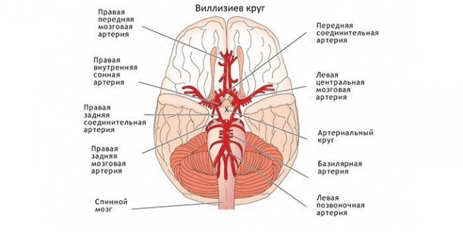
If any pathology has formed in one of the vessels, it can lead to disruption of the others, which ultimately leads to a stroke.
What is duplex scanning of the brachiocephalic arteries, and what is the method based on?
The apparatus for examining the BCA is based on the principles of echolocation . The working surface emits and then picks up ultrasonic pulses. The information is converted into a digital signal. This is how the image appears on the monitor.
The method is based on combining the advantages of B-mode - visual interpretation of the state of blood vessels and adjacent tissues and Doppleroscopy - qualitative and quantitative properties of blood flow. The Doppler spectrum can also be supplemented with color mapping.
Ultrasound diagnostics
The study of the main arteries using ultrasound is by far the most informative diagnostic method, allowing to identify not only physiological changes (wall deformations, width of the vascular bed, bends, the presence of atherosclerotic plaques), but also qualitative and quantitative indicators of blood flow.
Ultrasound scanning used to diagnose pathologies of the brachiocephalic arteries can be divided into 2 types:
- Doppler ultrasound (USDG);
- duplex scanning.
Both methods are based on the Doppler effect, the essence of which is to capture the reflected ultrasonic wave from moving objects. In the case of blood flow studies, red blood cells act as moving objects that reflect ultrasound. The change in the frequency emitted by the sensor (Doppler shift) occurs in direct proportion to the speed of red blood cells moving along the bloodstream, and if the red blood cells move towards the sensor, the frequency increases and the sensor records a positive shift, and if away from the sensor, the frequency decreases and a negative shift is recorded.
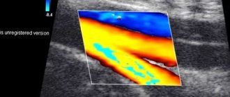
On the ultrasound monitor this is reflected in the form of a color image of multidirectional blood flows - red (positive shift) and blue (negative shift). The BCA ultrasound method has both positive and negative sides. A positive feature is the possibility of examining the transcarnial parts of the brain, that is, the arteries located inside the cranium, while for classical ultrasound examination (β-mode) this area is inaccessible.
The negative side is the inability to accurately determine the position of the vessel, and therefore diagnostics are carried out based on its probable location and changes in scanning depth. In rare cases, with an anatomically uncharacteristic location of the arteries, USDG cannot reflect blood flow, but this is not an unambiguous sign of its actual absence.
Duplex scanning of the brachiocephalic arteries is performed using the latest generation ultrasound scanners, combining classical β-mode ultrasound and Doppler ultrasound. The condition of the arteries and the quality of blood flow are assessed based on a two-dimensional (duplex scanning) or three-dimensional (triplex scanning) image. In this case, the vessel can be displayed in the transverse plane and in length.
The use of extracranial ultrasound (examination of vessels located outside the cranium) in duplex mode allows one to obtain the following detailed information:
- condition of the vessel wall;
- thickness, structure and number of atherosclerotic plaques;
- size of the vascular lumen;
- blood flow speed.
Important! Color Doppler mode reflects information about the quality of blood flow, and spectral mode is used to obtain quantitative information.
What does the ultrasound scan of the BCA show?
First of all, a duplex examination of the head is prescribed for those patients who have suspicions of atherosclerotic processes, aneurysms and deformations and other pathologies, and is aimed at identifying functional and structural arteriovenous disorders. During the examination, a specialist can recognize the presence of plaques, blood clots, thickening or reduction of vascular walls, a violation of the normal anatomical integrity of the walls, see tortuosity, surrounding tissue, and the speed of blood flow.
Ultrasonic scanning of the BCA shows:
- lumen of blood vessels;
- blood clots, plaques, detachments;
- stenosis, wall expansion;
- ruptures, aneurysms, deformations.
Using ultrasound scanning of the BCA, you can diagnose:
- vascular pathologies;
- vascular hypoplasia;
- violation of wall tone during VSD;
- atherosclerosis;
- arterial aneurysms;
- fistulas between vessels;
- angiopathy;
- thrombosis;
- vascular injuries;
- varicose veins.
The cerebral vessels are a complex system that is capable of self-regulation and maintaining cerebral blood flow.
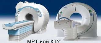
Only comprehensive diagnostics, which includes ultrasound scanning, CT, MRI, allows you to accurately and timely choose treatment, and then evaluate its effectiveness. Ultrasound scanning helps to assess the anatomy of the vessels of the neck and head, determine the characteristics of blood flow, and assess the condition of the walls and lumen . This way, atherosclerotic plaques, blood clots, tortuosity of arteries and their dissection can be diagnosed at an early stage.
What are the differences from ultrasound?
An ultrasound examination primarily measures the patency of blood vessels, responding to the movement of red blood cells in the blood. The image on the screen is one-dimensional; based on the state of patency, one can judge the presence or absence of vasoconstriction, atherosclerosis and other causes of stenosis. The examination is usually cheaper than ultrasound.
Duplex scanning of the vessels of the head and neck, together with conventional Doppler sonography, visualizes the vessels in B-mode, shows the localization of atherosclerotic plaques and thrombotic masses on the inner walls of the veins, their morphological features and the extent of the process. The picture is two-dimensional, in combination with color Doppler mapping (color Doppler mapping) - color.
Such capabilities of ultrasound scanning are its undeniable advantage, because the method not only saves time on diagnosing the disease, but also gives a broader picture of changes in the vessels . However, such research is correspondingly more expensive.
Why is the procedure prescribed?
Doppler ultrasound of the brachiocephalic vessels is necessary to assess various parameters of the main arteries, namely for:
- determining the degree of passability;
- identifying places of blockage;
- measuring the traversable diameter of the vessel;
- detection of atherosclerotic plaques and blood clots;
- studying the anatomical structure of the vascular system;
- determining the size of the vessel;
- detecting hypoplasia or dilatation.
Ultrasound scanning of the brachiocephalic vessels helps accurately diagnose abnormalities such as aneurysms or wall ruptures.
Features of diagnosing atherosclerosis
The initial sign of atherosclerosis, which an ultrasound examination can show, is not even a plaque, but a thickening of the wall of the carotid artery by just a fraction of a millimeter . With duplex scanning, this indicator is well determined. The thickness of the intima-media complex (the so-called IMM) is also called. IMT is taken into account to assess the effectiveness of treatment.
An increase in IMT of more than 1 mm is most often associated with risk factors such as smoking, arterial hypertension, diabetes, increased cholesterol, etc.
As the disease progresses, plaques begin to form. Usually they are localized in the so-called. Carotid bifurcation is the site of division of the common carotid artery into internal and external. The presence of a plaque in this segment is a serious risk factor for stroke and myocardial infarction. Therefore, it is very important to promptly identify atherosclerotic changes in the early stages .
Duplex scanning reveals the location of the plaque, as well as its shape, size, structure and degree of stenosis (narrowing of the lumen). When the lumen is already completely closed - this is occlusion .
During the examination of the BCA, tortuosity of the arteries is often revealed due to their elongation. Arteries lengthen due to atherosclerosis and high blood pressure. Tortuosity of the vertebral arteries usually occurs due to defects in the cervical spine. If tortuosity leads to compression of the lumen, this can cause disruption of cerebral blood flow.
Ultrasound scanning is also used to examine patients with traumatic vascular lesions: wall dissection or similar. The main symptom of this disease is a severe headache that cannot be relieved with conventional painkillers.
What are BCA and BCV?
Blood supply system to the head and neck
BCA stands for B
rachio
Cephalic
Arteries
,
and
BCV
-
as B rachio Cephalic Veins
.
“Brachion” means “shoulder” in Greek, and “kephale” means “head”.
The BCA is a set of arteries that branch from the cardiac aorta and supply blood to the brain, soft tissues of the head and upper extremities.
The brachiocephalic arteries are located in pairs on the right and left.
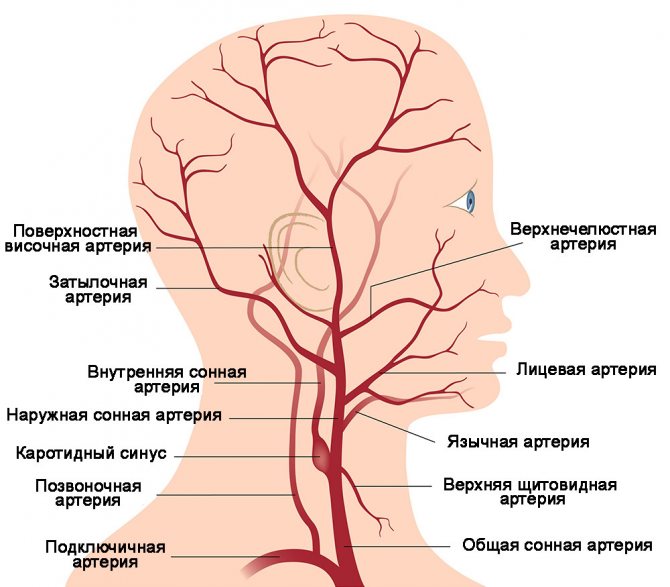
The brachiocephalic arteries include:
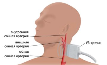
- Carotid arteries , which branch into internal and external in the region of the thyroid gland
- Vertebral arterieslocated in special openings of the cervical vertebrae
- Subclavian arteries , supplying oxygen and nutrition to the upper limbs
- Two brachiocephalic trunks (right and left), which are common vessels for each pair of carotid and subclavian arteries diverging from it
BCV , accordingly, is a set of veins through which there is a reverse outflow of blood from the brain, soft tissues of the head and upper extremities. The brachiocephalic veins, like the arteries, are also located in pairs on both sides of the body.
The brachiocephalic veins include:
- Internal and external jugular veins , which carry waste blood from the brain and head tissues and connect with the corresponding (right and left) subclavian veins in the clavicle area
- Subclavian veins , which carry blood from the upper extremities and jugular veins to the vena cava and then to the heart
Advantages of the method
The advantages of BCA ultrasound are:
- high information content;
- efficiency of research;
- safety and possibility of repeated implementation;
- painlessness of the procedure.
Ultrasound scanning of the BCA is more informative than conventional ultrasound scanning of the brachiocephalic vessels.
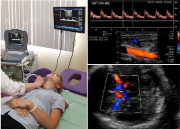
During the examination, an image is formed on the monitor, similar to a conventional ultrasound, but against its background the vessel in which the blood flow is formed is clearly visible. Due to the advantages of ultrasound scanning, BCA is considered the gold standard for diagnosing pathologies. A timely vascular ultrasound can save lives and prevent possible disability.
Learn more about the study and how duplex scanning of the vessels of the head is done from the video:
What does ultrasound examination of the vessels of the neck and neck examine?
Duplex scanning of blood vessels examines:
- vascular lumen, wall mobility;
- blood clots, plaques, detachments;
- narrowing, expansion of walls;
- deformations, ruptures, aneurysms.
Scanning of the extracranial sections of the brachiocephalic arteries in 99% of cases reveals circulatory disorders in the head area, leading to strokes: arterial hypertension, thrombosis, vasculitis. Timely ultrasound of the neck vessels will save your life and prevent the risk of disability!
Indications for use
The vessels of the brachiocephalic region include intracranial (intracranial, directly supplying the brain and surrounding tissues) and extracranial (extracranial, which capture the tissues of the neck, face, back of the head, etc. in the blood supply network). With duplex scanning, both the first and second groups are assessed.
Indications for prescribing duplex scanning of the BCA are:
- headache;
- dizziness;
- violation of movement coordination;
- blood pressure problems;
- fainting;
- elevated cholesterol levels;
- impaired sensitivity (numbness) of the limbs;
- blurred vision;
- flickering spots in the eyes;
- memory impairment and decreased concentration;
- preoperative examination.
Direct indications for the study are the following pathologies:
- atherosclerosis;
- VSD;
- hypertension;
- heart pathologies;
- neck injuries;
- compression of arteries and veins and other vascular injuries;
- vasculitis;
- blood diseases;
- suffered a stroke or heart attack.
Duplex scanning is also an important study for patients over the age of 40 years, with a history of stroke - acute cerebrovascular accident - and TIA - transient ischemic attack, with arterial hypertension, diabetes mellitus, elevated blood lipid levels, high body weight and planned myocardial surgery .
What does duplex scanning of brachiocephalic vessels: arteries and veins show?
What does a duplex scan of the brachiocephalic arteries (BCA) show?
Duplex scanning of brachiocephalic vessels
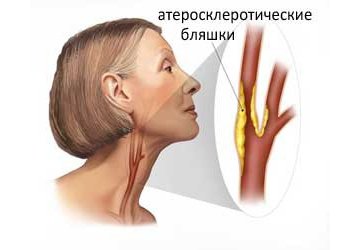
90% of all arterial pathologies are associated with atherosclerosis. Atherosclerosis is a disease characterized by the gradual deposition of cholesterol on the walls of blood vessels, leading to their narrowing, the formation of plaques, and in severe cases to complete blockage or thrombosis. Atherosclerosis is the “culprit” of such dangerous phenomena as stroke or heart attack .
With duplex scanning of the brachiocephalic arteries, it is possible not only to determine the presence of atherosclerosis, but also to assess the degree of narrowing of the lumen of the arteries, identify and measure the size of atherosclerotic plaques, their nature (stable-unstable), stage of formation and exact location.
In addition to atherosclerosis, duplex scanning of the brachiocephalic arteries makes it possible to diagnose a list of other, no less dangerous pathologies:
- vasculitis – inflammation of the vascular wall
- aneurysms - “protrusions” of the walls of blood vessels like a hernia. Aneurysms are dangerous because of their unpredictability, since if the walls of the vessel are significantly thinned, they can rupture, causing internal bleeding.
- dissection of the walls of the arteries - a longitudinal tear of the vessel wall with the formation of a blood clot in this place. Dissection is often the cause of stroke at a young age.
- mechanical compression of the arteries due to the growth of tumors or due to displacement of the cervical vertebrae
- congenital anomalies : atypical location, excessive tortuosity, loops, too narrow a diameter - which interfere with normal blood flow
- etc.
What does a duplex scan of the brachiocephalic veins (BCV) show?
Typically, pathologies of the brachiocephalic arteries cause more concern than pathologies of the brachiocephalic veins, however, the severity of the consequences when the brachiocephalic veins are affected cannot be underestimated.
In particular, the jugular veins (internal and external) are responsible for the outflow of blood from the cranial cavity. If the outflow is disrupted, the blood stagnates, causing certain brain damage.
With venous stagnation, the following may be observed: severe headaches, increased intracranial pressure, bettolepsy (short-term loss of consciousness combined with coughing attacks), small focal brain lesions, in rare cases, psychosis with auditory or visual hallucinations, etc.
Brachiocephalic veins, as well as arteries, are susceptible to pathologies such as atherosclerosis, vasculitis, aneurysms, dissections, congenital anomalies, mechanical compression due to growing tumors , etc., which are easily detected by duplex scanning.
In addition, duplex scanning is effective in diagnosing venous thrombosis , when a detached blood clot blocks the lumen of the vessel, disrupting the outflow of blood. Jugular vein thrombosis is usually accompanied by fever, swelling in the neck, headache, fainting, numbness of the extremities, etc.
Contraindications
The use of the device is absolutely harmless and does not have any effect on the human body. There are no restrictions on the use of this modern technique , and for any age group of patients.
In some cases, calcified atherosclerotic plaques can obstruct the ultrasound beam and interfere with diagnosis.
Do not forget that the doctor’s professionalism and good equipment play one of the key roles in deciphering the results of duplex scanning, so if a medical organization does not have professionals in this field or suitable equipment, it is better to use other diagnostic methods.
Indications
Such diagnostics are considered to be universal.
It helps to identify atherosclerosis and many other pathologies. But there is no thorough visualization of the anatomical structures during the procedure. Examination of the brachiocephalic arteries may be prescribed when:
- hypertension. It is accompanied by a stable increase in blood pressure;
- cervical osteochondrosis. This disease is characterized by compression of vessels by osteophytes that increase in size;
- diabetes mellitus, accompanied by a disruption of the process of insulin synthesis in the body.
Preparation for the procedure

Preparation before the study consists of excluding from the menu foods and dishes that can affect the tone and filling of blood vessels, which will distort the results of the study.
On the day of the study, you should not drink tea, coffee, energy drinks, Coca-Cola, alcohol, and do not indulge in overly spicy and salty foods . Directly before ultrasound examination of the BCA, you should not be in stuffy or smoky rooms, as this can also change the blood flow to the vessels.
It is better to refrain from taking vitamins and nootropics the day before the study. Be sure to consult a specialist if you are taking medications that affect the functioning of the cardiovascular system.
How is it carried out?
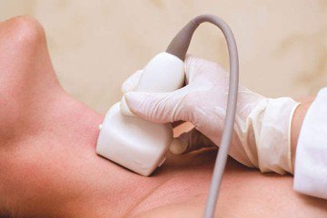
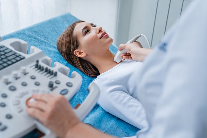
The patient lies on his back on a couch near the device, and the doctor places a cushion under his neck. The head should be turned in the direction opposite to the device. The doctor lubricates the surface of the skin with a gel that facilitates the passage of the ultrasound signal.
Using the sensor, the doctor will examine segment by segment in the neck area, observing the change in signal on the monitor. He may press the sensor lightly on the vessels or ask you to stop breathing for a short time.
If pathological changes are detected, a more thorough examination of all vessels extending from these branches is carried out.
Functional tests can be carried out to assess changes in indicators in horizontal and vertical positions of the body: lying down, sitting, and standing.
no uncomfortable sensations during the examination : the procedure feels no different from the usual ultrasound scan familiar to everyone. The study lasts 20-30 minutes.
Useful video about how transcranial duplex scanning of cerebral vessels is performed (through the cranial bone):
Duplex scanning technique
Duplex ultrasound scanning is not much different from ultrasound of other organs. The patient lies with his back on the couch, his head is turned to the side, and for convenience, sometimes a cushion is placed under the shoulders and neck. Scanning of the arteries is carried out using a sensor, which is moved along the skin of the neck in the planes required for examination - longitudinal - lateral, transverse and posterior - lateral.
Traditionally, the examination begins with the carotid artery; sometimes this vessel may be located differently than usual, so the diagnostician’s task is to fully examine it, regardless of the location. The carotid artery is examined before entering the cranial cavity; the sensor must be placed both directly above the vessel and at an angle to the surface of the neck. Stenoses, the size of formations, and the condition of the walls are well determined if the vessels are examined not only in the longitudinal, but also in their transverse direction.
The norm of the vessel lumen, blood flow speed, mobility, echogenicity are indicated in the device data, so the doctor only needs to correlate the readings obtained during the examination with the physiological ones. In some cases, ultrasound scanning may not be performed correctly, but this is usually due to anatomical deviations in the structure of the patient’s neck or the low resolution of the device. The experience of the doctor is also important, who can select a convenient sensor position that records all external and internal surfaces of the vessels in the optimal ratio.
Based on the pathologies identified during the examination and interpretation of the data, the doctor may decide to consult with other specialists or prescribe additional examinations of other organs and systems. A complete and comprehensive examination of the body will give a real idea of the pathological processes occurring in the body and will allow you to select the optimal treatment regimen. Duplex scanning performed at the beginning of the development of changes in blood vessels allows the patient to receive timely therapy, which significantly reduces the risk of acute circulatory disorders at almost any age.
Decoding the research results
The scanner will record the necessary indicators, and the doctor will enter them into the scanning protocol. Decoding the Doppler spectrum and blood flow charts will take no more than 10 minutes , after which you will receive the results.
The result of the scan is a printed sheet with a list of the examined vessels and a description of their sizes and condition. The decoding makes it possible to determine whether the vessels correspond to the anatomical norm , whether there is pathology, etc. Based on the decoding, your attending physician, if necessary, prescribes treatment.
Decoding is carried out by comparing indicators:
- nature of blood flow;
- its speeds: systolic (max) and diastolic (min);
- wall thickness;
- pulsator index (so-called PI) is the ratio of the difference between max and min speeds to the average (the sum of max speed and two min, divided by 3);
- resistive index (so-called RI) is the ratio of the difference between max and min speeds to min;
- systolic-diastolic ratio: max speed divided by min.
Based on the last 3 indices, the patency of the vessel is judged.
Blood flow is assessed in the external and internal carotid arteries, common (ECA and ICA, CCA), supratrochlear (SBA), main (OA), vertebral (VA) and in its segments, each of which has its own designations, for example, Vo, V1, V3 etc. Also in the anterior, posterior, middle cerebral arteries (ACA, PCA, SMA), subclavian (RCA), anterior and posterior communicating (ACA, PCA) arteries.
Normal indicators
Different vessels of the brachiocephalic zone have their own individual standards based on the results of duplex scanning. For the common carotid artery, a diameter of 4-7 mm, a systolic blood flow velocity of 50-105 cm/sec, a diastolic velocity of 9-36 cm/sec, and a vessel resistance index of 0.6-0.9 are considered normal.
The following values are acceptable for the branches of the common carotid artery:
- diameter of the internal branch – 3-6.5 mm; outer branch – 3-6 mm;
- systolic blood flow velocity of the internal branch – 33-100 cm/sec; outer branch – 35-105 cm/sec;
- diastolic blood flow velocity of the internal branch – 9-35 cm/sec; external branch – 6-25 cm/sec;
- resistance index of the internal and external branches is 0.5-0.9.
Normal parameters of the vertebral arteries:
- diameter – 2-4.5 mm;
- systolic blood flow velocity – 20-60 cm/sec;
- diastolic blood flow velocity – 5-25 cm/sec;
- resistance index – 0.5-0.8.
How affordable is it?
Such diagnostics require the use of expensive specialized equipment and specially trained medical personnel, so the prices for the study are not very liberal .
In large cities of the Russian Federation, the average cost of a duplex is from 2,000 to 5,000 rubles. In areas more distant from the capital, you can do the procedure for 800-1500 rubles. Abroad, you can undergo color duplex scanning of the main arteries of the head and vessels of the neck for $500-600 and more.
Let's summarize. Ultrasound scanning of the BCA is a special type of ultrasound diagnostics of vessels that provide nutrition to the brain, other organs of the head, neck, and upper limbs .
This is an accessible, safe, detailed and informative study, which in ten minutes can show the condition of the blood vessels and identify the cause of some unpleasant symptoms. An annual examination will allow you to predict the development of a cerebral stroke by 90%.