What does an ultrasound of the head and neck show?
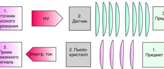
Ultrasound diagnostics is one of the most accessible and informative research methods and is widely used in most areas of medicine. The essence of the method is based on the ability of ultrasonic waves to penetrate into the body and be reflected from internal organs. The reflected signal is captured by the sensor and then transformed into an image on the monitor, allowing the doctor to literally look inside the patient’s body.
Ultrasound of the head and neck is aimed at examining the condition of the vessels, arteries and arterial ring in the neck and brain: vertebral arteries and veins, internal and common carotid arteries, basilar arteries, internal and anterior jugular veins, facial vein, subclavian artery, as well as some other vessels. Modern devices also allow you to listen to the noise of blood flow.
During the examination, the following is determined:
- vascular patency;
- lumen diameter;
- location of the lumen;
- the presence or absence of plaques and blood clots;
- condition of tissues around blood vessels and arteries;
- compliance of the blood flow with the norm.
Ultrasound of the head can detect brain diseases, which in the early stages are often asymptomatic. Moreover, their early detection significantly increases the chances of successful treatment. Also, using ultrasound, you can identify a predisposition to ischemic stroke. If you have such a predisposition, regular examinations and consultations with a doctor make it possible to prevent a stroke in 80% of cases.
What does triplex ultrasound of the vessels of the head and neck show?
Triplex scanning of the vessels of the head and neck allows the doctor to:
— assess the anatomical location of the vessels and their patency;
— determine the presence of stenoses (narrowings), kinks of blood vessels and their significance;
— identify atherosclerotic plaques, blood clots, vascular development abnormalities and their significance;
— assess the condition of the vertebral arteries;
- assess the state of venous blood flow in the vessels of the neck;
— find out the causes of headaches (increased intracranial pressure, vasospasm);
— diagnose the presence of cerebral aneurysms;
— quantitatively assess blood flow in the arteries of the head and neck, calculate total cerebral blood flow;
- see in color areas of vessel narrowing, turbulence in blood flow, areas of lack of blood flow
Indications for ultrasound of the head and neck
An ultrasound of the neck and head is prescribed if the following symptoms are present:
- frequent headaches;
- feeling of heaviness in the head;
- memory impairment;
- dizziness and fainting;
- noise in ears;
- decreased visual acuity;
- the appearance of “goosebumps” before the eyes;
- unsteadiness of gait;
- insomnia.
All these symptoms may indicate the presence of dangerous diseases and require immediate medical attention. In addition, ultrasound of the head and neck is used as a routine diagnosis for patients at risk. These include long-term smokers, patients with diabetes, patients with cervical osteochondrosis, hypertensive patients, people who have had a heart attack or stroke, and patients with high cholesterol levels. This examination can also be prescribed after injuries to the cervical spine.
The examination is prescribed by the attending physician and requires basic preparation, which is usually notified in advance. So, three days before the ultrasound, you need to give up alcohol, coffee, strong tea and energy drinks - these products can distort the picture of the study. It is not recommended to smoke directly on the day of the examination. Taking cardiovascular medications can affect the information content of the examination, but refusal of them should be discussed with the doctor, as this may lead to a worsening of the patient’s condition.
Ultrasound of head and neck vessels
This is a modern ultrasound diagnostic method, which makes it possible to most accurately determine the condition of the neck vessels that supply the brain, shoulder girdle and arms.
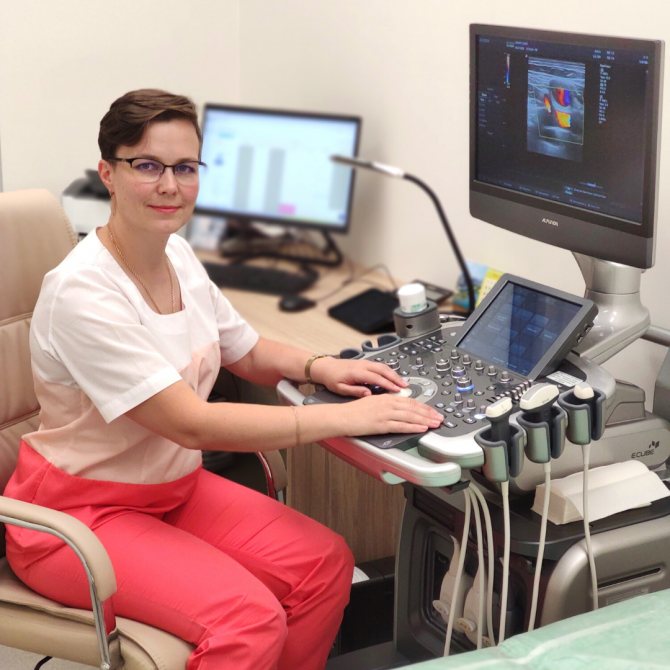
The article was prepared by our ultrasound diagnostics doctor Irina Alekseevna Romadova.
When a person is prescribed a study of the brachiocephalic arteries (BCA), he has questions:
— what will he have to deal with?
- How effective is this method?
- isn't it harmful?
— and won’t this procedure cause discomfort?
What is ultrasound of the vessels of the head and neck.
Duplex ultrasound scanning of the brachiocephalic arteries (USD BCA) is a modern ultrasound diagnostic method that makes it possible to most accurately determine the condition of the neck vessels that supply the brain, shoulder girdle and arms.
These vessels are called brachycephalic. These include the carotid, vertebral, subclavian arteries and their connection, which forms the brachiocephalic trunk.
The BCA study allows the doctor to determine the presence of a violation of the blood supply to the brain , and to predict the likelihood of worsening the disease in the future.
Types of vascular studies
Vascular examination can be carried out in three ways:
- Ultrasound Dopplerography or Dopplerography . The research is based on the Doppler effect, named after the Austrian scientist Christian Doppler who discovered it.
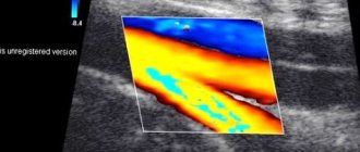
- Ultrasound scanning or duplex scanning of the vessels of the neck and head is a combination of both the Doppler effect and B-mode (regular gray ultrasound image). That is, the doctor can evaluate the movement and speed of blood flow , as in Dopplerography, as well as the internal walls of blood vessels , deposits on them, the presence of atherosclerotic plaques, thickening, and developmental anomalies. With this method, two functions are examined at once, hence the name duplex. Ultrasound scanning shows a complete picture of the movement of blood through the arteries and veins, which makes it possible to make a more accurate diagnosis.
- Triplex scanning is a type of duplex assessment. However, it provides even more information because it uses the additional technique of color Doppler mapping. A color image of the movement of blood in the vessels makes it possible to more accurately determine changes in blood flow.
Unlike conventional ultrasound, which shows organs in a static state, Doppler ultrasound shows the movement of blood flow and its speed. This is made possible by the difference in speed at which Doppler ultrasound reflects and receives waves.
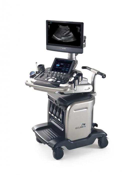
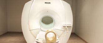
Undoubtedly, duplex scanning is the most modern research that provides a complete picture of changes in blood vessels. The ultrasound machine in our clinic belongs to the expert class and is equipped with all vascular programs. This makes it possible to most effectively visualize and evaluate pathological changes in the vascular bed.
Doctor's qualifications
We all know that the results of the study directly depend on the qualifications of the doctor performing the study. Our doctor Irina Alekseevna Romadova completed a full course of training in ultrasound angiology (examination of all vessels) at the Northwestern State Medical University named after I.I. Mechnikov, and masters all methods of vascular research. This is undoubtedly a big plus for our patients , because if there is damage to various vessels, then she can evaluate them all, besides, Irina Alekseevna is a former surgeon and is well versed not only in physiology, but also in anatomy .
Safety
Ultrasound examination is completely safe and painless for the patient. The use of this technique has no restrictions or contraindications. Ultrasound scanning can be used repeatedly, both for diagnosis and for monitoring the course of the disease at various stages of treatment.
What pathologies
The most common pathologies diagnosed with ultrasonography are:
- atherosclerotic lesions of the vascular wall
- vascular obstruction
- aneurysms
- deformations and abnormalities of vascular development
- dilation and incompetence of the venous bed
- thrombosis
- damage to the walls of blood vessels
The sensitivity of the method is very high , with ultrasound even minor, initial manifestations of atherosclerosis in the walls of blood vessels are visible, the identification of which will allow timely measures to be taken and prevent the formation of atherosclerotic plaques .
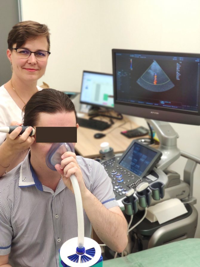
In some situations, it is necessary not only to examine the vascular bed, but also to evaluate the elasticity of the vessels, their elasticity, and ability to respond to changes in blood pressure and oxygen content in the atmosphere ( resistance ). For these purposes, the Carbonik , which is equipped in the ultrasound room at the Smart clinic. It has been clinically proven that with the help of a breath test using the Carbonik device, the most reliable information about changes in the resistivity of the vascular bed is obtained.
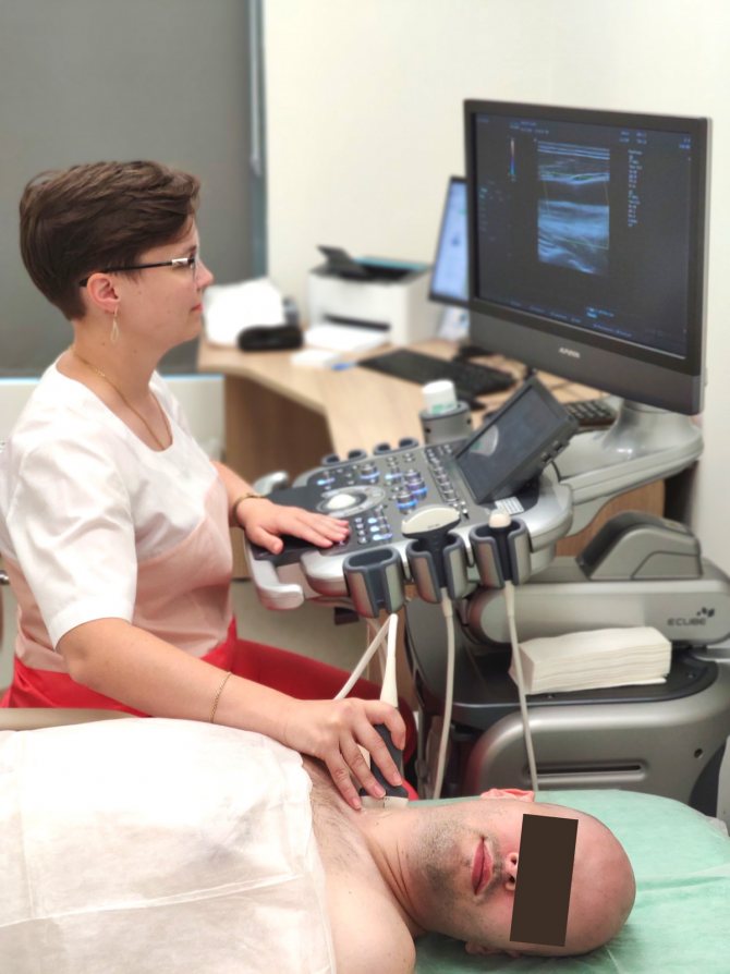
This photo shows the process of conducting a study of cerebral vessels with the Carbonik apparatus.
Who is this procedure indicated for?
As a measure to prevent serious complications of cerebrovascular accidents, such as stroke, BCA ultrasonography is recommended to be performed on individuals at risk at least once a year.
- People over 40–45 years of age.
- Patients with diabetes mellitus.
- Persons with a family history of stroke.
- If abnormalities are detected in the lipid profile.
- Patients with pathology of the heart and vascular system.
- Smoking and alcohol abuse.
- Patients with osteochondrosis of the cervical spine.
- Autoimmune vascular disease (hemorrhagic vasculitis)
This study is also indicated for all people with complaints indicating changes in the vessels of the neck.
- constant or recurrent headaches;
- migraine
- dizziness;
- sensation of noise or ringing in the ears;
- absent-minded attention;
- impaired coordination of movement;
- decreased vision;
- memory change;
- blood pressure problems;
- fainting;
- neck injuries;
- condition after a heart attack or stroke.
Preparing for the study
BCA ultrasound does not require , but on the day of the examination it is better to exclude from the diet foods that can change the tone of the vascular wall, such as coffee, strong tea, alcohol, energy drinks, cola. It is advisable to refrain from smoking two hours before the test. If you are taking medications that affect the functioning of the cardiovascular system, be sure to tell your doctor.
Carrying out the procedure
The procedure takes about 30-40 minutes, the patient lies on his back, the chin should be raised up and slightly turned in the direction opposite to the one being examined. The doctor may press a little on the patient’s neck during the examination, but in general this does not cause any discomfort. Our clinic has a “ platinum ” version of the device, including a device for heating the gel , which makes the procedure even more comfortable. The ultrasound scanner will record all the indicators, and the doctor will enter them into the study protocol.
Summary: Duplex ultrasound scanning of the brachiocephalic arteries is accessible and effective diagnostic method. In addition to high resolution, the method is absolutely safe . The Smart Clinic has an expert level ultrasound machine installed, and our doctor, Irina Alekseevna Romadova, is an experienced specialist, which together gives our patients timely and effective diagnosis, and therefore, subsequently, the correct treatment.
How is the examination carried out?
To perform an ultrasound of the head and neck, the patient is placed on a couch on his back. The neck area is freed from clothing and jewelry. To conduct the examination, the head is tilted back and turned away from the sensor. A gel is applied to the skin, this is necessary to remove air from under the sensor. The doctor moves the sensor over the area under study, stopping at the places where the vessels and arteries are projected. To increase the information content, functional tests may be required: the doctor can press the vessels with fingers or a sensor, ask the patient to breathe deeply. In some cases, vascular medications may also be required. The examination is painless and usually does not take more than 30 minutes. The big advantage of ultrasound is that it is harmless and has no contraindications. The examination is often prescribed even for newborn children and can be carried out as often as necessary.
Ultrasound involves conducting an examination in parallel with decoding. Such indicators as the maximum and minimum speeds of blood flow in the vessels, the ratio between the maximum and minimum speeds, and the nature of the blood flow should be recorded. The resistance index is also calculated for vessels. The obtained data are compared with the norm, which allows us to draw a conclusion about the patency of the vessels and the state of their lumen.
The diagnostic method is used not only to make a diagnosis, but also to monitor the dynamics of the disease and evaluate the effectiveness of the therapy.
You can undergo an ultrasound of the neck and head in Volgograd, Volzhsky and Mikhailovka at Dialine clinics. We use the most modern ultrasound machines, which, combined with the extensive experience and professionalism of our employees, allows us to carry out highly accurate diagnostics. You can sign up for an examination by phone or through the website. For maximum convenience of receiving medical services in our clinics, register in your personal account.