MRI of the cerebral arteries is a progressive technology that allows you to obtain a three-dimensional image of the vascular network and identify abnormalities and pathologies in the functioning of the arteries.
For a long period of time, neurologists made diagnoses based on clinical findings and examination results. However, after the advent of magnetic resonance imaging, disease diagnosis has become easier and more accessible. MRI of the arteries of the head allows not only to identify pathology, but also to determine the stage of its development. This will allow the doctor to prescribe treatment in a timely manner and reduce the likelihood of complications.
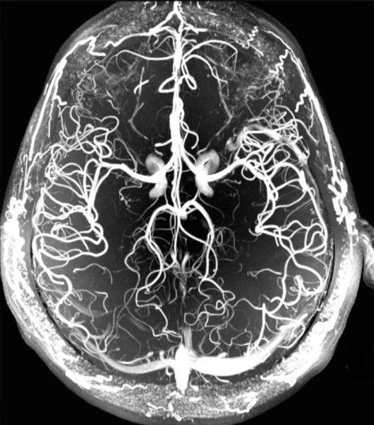
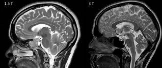
MRI image of cerebral arteries
Magnetic resonance imaging allows not only to see the structure of the arteries, but also to evaluate the lumen of the vessel and visualize the vascular wall. Thanks to MRI, diagnosis of diseases has become possible at the earliest stages of their occurrence.
Abnormalities of the cerebral arteries
Pathologies of the arteries of the brain can lead to a number of serious conditions. Among the most dangerous diseases that MRI shows in the arteries of the brain are the following:
Aneurysm
With this pathology, an expansion of the lumen of the artery appears in the affected area. Previously, this disease was diagnosed using X-ray angiography. However, the safest method today is MRI. Timely detection of an aneurysm can prevent massive hemorrhage into brain tissue.
Angioma
An angioma is a neoplasm of vascular tissue, the size and location of which can vary. On MRI images, the pathology appears as a multinodular formation.
Atherosclerosis
In atherosclerosis, fats are deposited on the inner walls of blood vessels, forming plaques. If treatment is not started in time, hypoxia of brain tissue may develop.
When conducting an examination, three stages of the disease can be identified:
- There are a few inclusions of cholesterol on the walls of blood vessels.
- Cholesterol becomes more and more, platelets are added to it, plaques are formed, narrowing the lumen of blood vessels.
- Calcium ions settle on the plaques.
In the third stage, a person often experiences severe headaches, cognitive dysfunction and memory impairment are possible.
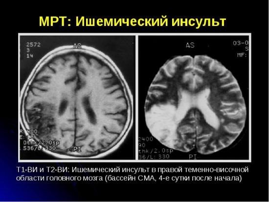
Stroke
This disease is an acute disorder of cerebral circulation, in which an area of the brain ceases to be supplied with blood, resulting in tissue necrosis. There are several types of pathology:
- Hemorrhagic. In this type of stroke, an artery in the brain ruptures.
- Ischemic. A blockage of the artery lumen with a thrombus occurs.
- Lacunar. When blood vessels become blocked, cysts can form.
- Cardioembolic. The cause of blockage of blood vessels is cardiac emboli.
If symptoms of a stroke appear, an urgent MRI is necessary, which will show the extent of damage to surrounding tissues. According to statistics, only 10% of people can fully recover from a stroke; 90% of patients develop serious complications. It is timely diagnosis that will help significantly facilitate rehabilitation after illness.
MRI of the cerebral arteries allows one to accurately determine the type of stroke and the location of circulatory disorders.
If the examination was done in the first hours after a stroke, the lesions will be clearly reflected in the images and doctors will be able to begin emergency treatment to resolve the blood clot. If a stroke is detected after some time, the edema resolves, the sulci of the cerebral cortex and the lateral ventricles expand.
Hypertension
Hypertension is a disease of the cardiovascular system, which is characterized by increased blood pressure caused by impaired vascular regulation and cardiac pathologies. In the cerebral arteries, MRI reveals indirect signs of hypertensive angiopathy.
Arteriovenous malformation
Malformation occurs when several vessels of different diameters become entwined together. One artery feeds this node, and a vein drains blood. MRI requires finding these vessels. During the study, a contrast agent may occasionally be used to help determine the location of the malformation.
The main advantages of MRI of neck and brain vessels
In addition to the high accuracy of the study, MRI of neck vessels and MRI of cerebral arteries have a whole list of advantages. These include:
- Absolute harmlessness and non-toxicity of the medical device for the health of the patient.
- No contact with surgical or any other medical equipment.
- Maximum ease of assessment for the patient.
- A narrow list of contraindications.
- The ability of a tomograph to detect deviations from the norm and different types of diseases at the initial stage of their development.
- The ability to be tested repeatedly without a long recovery period.
You need to know that examination of the main organ in the human body using resonance equipment is almost always done together with an MRI of the neck and its parts. This technique is required because the filling of blood vessels in the brain is directly dependent on stable blood flow in the arteries below. If the neck was subject to trauma or injury, then the described case may lead to a lack of oxygen supply, which could very well lead to death.
With the help of the ECP, our clients can apply for an appointment with a treating specialist - without leaving the comfort of their home! All you need to do is visit our official website, where you can find a lot of useful information: price lists of clinics, verified addresses of medical organizations and the number of available places for the coming days. Also, with the help of our company, you have the opportunity to get acquainted with truthful and honest reviews of real patients of medical centers and make a choice of clinic based on the assessment. Our managers are always ready to listen to you and give detailed answers to your questions.
MRI of the cerebral arteries: for what symptoms is it done?
Indications for MRI of the cerebral arteries may include the following symptoms:
- frequent, severe headaches;
- dizziness, loss of coordination;
- head injuries and bruises;
- increased intracranial pressure;
- speech disorder;
- cognitive impairment;
- noise in ears;
- sudden deterioration of vision.
When such conditions occur, patients most often turn to a neurologist. After asking the patient about troubling symptoms, the doctor may prescribe a number of examinations, including an MRI of the arteries of the brain.
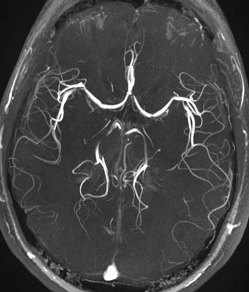
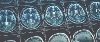
MR image of cerebral arteries
The study can be carried out not only to diagnose the disease, but also to determine the effectiveness of treatment for various pathologies.
MRI of the arteries of the head - indications and contraindications
MRI of the cerebral arteries is prescribed to identify a number of diseases:
- dissection of the walls of blood vessels;
- cerebral hemorrhages;
- senile dementia;
- congenital vascular pathologies;
- vascular malformations;
- arterial stenosis;
- atherosclerosis;
- thrombosis;
- cerebral ischemia;
- aneurysm (expansion of artery walls);
- neoplasms.
Like any other diagnostic procedure, magnetic resonance imaging has its contraindications.
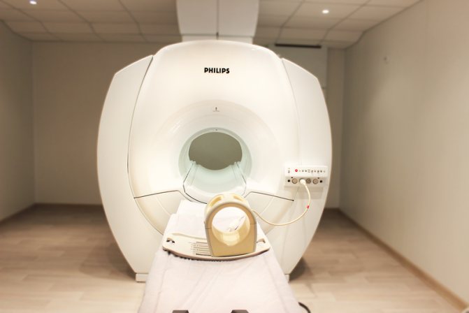
Modern tomograph Philips Intera 1.5 T
The examination cannot be carried out in the following cases:
- first trimester of pregnancy;
- the patient has a pacemaker, neurostimulator, vascular clips or medical devices and products made of ferromagnetic materials;
- the patient's weight is more than 130 kilograms;
- waist circumference more than 150 centimeters;
- inability to maintain a stationary position for various reasons;
- children under 5 years of age.
If you suspect possible contraindications, you must inform your doctor about them, who will help determine whether the procedure is possible in each specific case.
If it is necessary to use a contrast agent, you must ensure that there are no previous severe allergic reactions. MRI of the cerebral arteries usually does not need to be performed with contrast, but if such a need arises, you need to make sure there is no pregnancy.
Preparing for an MRI of the head with intravenous contrast
No special preparation is required for contrast MRI of the brain, unless the woman is breastfeeding. Chemicals introduced into the blood pass into the milk. When it is used in the body of a newborn, undesirable reactions develop: flatulence with bloating, diarrhea, increased nervous excitability, problems with falling asleep, etc. If the child has an individual intolerance to contrast components, the negative impact of the latter increases. After injection of the drug, the baby is weaned from the breast for 24 hours. For this period of time, the mother needs to prepare a sufficient amount of food.

Before an MRI with contrast, a nursing mother should prepare food for the baby for 24 hours
For MRI of the brain with a contrast agent, patients are advised to come with a light snack, since excess food in the stomach or the absence of it can cause nausea. For the procedure, you need to choose comfortable clothes without metal fittings. Some diagnostic clinics have disposable shirts (it’s best to check this question when making an appointment).
You need to have with you:
- identification document for drawing up a contract;
- a referral from the attending physician, which indicates why this study is necessary (presumptive diagnosis);
- an extract from medical records or an outpatient card;
- results of previous examinations of the head and neck area, if any.
Electronic devices, wallets, and bank cards cannot be brought into the room where the tomograph is located. Jewelry, glasses, watches, dentures, hearing aids, hair clips and other items that the device can attract and damage are left in the locker room. Women should not use decorative cosmetics, creams, or hairsprays to avoid artifacts that reduce the quality of images.
MRI of cerebral arteries with contrast
In most cases, modern high-field tomographs will help provide a detailed picture of the structure of the arteries and veins of the brain. But sometimes doctors resort to improving image quality by introducing a special coloring agent - contrast. This is done in order to see a clearer picture when damage or changes in the arteries may be very small in size.
Modern contrast agents are made from the metal gadolinium and are safe. The drug is administered intravenously and stains damaged tissue. In this case, the patient usually does not feel anything unusual. In rare cases, a feeling of warmth may occur at the injection site or area of examination.
Contrast drugs practically do not cause allergies and do not require a preliminary allergy test if the patient has not previously experienced acute allergic reactions or severe renal failure.
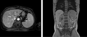
What does an MRI show?
Doctors recommend doing an MRI to confirm diseases of the brain and spinal cord, internal organs (with the exception of hollow organs), articular apparatus, spinal column, and organs of the reproductive system. Scanning is used to determine:
- spinal hernias;
- inflammatory processes;
- benign and malignant neoplasms;
- vascular pathologies (diseases of the cardiovascular system, changes in blood flow);
- presence of bone fragments;
- infectious processes of the osteoarticular apparatus;
- traumatic lesions;
- liquid in cavities.
The procedure is not relevant for identifying problems of the stomach, intestines, lungs (contain air), as well as bone structures.
The accuracy of MRI can be improved by using a contrast agent. A gadolinium-based agent is used as a contrast, administered intravenously. The drug accumulates in areas with increased blood flow (inflammation or tumor). MRI with contrast determines the pathological process in the initial stages of the disease. The substance introduced into the bloodstream is absolutely harmless to the body and is eliminated within 24 hours.
How is an MRI of the cerebral arteries performed?
After the patient enters the office, he must notify the doctor about contraindications, if any. He needs to remove all metal objects. If the procedure will be carried out in clothing, you must make sure that there is no metal on it.
The patient is asked to lie on the couch and the position of the head is stabilized. This will reduce the risk of deterioration in the quality of the study, since the slightest movements of the head may result in blurry images.
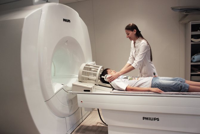
Preparation for MRI of cerebral arteries
Patients who are afraid of being in a confined space can take sedatives first. In case of deterioration of health, the patient can always contact the doctor during the procedure via two-way communication.
Modern high-field tomographs produce quite loud sounds during operation. There is no need to be afraid of this - such sounds indicate normal operation of the device. To reduce discomfort in the medical room, each patient is given headphones, in which pleasant music plays during the procedure, drowning out the noise from the operation of the tomograph.
After the end of the study, the patient is given images recorded on a disk or flash drive, as well as a written report from the doctor, within half an hour.
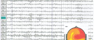
Interpretation of MRI of cerebral arteries
After the patient has completed the examination, the results are deciphered, consisting of several stages:
- The tomograph transfers many of the images taken to a special computer. The number of images can reach a thousand, and the slice thickness is 1-3 millimeters.
- The radiologist carefully examines the images and draws up a written report that describes in detail the anatomical structures, possible deviations in structure and functioning.
If a person does not have any problems with the arteries, then in the pictures they will be highlighted evenly, there will be no nodes or voids. If a tumor or cyst is present, dark spots will appear on the images.
The radiologist describes what he sees in the images, and the final diagnosis is made by the attending physician based on the MRI report.
At the medical office, you can undergo a magnetic resonance imaging procedure of the arteries of the brain on any day and time convenient for you. The center’s doctors have extensive experience in quickly and clearly interpreting the images received.
MRI of the cerebral arteries is a safe procedure that shows the slightest manifestations of the disease in the early stages of their occurrence. The study does not cause discomfort, is quick and painless, and its effectiveness is not comparable to any other type of study of the cerebral arteries.
What can an MRI show in vascular mode?
MRI can reveal any vascular changes. Scan results reveal:
- atherosclerosis
- aneurysms
- strokes
- thrombosis
- encephalopathy
In most industrialized countries, cerebrovascular diseases account for up to 15% of total mortality. Mortality from cerebrovascular accidents in Russia ranks 3rd in the statistics of overall mortality. Mortality from cerebral strokes exceeds that from myocardial infarction by 2-3 times.
Screening visualizes intracranial and extracranial vessels, which makes it possible to detect diseases in the early stages, which significantly affects the effectiveness of treatment. Magnetic resonance angiography provides a comprehensive assessment of cerebral and vascular activity, as well as the state of the central nervous system.