Various types of tumors in the brain account for approximately 6% of all tumor formations in the human body. They occur in approximately 10 - 15 cases per 100 thousand population.
ALENA PARETSKAYA
Pathophysiologist, immunologist, member of the St. Petersburg Society of Pathophysiologists ALEXANDER SERYAKOV MD, professor, oncologist, hematologist, radiologist (radiation therapist) of the highest category, leading oncologist of the SM-Clinic holding »
What is a brain tumor
“Brain tumors,” says oncologist Alexander Seryakov, “are intracranial neoplasms, including both tumor lesions of the brain tissue itself, as well as nerves, membranes, blood vessels, and endocrine structures of the brain.
Such formations can be benign or malignant in nature. Benign tumors put pressure on certain areas of the brain directly adjacent to the tumor and can lead to disruption of the functioning of individual structures in the brain itself.
A malignant tumor is distinguished by very rapid growth and the ability to grow into the surrounding tissues. Glioma is one of the most common types of malignant brain tumor. Its most aggressive type is glioblastoma. It grows quickly and has no clear edges. It is difficult to treat, with a high relapse rate.
Other malignant brain tumors include:
- meningiomas – tumors of the meninges;
- neuroepithelial tumors (gangliomas and astrocytomas);
- Neuromas are special tumors from the membranes of nerve cells.
Causes of tumors of the cerebral hemispheres
One of the most common and undeniable causes of the development of intracerebral tumors is radiation exposure. Many scientists consider electromagnetic waves emitted by mobile phones to be a risk factor.
Provocateurs of the origin and development of CNS tumors can be:
- disruptions or disturbances in the functioning of the immune system (sometimes the disturbances are hereditary in nature or are the result of immunosuppressive therapy);
- diseases associated with impaired immunity (for example, the HIV virus).
An altered immune system contributes to the occurrence of lymphomas (formations from cells of the immune system - lymphocytes), which originate in the lymph nodes, spinal cord or brain.
Consumption of the sugar substitute aspartame and exposure to vinyl chloride (a colorless gas used in the manufacture of plastic products) also contribute to the development of intracerebral tumors. There is a polyetiological theory of tumor growth, which indicates the important role of external environmental influences along with dysontogenetic factors. But today there is no scientific evidence of the dependence of the development of these formations on geographical conditions or territorial features.
Not least important is hyperplasia, which occurs due to inflammatory processes, injury or intoxication. Also influenced by:
- human hormonal imbalances;
- anaplasia;
- tissue dedifferentiation;
- hereditary tuberculous sclerosis;
- Recklinghausen's neurofibromatosis.
Possible factors influencing the development of intracerebral tumors include the patient's gender, age, hormonal disorders, and heredity (family history).
Causes of brain tumors in adults
Scientists have not yet determined the exact cause of tumors, but there are assumptions about the connection of their growth with the effects of radioactive radiation, toxins penetrating the body, and environmental pollution.
Children may develop congenital neoplasms - one of the reasons is considered to be intrauterine development disorders. A possible factor may be traumatic brain injury, which can also intensify an already existing process.
There is evidence that some brain tumors can develop after radiation therapy prescribed for the treatment of other pathologies, immunosuppressive treatment, and HIV infection. There is a hereditary predisposition to certain types of brain cancer. But for many people the cause remains unknown.
About 10 - 30% of brain tumors are of metastatic origin - these are cells that are carried by the blood flow (less often lymph) into the brain vessels, tissues, and membranes. About 60% of such tumors are multiple.
Most often it metastasizes to the brain:
- in men – lung damage, colorectal cancer, kidney damage;
- in women - breast cancer, melanoma, colorectal and lung cancer.
Intracerebral metastases occur with cancer of the uterus, digestive tract or prostate.
Causes of brain cancer
Despite numerous scientific and clinical studies, the exact causes of brain cancer have not been established.
Here are the key risk factors for brain cancer:
- previous radiation therapy;
- genetic diseases: Turcotte syndrome, Gorlin syndrome, etc.;
- HIV infection;
- hazardous industries: chemical, oil industry;
- head injuries;
- smoking;
- metastases to the brain in secondary (metastatic) brain cancer.

Signs of a brain tumor in adults
Focal symptoms are often among the first to appear.
They arise due to the pressure of the tumor on the surrounding tissues and chemical reactions to foreign cells, hemorrhages, blockage of blood vessels with a tumor embolus, compression of tissues and nerves. As the tumor grows, symptoms from neighboring areas appear, then general brain symptoms. If the tumor is large, a so-called mass effect may occur (the main structures of the brain are displaced, causing the cerebellar region to become wedged into the opening of the skull). One of the early signs is headache. It is usually local and occurs due to irritation of blood vessels, nerves, and meninges. There may be diffuse pain throughout the head, which is typical of meningeal lesions. The nature of the pain is paroxysmal, deep, intense or bursting.
Another sign is vomiting, which is not associated with food intake. It may or may not be at the peak of the headache.
Dizziness occurs in the form of dips, rotation of the body or objects around.
Possible muscle weakness, muscle hypertonicity, uneven on one and the other side of the body, changes in tendon reflexes. Muscle-joint sensations may also suffer - sensations during movement, pressure, vibration.
For many tumors, convulsive syndrome is typical - sometimes it becomes the first sign of brain damage. There may be absence seizures or tonic-clonic seizures, Jacksonian epilepsy. Some people also experience an aura before attacks. As the tumor grows, there may be partial seizures or decreased activity of the lesions.
Some people may have mental disorders. Sloppiness, lack of initiative, aggression, euphoria or causeless gaiety, apathy, complacency can also be signs of brain damage. Tumor growth increases the severity of symptoms. Visual hallucinations, severe memory disturbances, attention problems and thinking problems may occur.
Possible problems with vision, congestion in the area of the optic nerve - these are floaters before the eyes, blurred vision, fog, blurred vision. Fields of vision may be lost.
Additionally possible:
- hearing loss;
- aphasia;
- ataxia;
- eye movement disorders;
- hallucinations (auditory, gustatory);
- autonomic dysfunctions.
If the hypothalamic-pituitary region is affected, hormonal metabolism suffers.
Symptoms and stages of brain cancer
The clinical signs of primary and secondary (metastatic) brain cancer are similar and are determined by tumor compression of different brain structures, as well as growth into these structures. There are focal and general cerebral symptoms.
Focal symptoms of brain cancer depend on the location of the tumor and include:
- disorders of skin and other sensitivity;
- violation of determination of body position in space; coordination of movements;
- deterioration of motor activity - paresis and paralysis;
- speech and vision disorders;
- memory impairment;
- autonomic disorders - fluctuations in pulse and blood pressure;
- epileptic seizures;
- hormonal disorders;
- intellectual and behavioral disorders; absent-mindedness;
- hallucinations, psychoses, sleep disorders, depression, encephalopathy, etc.
General cerebral symptoms of brain cancer occur when the main structures of the brain are displaced as a result of increased intracranial pressure. General cerebral symptoms include:
- constant intense headache, difficult to treat with painkillers and, on the contrary, goes away with a decrease in intracranial pressure;
- dizziness, vestibular disorders, nystagmus (usually due to compression of the cerebellum);
- nausea and vomiting to the point of inability to drink water or eat food.
There are 4 stages of brain cancer.
At stage 1, the growth of tumor cells is sharply slowed down or almost absent. Transformation of malignant cells into benign ones is possible. Symptoms are mild. Headache, weakness, dizziness predominate.
At stage 2, cells grow faster than at stage 1, the tumor can spread to nearby tissues, compress them, which causes focal symptoms.
At stage 3, there is rapid growth and spread of the tumor to nearby brain structures. The disease is manifested by increased intracranial pressure, hydrocephalus and the appearance of cerebral symptoms.
At stage 4, the tumor grows very quickly, grows into other brain structures, displaces them, which is why it manifests itself in a large number of focal and general cerebral symptoms. At stage 4, the tumor actively metastasizes to other parts of the brain; to other organs and tissues.
Stages of brain tumor in adults
A typical stage for a cancerous lesion determines treatment options, prognosis, and typical signs and symptoms.
There are 4 stages in total - from the mildest to the extremely severe. The first stage is when one of the parts of the brain is affected by a small neoplasm, a nodule, there is no penetration into neighboring tissues, there is no pressure on the surrounding areas of the brain.
The second stage - the tumor grows slowly, but there is penetration into neighboring areas;
The third stage - the tumor changes its structure, cells divide faster, neighboring sections and tissues grow;
Stage four – the tumor is large, grows into neighboring brain structures, and distant metastases are possible.
Classification of cerebral hemisphere tumors
There is no uniform classification of intracerebral tumors of the cerebral hemispheres. This presents difficulties not only when analyzing thematic literature, but also when diagnosing these diseases. You can find synonymous terminology or vice versa, scientists use the same term to call completely different processes or diseases.
Classification of intracerebral tumors presented by B.S. Khominsky in 1967:
- Neuroectodermal tumors.
- Tumors from mesenchymal derivatives.
- Pituitary adenomas.
- Metastatic tumors.
- Tumors from the remnants of the pituitary tract.
- Teratomas and teratoid tumors.
- Heterotopic tumors of ectodermal origin.
Neuroecdermal tumors constitute the largest group of central nervous system tumors—approximately half of all intracerebral tumors.
This group includes:
- Astrocytomas are benign tumors formed by astrocytes. Sometimes cysts of various sizes form in astrocytomas; they are localized in different areas of the brain - in children they are more often observed in the cerebellum, in adults - in the cerebral hemispheres.
- Astrocytomas with cell atypia (malignant tumors).
- Oligodendroglioma or oligodendrocytoma (a tumor arising from oligodendroglial cells, localized in the subcortical ganglia or cerebral hemispheres of the brain. Sometimes it contains cysts or lime, consists of small isomorphic cells. Most often it is a benign tumor, but differentiated malignants are also found.
- Ependymoma - consists of cells of the ependyma or suependymal region, observed in children or people at a young age, can be differentiated and malignant.
- Glioblastoma is a malignant tumor consisting of astrocytes, localized in the cerebral hemispheres or subcortical ganglia. Characterized by the ability to grow through the corpus callosum into the other hemisphere, they grow in the form of a clear node, with infiltration of the surrounding medulla. More often observed in male patients.
- Medulloblastomas are dysgenetic tumors of a malignant nature, arising from embryonic medulloblasts, most often found in children and account for 20 percent of all intracranial formations, localized in the cerebellum. Observed mainly in boys, it looks like a loose nodule of pink-gray color, consisting of undifferentiated cells located very close to each other. Can metastasize, but is susceptible to radiation.
- Pineal adenoma or pinealoma: arises from the cells of the pineal gland, is observed more often in children, is located in the pineal gland area and provokes early puberty and hydrocephalus. It can be malignant - pineoblastoma.
- Choroidpapilloma: formed from the epithelial tissues of the choroid plexuses of the ventricles of the brain. It is usually localized in the 4th ventricle and has an ovoid shape. It is observed more often in children or at an early young age. There is a variant of a malignant tumor - plexuscarcinoma.
- Haglionic cell tumors are dysontogenetic tumors that are very rare. This group consists of gangliocytomas (tumors consisting of nerve cells), ganglioneuromas (formations of nerve cells and fibers), gangliogliomas (tumors formed of nerve and glial cells), neuroblastomas (formations of neuroblasts).
- Neuroma: consists predominantly of Schwann cells of the roots of the brain or spinal cord. The tumor is benign, covered with a capsule-shaped membrane, and is more often observed in women. In the brain, neuromas form from the roots of the auditory nerve. May be of multiple nature.
- Neuroectodermal tumors of complex structure. They are divided into two groups - glial-mesenchymal and consisting of ependymoastrocytoma and oligodendroastrocytoma.
Meningiomas or tumors of mesenchymal derivatives constitute a group of tumors less common than neuroectodermal ones. They originate from the endothelium of the meninges (arachnoidendothelioma). The tumors are mostly benign, take on a nodular shape, and are localized on the basal surface, less often in the ventricle of the brain. They have different structures. It can be malignant - meningeal sarcoma.
Brain sarcoma is localized inside the brain structures and develops in the form of a node of connective tissue of the brain.
Angioreticuloma is formed from vascular connective tissue, the tumor is benign, progresses slowly, and does not have a capsule-like membrane. It often provokes the appearance of a cyst with a yellowish protein liquid. Localized under the cerebral cortex in the cerebellum or cerebral hemisphere, it often grows into the meninges.
Pigmented tumors, melanomas and melonoblasts develop from pigment cells. Melanoma metastases to the brain are often observed.
Pituitary adenomas develop from the anterior pituitary gland. As it progresses, it can impair the optic nerves and cavernous sinuses, and often form cysts. They are localized in the sella turcica, contributing to its enlargement.
Metastatic tumors are malignant tumors. They metastasize to the lungs or mammary glands, and less often provoke malignant tumors in the uterus. They give metastases to the brain, both multiple and single. Histological examination of metastases shows similarities with primary cancer tumors.
Diagnostics
The first examination is carried out by a neurologist.
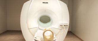
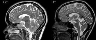
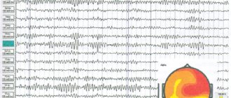
The doctor evaluates general complaints, conducts an examination, examines muscle tone, reflexes, communication, emotions and cognitive functions. EEG and echo-encephalography are also performed. A visit to an ophthalmologist is indicated to assess the condition of the fundus, acuity, and visual fields. Suspicion of a tumor formation is an indication for a CT or MRI of the brain. Angiography of cerebral vessels, PET scan and other complementary examinations may be prescribed. In some cases, a biopsy can help determine the type of cancer, but it is not possible in all cases.
Diagnosis of brain cancer
Brain cancer is diagnosed by a neurologist or neurosurgeon. Often, patients first turn to a therapist or cardiologist.
The specialist interviews the patient and examines him, including determining the activity of key tendon reflexes; tests skin and tactile sensitivity, etc.
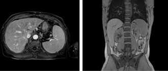
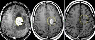
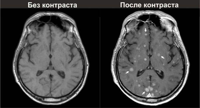
If brain cancer is suspected, instrumental studies are prescribed: fundus examination, brain CT, brain MRI, positron emission tomography (PET tomography), etc. Contrast-enhanced MRI is the most accurate instrumental method for diagnosing brain cancer.
To increase the effectiveness of complex treatment during surgical removal of a tumor, a biopsy is taken, followed by a histological examination of the resulting material.
Modern methods of treatment
“The volume of treatment is selected depending on the course of the brain tumor,” says oncologist Alexander Seryakov.
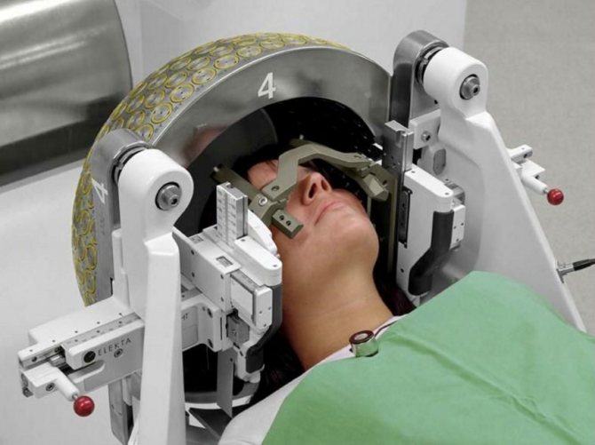
– The most effective is surgical removal of the tumor. The use of surgical microscopy and navigation allows for radical removal of the tumor and minimizes injury to healthy tissue. If the tumor cannot be completely removed, after its partial removal, the patient is prescribed radiation, as well as chemotherapy, targeted (“targeted”) or immunotherapy. For small malignant tumors (up to 3 cm), stereotactic radiosurgery is possible (using Cyber-Knife linear accelerators or Gamma-Knife gamma machines). To remove tumor tissue, many beams of radiation are used at once, which are directed at one point or collected into one beam. Its direction will constantly change during the irradiation session, but the beam necessarily passes through the tumor tissue.
For large tumors, and especially in cases where it is impossible to surgically remove the tumor, classical external beam radiation therapy is used. In some cases, it is used after surgery to reduce the risk of relapse (regrowth).
Chemotherapy is carried out with cytostatic drugs, taking into account the type of tumor. The effectiveness of chemotherapy increases significantly if it is combined with radiation therapy.
Targeted therapy in combination with radiotherapy and chemotherapy improves survival of patients with aggressive brain tumors. Most often used in the treatment of glioblastomas.
Currently, methods of using immunotherapy for malignant tumors (antitumor vaccines, immunotherapy drugs) and its integration into existing standards of treatment are being considered.
Diagnostic methods
To make a diagnosis, doctors prescribe various types of scans (CT, MRI), laboratory tests, and functional tests.
The use of the latest generation computerized complexes allows specialists to take tumor tissue for examination under a microscope without resorting to a full-scale operation. At the same time, the risk of complications is reduced to a minimum even when the tumor is located in a hard-to-reach place.
Detailed information about modern diagnostic methods and capabilities.
Prevention of brain tumors in adults at home
Prevention of brain tumors, according to oncologist Alexander Seryakov, generally includes:
- healthy lifestyle;
- optimal physical activity (preferably in the fresh air);
- complete rest;
- giving up bad habits (for smokers and alcohol abusers, the likelihood of developing brain tumors increases by almost 30%);
- a diet rich in fruits and vegetables;
- limiting stressful situations (or changing your attitude towards negative circumstances).