Magnetic resonance imaging and electroencephalography are two common methods for examining the brain. These are two different diagnostic procedures. They differ not only in the principle of operation, but also in what pathologies can be detected as a result of their implementation. Let's find out which is better: EEG or MRI of the brain.

Comparison of operating principles
Magnetic resonance imaging is based on the active response of hydrogen atoms to radio frequency radiation under conditions of a high or ultra-high magnetic field. The human body is mostly made up of water, with each molecule containing two hydrogen molecules. Therefore, it is clearly “visible” by the tomograph.
By registering responses from different areas, the computer converts them into a graphical form of information and assembles a single picture from them. It clearly shows all the structures of the brain and its blood vessels. Hard tissues—the skull—are less visible. This is due to the fact that bones contain fewer hydrogen atoms than soft ones.
When an ultra-precise picture is required to diagnose pathologies, the doctor - an MRI specialist - injects the patient with a contrast agent. It is based on gadolinium, which actively responds to the influence of a magnetic field.
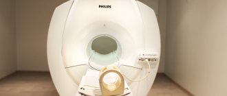
To perform an MRI of the brain, the patient is placed on the machine table and pushed into the tunnel. If necessary, his head is first fixed and a sedative is administered: he must lie motionless inside the tomograph. The scanning time is about 30 minutes, if with contrast - about an hour. The procedure is painless, but not very pleasant psychologically, because the patient has to stay in a small and confined space for a long time.
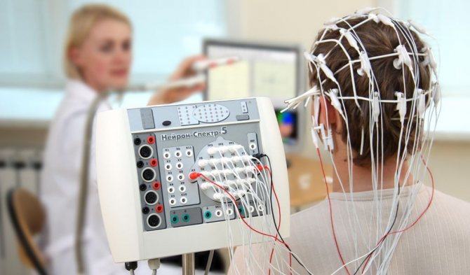
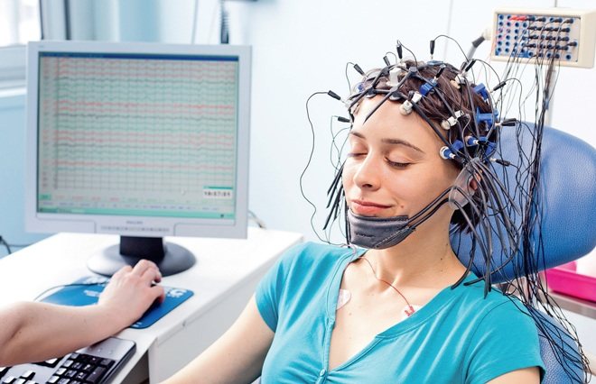
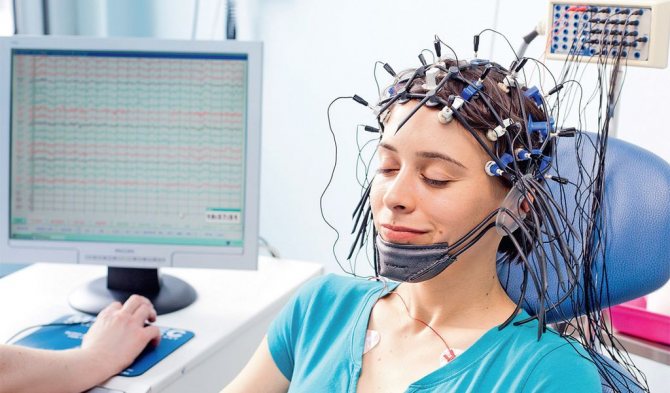
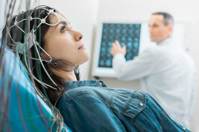
The principle of operation of electroencephalography is based on the capture of electrical impulses in the brain by a special device. After all, it is with their help that the nervous system transmits signals along the nerves. To conduct an EEG, a helmet with suction cups sensitive to electrical impulses is put on the patient's head. The examination time is only a few minutes. Diagnostics are carried out in an open space; complete immobility is not required. But there is an important condition - the patient’s emotional calm. Otherwise, the device will give an incorrect idea of the functionality of the brain.
The way EEG and MRI obtain information about the state of the brain is different, and therefore these studies show different results from each other.
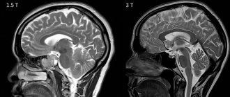
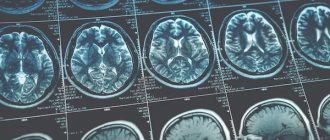
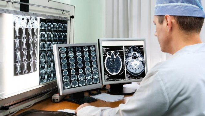
Magnetic resonance imaging allows you to visualize the structure of the organ, identifying pathological areas. Experts make a conclusion based on studying photographs taken from different points and in different planes. MRI helps to create a three-dimensional image and show layer-by-layer slices for a detailed examination of the area of interest of the head.
The result of electroencephalography is a graph - an electroencephalogram of electrical oscillations, displaying the activity of neurons in the brain. It is written down on paper, which is subsequently examined by the doctor. This means that EEG differs from MRI in that it allows us to detect not structural, but functional disorders of the brain.
Advantages and disadvantages
The main general advantage of these research methods is their painlessness.
EEG
Speaking about EEG, one cannot fail to mention the affordable price - everyone can afford this type of diagnosis. An additional advantage is that the examination time is short, the patient does not need to remain completely immobile, so this method is easier psychologically.
The disadvantages include the following: EEG is very difficult to detect a number of brain diseases, such as tumors and inflammation; During the procedure, the patient must maintain absolute emotional calm.
MRI
The advantage of magnetic resonance imaging is the ability to examine the structure of the brain and recognize a tumor at an early stage. The most obvious disadvantages of the method: the inability to see mental abnormalities; the need to remain motionless for a long time; high cost of diagnostics; Not every clinic has a tomograph; a greater number of contraindications for the study.
What is safer for pregnant women
The described methods of studying the brain are allowed to be used during pregnancy, since diagnosis occurs without radiation. But in the first trimester, such procedures are prescribed only if there is a threat to the mother’s life, and the examination is absolutely necessary to save her.
Diagnostic value analysis
MRI and EEG reveal different, complementary information. Therefore, their indications are different. Magnetic resonance imaging is prescribed in the following cases:
- Traumatic brain injury;
- Having had a stroke or heart attack;
- Suspicion of brain tumor and metastasis;
- Symptoms of demyelination and degeneration of brain tissue;
- Diagnosis of multiple sclerosis;
- Vascular examination (MR angiography);
- Postoperative control.
MRI can detect the following brain pathologies:
- Hematomas and contusions;
- Tumor neoplasms;
- Brain damage due to stroke;
- Multiple sclerosis;
- Vascular pathologies (aneurysms, vasculitis, atherosclerosis);
- Foci of infection in brain tissue.
An encephalogram is done in cases where it is necessary to identify the functional state of the brain. EEG indications include neurological disorders:
- Insomnia and frequent awakening during sleep;
- Headaches, dizziness;
- Panic conditions and nervous system disorders;
- Endocrine disorders;
- Stuttering;
- Autism;
- Recovery period after a stroke.
An encephalogram of the brain helps to identify:
- Foci of epileptic seizures;
- Cause of hypertension and hypotension;
- Causes of disturbed sleep;
- Mental disorders;
- Psychopathic reaction.
EEG makes it possible to determine brain areas in which there are obvious disturbances. If the examination does not help make a diagnosis, then it may be the basis for prescribing an MRI, CT or MSCT. But unlike MRI, an electroencephalogram shows whether the patient is faking his condition or whether he is really sick. This cannot be determined during MRI, despite the fact that both research methods are accurate.
What does EEG reveal?
The procedure is necessarily prescribed for the following brain pathologies and patient complaints:
- frequent dizziness;
- loss of consciousness with stable blood pressure;
- speech disorder;
- pain after a stroke;
- hormonal disorders affecting brain function;
- panic attacks;
- comprehensive examination for cerebral palsy or autism;
- chronic insomnia.
Using an EEG, the doctor determines which areas of the brain are affected by injury or infection. This facilitates further diagnosis and becomes the basis for CT or magnetic resonance imaging.
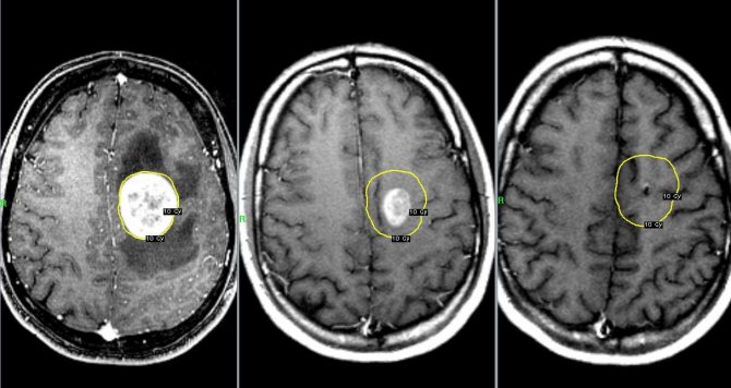
MRI differs from EEG of the brain in the clarity of the image for tumors. While the electroencephalograph is operating, the device receives signals indicating the possible presence of formations. But they do not show the structure, the place of formation. But the procedure helps to understand how the pathology affected the functioning of the brain: often with oncology, patients do not feel pain, but complain of insomnia, irritability, and changes in behavior.
Review of contraindications
Each research method has contraindications. For EEG this is:
- Damage to the scalp (absolute contraindication);
- Violent patients. This is a relative contraindication: an EEG can be done if a sedative is administered.
For MRI this is:
- Pregnancy in the first trimester;
- The patient's body weight is above 130 kg;
- Metal structures in the study area. We are talking only about steel; titanium and other metals that are not ferromagnetic are not contraindicated;
- Gadolinium intolerance (with contrast-enhanced MRI);
- Claustrophobia and other mental disorders in which the subject cannot lie without moving (in this case, the patient can be given a sedative);
- Having a pacemaker or device installed in the middle ear (the only absolute contraindication of all).
It can be seen that in case of a traumatic brain injury, one can only undergo examination with a tomograph, while in the presence of a pacemaker, only an EEG can be done. Electroencephalography also does not cause attacks of claustrophobia. Another advantage of the method is the absence of weight restrictions, since the patient will not be on a table, but on a regular chair or couch.
Both methods do not require preparation for the study, but with the caveat that the patient must come to the EEG in a good mood and well-rested. Otherwise, the diagnosis may give incorrect results.
Pregnancy is not a contraindication for MRI and EEG, since human organs do not receive radiation during diagnostic procedures. Both methods are safe for both mother and baby.
Diagnostic capabilities of encephalography
An EEG or encephalogram is a unique method of examining the human brain using a special apparatus. It is able to capture the activity of certain groups of neurons, translating them into a signal. The patient is offered a special cap with fixed electrodes, which fits tightly to the head. An additional layer of gel amplifies the signals, allowing the slightest deviations and discharges to be detected.
To get the correct diagnosis, you must follow simple rules:
- during EEG diagnostics, remain as calm as possible;
- take a comfortable position sitting or lying on the couch;
- talk or open your eyes only at the request of the doctor.
When taking an encephalogram, the diagnostician uses various stimuli: bright light, voice, and asks to breathe frequently or deeply. The result of the encephalograph remains in the form of graphs with different amplitudes. The specialist deciphers fluctuations that occur at certain periods of time. Despite its apparent complexity, the device helps to identify mental illnesses and serious abnormalities in epilepsy.
Diagnostic cost comparison
EEG and MRI are paid diagnostic procedures, as they are carried out using innovative, expensive equipment. Let's look at the prices offered by clinics for brain research:
- EEG – from 1600 to 5700 rubles, depending on the newness of the equipment and the reputation of the clinic.
- An MRI costs an average of 5,000 rubles, and when examining the blood vessels of a person’s brain or detecting tumors, MR diagnostics can cost 7,000-8,000 rubles, depending on the amount of contrast agent administered.
- An EEG and MRI with the administration of a sedative will cost 2000-6000 rubles more.
- The cost of the procedures will increase by another 500-1000 rubles if the results of the study are recorded on electronic media (disk, flash drive).
The legislation provides for some cases for free EEG and MRI under the compulsory medical insurance policy. In other situations, the patient pays for the procedures.
There are many methods for diagnosing brain diseases, so the question is which is more effective: MRI and EEG. This is a case where there is no clear answer, because these studies are designed to study the organ from two different “sides”. Electroencephalography will accurately identify functional capabilities and only suggest the presence of pathologies in different brain structures. And magnetic tomography accurately diagnoses the localization of brain tissue damage: the presence of a tumor, necrosis, inflammation. But tomography only suggests the presence of mental and cognitive disorders. Thus, these are two procedures that do not replace, but complement each other.
Information content of methods
When neurological symptoms appear, the doctor chooses between two methods depending on the disease.
Each diagnostic option is informative for some pathologies and irrelevant for others.
MRI helps identify:
- injuries;
- hemorrhages;
- circulatory disorders;
- sclerosis of nervous tissue;
- vascular damage and abnormalities;
- local inflammation;
- brain tumor.
Signs of the listed conditions are visible in the images; to increase the information content, a contrast study is carried out.
The diagnosis of the disease is made on the basis of the destruction of nervous tissue or blood vessels, its thickening or hypoplasia.
EEG is prescribed for long-term physiological disorders; emergency research is rarely performed. Based on the resulting graph, disorders of brain activity in certain areas are calculated.
An encephalogram will help in diagnosing:
- epilepsy;
- hypertension;
- mental disorders;
- sleep disorders.
Oncology and stroke are determined by indirect signs of EEG. In this case, an MRI is more informative, but if it is impossible, an encephalogram is taken.