In what cases is ultrasound examination of the blood vessels of the legs prescribed?
Ultrasound examination helps to understand the causes of symptoms such as:
- Pain, heaviness in the legs.
- Numbness, unpleasant sensations: tingling, chilliness, “pins and needles.”
- Change in skin color: pallor, cyanosis, dark spots.
- Swelling on the feet and legs.
- Feeling cold, feet quickly freeze even when warm.
- Signs of atrophy: decreased circumference of the legs, decreased tone, muscle strength.
- Leg cramps.
- Rapid onset of fatigue and pain in the legs while walking: you often have to stop and rest.
- Pain that intensifies when you raise your legs up and subsides when you lower them from the bed.
- Dilated veins under the skin, spider veins.
A woman should undergo an ultrasound examination of the vessels of the lower extremities during pregnancy if she begins to experience pain, swelling in the legs, or dilated veins become visible under the skin.
Ultrasound of leg vessels is prescribed to people who suffer from arterial hypertension, diabetes, atherosclerosis, have had a heart attack, or vascular surgery. It will not be superfluous to undergo a study if you are obese or have a long history of smoking.
Why do you need ultrasound of the vessels of the lower extremities?

We monitor heart function every year at a medical examination: we undergo an examination by a cardiologist, take a general blood test and do an ECG. But the work of the entire circulatory system depends not only on the tone of the heart muscle, but also on the condition of the veins and arteries. If the blood channels of the upper extremities are sometimes checked by ultrasound of the heart, then the lower ones are rarely examined. Therefore, diagnosis is incomplete without ultrasound examination of the large femoral artery and the vessels branching from it.
The diagnosis determines:
- blood flow speed, wall thickness,
- the condition of the valves, the presence or absence of thrombotic formations and cholesterol plaques,
- development of inflammatory processes.
In this article we will look at the cases in which vascular ultrasound is prescribed and why it is important to undergo a scheduled examination yourself.
When is it necessary to do ultrasound diagnostics of the veins and arteries of the legs?
The therapist sends for diagnosis when the patient feels:
- swelling of the legs,
- heaviness in the legs,
- cramps and numbness,
- lameness or pain when walking.
These symptoms are the most common, but the doctor may prescribe an ultrasound in other cases, for example, with spinal injuries. In this case, it is necessary to examine the arteries and veins of the legs to check the blood flow: intervertebral discs can pinch the vessels.
If nothing worries you, doctors recommend regular diagnostics for related problems:
- stroke - the walls of the blood channels become thinner after illness;
- diabetes - arteries become less permeable due to increased sugar and poorly nourish organs and tissues;
- bad habits - veins narrow and are in a state of spasm due to cigarettes, alcohol and poor nutrition.
If you do a vascular ultrasound in Minsk at least once a year, you can prevent the development of varicose veins, thrombosis, atherosclerosis, and cancer: the initial stage of the disease can be seen on ultrasound.

How to decipher the results of the study?
On the doctor’s report they note:
- The presence or absence of cholesterol plaques and blood clots.
- Blood circulation speed.
- Contour of the walls and lumen of blood vessels.
Doctors compare these data with reference data and calculate indicators based on them, for example, the ankle-brachial index.
It is impossible to understand the conclusion on your own, so to decipher it, sign up for a consultation with a phlebologist - a doctor who specializes in the treatment of vein diseases. He will talk about the structure of the blood channels and pay attention to problems, such as thin walls or slow blood flow. He will also give advice on the prevention of vascular diseases and prescribe treatment if necessary.
An ultrasound should be done if the doctor prescribes it according to indications, and in case of concomitant diseases, experts recommend checking the legs twice a year.
To promptly recognize pathologies of arteries and veins at the initial stage, include diagnostics in your routine medical examination: preventing a disease is easier than treating it.
Sign up
How is ultrasound examination of the blood vessels of the legs performed?
In the ultrasound diagnostic room, you will be asked to remove your clothes from your legs and lie down on the couch. The doctor will apply a small amount of a special gel to your skin and place an ultrasound probe. After the vessels in your legs have been examined while lying down, you will be asked to stand up and the examination will be repeated. In an upright position, it is more difficult for blood to rise through the veins of the legs, since gravity acts in the opposite direction.
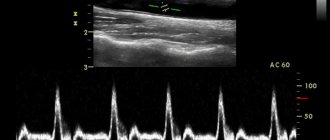
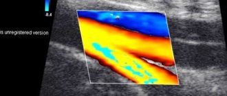
In order to study the speed and other parameters of blood flow, ultrasound is combined with Doppler ultrasound, duplex, triplex scanning. You may hear the machine make a whistling sound as it records the pulse in your legs. Different blood flows on the screen will be colored blue and red. After the study is completed, the ultrasound doctor will give you a form with a protocol. It should be taken to the attending physician - phlebologist or vascular surgeon.
Indications for ultrasound examination of veins and arteries of the lower extremities
The study is necessary in the presence of diseases such as:
- diabetes mellitus type I and II
- overweight and obesity
- phlebeurysm
- arterial hypertension
- previous stroke or myocardial infarction
- previous surgical interventions on the legs
- obliterating atherosclerosis
- hyperlipidemia
- history of pulmonary embolism
An ultrasound scan may be prescribed to a patient if certain symptoms occur. The main list includes:
- numbness of the limbs
- appearance of peeling
- changes in skin color or severe pallor
- feeling of coldness in the fingers and feet at any ambient temperature
- tingling in the absence of external stimuli
- pain in the limbs during physical activity and its absence
- tenderness of the saphenous veins and their expansion
- non-healing ulcers
- “crawling” goosebumps
- presence of spots on the skin and changes in pigmentation
- uncontrollable muscle cramps
- swelling in the feet and legs
Diagnosis of the veins and arteries of the lower extremities is also indicated for smoking patients over 15 years of age, persons over 45 years of age, those with a hereditary predisposition to varicose veins, and pregnant women.
How is ultrasound examination of the veins and arteries of the lower extremities performed at the Istok Health Clinic?
- The patient frees his legs from clothes and sits on the couch.
- A special gel is applied to the areas of interest, which serves as a conductor for transmitting data to the ultrasound machine. As the sensor moves along the legs, the monitor displays a picture in the form of different sections of veins and arteries.
- When examining the popliteal and popliteal veins, the specialist may ask the patient to lie on their stomach or sit up on a couch.
- If the doctor needs to examine varicose veins, then additional tests may be performed on the patient - breathing and compression tests.
- Doppler ultrasound is performed strictly on both legs, even if only one of them is bothering you - this measure is necessary to compare blood flow indicators.
Depending on the symptoms and existing diseases, ultrasound examination takes about 30-40 minutes. In conclusion, the patient is given a protocol displaying the results of the vascular examination.
Doppler ultrasound of the vessels of the upper and lower extremities
The procedure is carried out to obtain a quantitative and qualitative level of blood flow in the legs. Based on ultrasound, varicose veins of the lower extremities, the degree of pathological narrowing of the arteries (stenosis), and abnormalities in the functioning of the venous valves are determined.
Doppler ultrasound will determine whether the patient requires surgical treatment.
The study does not require preparatory manipulations and can be performed on patients of any age.
Symptoms indicating the need for this procedure:
- Frequent leg cramps.
- The skin on the legs is blue-pink.
- The appearance of "stars".
- Pronounced dilatation of veins.
- Feeling of heaviness in the legs, increasing in the evening.
- Swelling of the feet and legs.
- Trophic ulcers.
- The appearance of purple or brown seals.
When these symptoms appear, many (especially women) become upset because their legs do not look attractive. But the seriousness of this disease is that due to impaired blood flow, blood clots form in dilated areas of the veins. They can break off at any time, penetrate the pulmonary artery and lead to death.
There is a high risk of thromboembolism if the muscles of the lower extremities were relaxed for a long time (after the introduction of general anesthesia) and then returned to their previous state.
What is ultrasound examination (ultrasound)?
When it comes to vascular examination, ultrasound usually means duplex scanning - a method that combines Dopplerography and vascular scanning, resulting in a two-dimensional image of the vessel, on which you can see not only pathologies, but also the degree of their development. Another option for ultrasound examination is triplex scanning.
, which allows you to get an even more informative image in color. Unlike Doppler Doppler Ultrasound, these vascular ultrasound methods allow one to obtain not only information about the patency of veins and arteries, but also provide a detailed assessment of their condition.
When is an ultrasound performed?
Ultrasound is an accessible and relatively inexpensive research method, and it is prescribed for diagnosing diseases and pathologies of various kinds, however, ultrasound examination of blood vessels, which is more advanced and more expensive, is usually prescribed for already diagnosed vascular diseases to monitor their treatment. Ultrasound is often prescribed when ultrasound results are unsatisfactory or to clarify the details of the identified pathology. This is the most effective way to identify vascular pathologies of the lower extremities.
How is the ultrasound procedure performed?
The vascular ultrasound procedure does not require prior preparation. It is necessary to refrain from drugs and drinks that affect vascular tone, which can distort the test results. The area being examined is lubricated with gel so that oxygen does not interfere with the scanner’s reading. During an ultrasound, the sonologist moves a sensor over the surface being examined and records the readings in the conclusion. During a duplex study, a two-dimensional image of veins and arteries is obtained, which allows one to judge changes in their structure and find out the cause of problems with vascular patency.
What is examined during Doppler Doppler Doppler (US) of the veins and vessels of the lower extremities
During the procedure, the doctor evaluates the speed of blood flow, the patency and lumen of blood vessels, the functioning of the valves, the location and size of blood clots. The narrowing of the arteries and the thickness of the vascular walls are also examined. The arteries are examined in detail; with the help of ultrasound, the doctor can identify many diseases: Raynaud's disease, arterial insufficiency, obliterating atherosclerosis and endarteritis, as well as aneurysms.
If atherosclerotic plaques are detected during diagnostics using ultrasound, their size and structure must be assessed. In case of thromboembolism (blood rupture), an ultrasound scan of the blood vessels is performed urgently to find the detached blood clot and the site of blockage. This condition requires urgent intervention, as it is very dangerous to human health.
The advantages of vascular Doppler ultrasound include the ability to examine the extremities of bedridden patients, since portable devices can be used and there is no need to transport or disturb patients.
How to properly prepare for an ultrasound examination of the veins and vessels of the lower extremities
Ultrasound examination does not require preparation, except for hygiene procedures before visiting a doctor. The patient does not need to limit himself in food or water. An ultrasound examination lasts about 20-25 minutes.
How does an ultrasound scan of the veins and vessels of the lower extremities work?
Before starting the diagnosis, the patient must remove clothing covering his legs. The uzologist applies a special conductor gel to the examined areas of the limbs. Ultrasound examination can be performed from different angles, so the patient may be asked to lie on a couch or take an upright position. The examination begins with the lower veins, then the outer parts and structures of the arteries are examined. Be sure to check the structure of the popliteal, femoral, small, and posterior tibial veins. Data from the examination of the vessels of the extremities are displayed in the form of a graphic image (black and white picture) on the monitor. The doctor describes the characteristics obtained and draws up a conclusion.
How to prepare for ultrasound examination of leg vessels?
The procedure usually does not require special preparation and does not require adherence to strict diets, however, specialists at the Istok Health Clinic claim that in order to obtain the most accurate results, certain restrictions must be observed
- Postpone active physical activity one day before ultrasound examination.
- Avoid drinking any alcoholic beverages 24 hours before the test;
- On the day of ultrasound examination, do not drink coffee, cocoa, strong tea or energy drinks.
- Do not smoke tobacco products at least 3 hours before your appointment.
- Before going to the clinic, do not rub or warm your feet under any circumstances.
How to make an appointment for a limb examination
To sign up for a duplex scan of the arteries and veins of the extremities at the Health Workshop, you need to call the call center or fill out the form. The cost of a duplex is from 1800 rubles. We also perform Doppler ultrasound of the vessels of the upper and lower extremities.
Before the examination, we advise you to undergo an examination by our doctor. This is a free service and is included in the cost of scanning. At the appointment, the doctor will examine you, ask about your symptoms and give you a referral. This will help the ultrasound specialist make a more accurate diagnosis.