Without an ultrasound examination, it is impossible to imagine the course and management of pregnancy. This method allows you to answer a number of questions from obstetricians:
- fetal condition;
- presence of developmental defects;
- intensity of uteroplacental blood flow and others;
- structure of the “children's place”.
Pregnant women are often concerned about two questions: is everything okay with the baby and how many gestational weeks are there already? And hence the interest arises in how the gestational age is determined by ultrasound and how accurate it is.
Timing of pregnancy determination by ultrasound.
Visualization is difficult: what does this mean?
Pregnancy and the birth of a child are the most touching, exciting, love-filled, and unforgettable stage in the life of any woman. When the expectant mother learns about the birth of a new, small life inside herself, along with a feeling of great happiness, questions arise regarding the timing of conception.
The most important question is: “At what stage can a pregnancy be detected by ultrasound?”
Because, based on this, doctors will determine the state of development of the unborn child and calculate the date of the upcoming birth.
Calculation options
Even before the advent and widespread introduction of ultrasound in obstetrics, women were interested in the week of gestation and the estimated date of birth. There are many options for calculating the length of pregnancy, the accuracy of which varies.
- By ovulation. Measuring the period of “interesting position” begins with the date of expected maturation of the egg (day of missed menstruation minus 14 days). This calculation option is relevant for a regular cycle.
- By movement. It is believed that the first movement of the fetus is felt at the 20th week, and in case of repeated pregnancy - at the eighteenth. The method is irrelevant and has a large error.
- By date of conception. This countdown can occur during in vitro fertilization (embryonic gestation period).
- The degree of maturity of the cervix during examination will not allow you to determine the exact week of gestation, but will indicate readiness for childbirth.
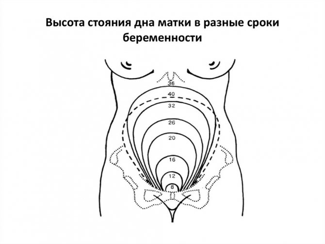
- According to the height of the uterine fundus. The method has indicative value and is used to assess fetal development by a gynecologist during examination.
Normal location of the uterine fundus
- By last menstruation. Based on this principle, the obstetric period is calculated, which is taken as the fundamental one. Pregnancy calculations begin from the date of the last menstruation. The day of birth is determined by adding 280 days to this number. The method is the basis for online calculation of gestational week using calculators.
It should be noted that the obstetric period is slightly longer than the embryonic (true) period, since it does not take into account the time of conception. However, its calculation is the simplest and most common.
- Using ultrasound diagnostics. Determining the gestational age by ultrasound can be carried out at any length of gestation and there are several methods for calculation.
When will the ultrasound show the future baby?

At what time does an ultrasound show conception? - this question worries every woman, in case of expecting a long-awaited conception, a delay in menstruation, or simply because of symptoms indicating pregnancy. But first, you need to know how the fertilization period is determined :
- According to the woman’s calculations, starting from the last day of the delay;
- Based on the results of a gynecological examination;
- But the most effective and more accurate will be ultrasound diagnostics.
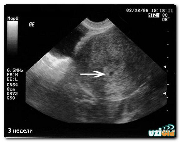
Ultrasound diagnostics in the case of a vaginal examination (that is, a condom is put on the sensor of the device and inserted into the vagina), provided that the examination is performed by an experienced doctor using a modern device, allows you to record the fact of successful fertilization for a period of 14 days after conception.
This means that on the ultrasound monitor, a device in the uterus, you can see a fertilized egg measuring 0.4 cm after a week of missed period or a month later according to the obstetric period. If you go for an ultrasound a little later, at 1.5 months, then already in such a short time, the device will record the heartbeat of the unborn child.
At what day of delay will an ultrasound show pregnancy? In the case of an abdominal examination (that is, diagnosis is carried out through the abdominal wall), pregnancy will be visible no earlier than 4 weeks of embryonic age, or at least 14-21 days after the 1st day of delay.
Accuracy of the method
The calculation based on the diameter of the fertilized egg is very arbitrary and is of an orientational nature. The purpose of the study in the initial period is to determine the fact of an “interesting situation”, and not an exact calculation of the week of gestation.
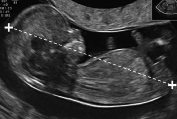
The coccyx-parietal size is the main indicator. It is used in ultrasound scanning to determine gestational age. The accuracy of the parameter is due to the fact that it has minimal dependence on external and internal factors (race, physical characteristics of parents, mother’s health, etc.).
However, the definition of pregnancy by KTP and the obstetric period may not coincide, since in the second case the counting starts from the last menstruation without taking into account the onset of ovulation and fertilization.
If there is a significant difference between the obstetric period and cardiotocography, this may indicate a serious pathology (fetal death, chromosomal abnormalities).
When a pregnant woman does not remember the date of her last period, the period is counted based on the results of measuring CTE.
Terms of pregnancy: embryonic and obstetric
The gestational age indicated in the maternal card, filled out when registering at the antenatal clinic, differs slightly from the actual time elapsed from the day of fertilization by about 2 weeks.
But there may be other options, for 1-1.5 weeks, depending on the length of the menstrual cycle. This is considered the norm, because we are talking about different periods: embryonic and obstetric:
- Embryonic - counted from the actual day of conception, which coincides with the date of ovulation. For women, it is not difficult to calculate the day of conception only if they keep an ovulation calendar.
- Obstetric period is the period that has passed since the last menstrual period, after which conception occurred.
It is this period, which is easier for a woman to determine than the embryonic period, that is considered a reference point for scheduling routine examinations and establishing the upcoming date of birth.
Sometimes, women wonder “can an ultrasound not show pregnancy”? - yes, maybe, in the case when the doctor cannot see the fertilized egg, a repeat examination is prescribed due to the probable risk of an ectopic pregnancy.
Advantages of detecting pregnancy in the early stages:
- Timely detection of ectopic pregnancy;
- Establishing the fact of successful conception and its duration;
- Determining the cause of delayed menstruation in the absence of a positive pregnancy test;
- Determination of the number of conceived embryos;
- Recording possible threats to pregnancy.
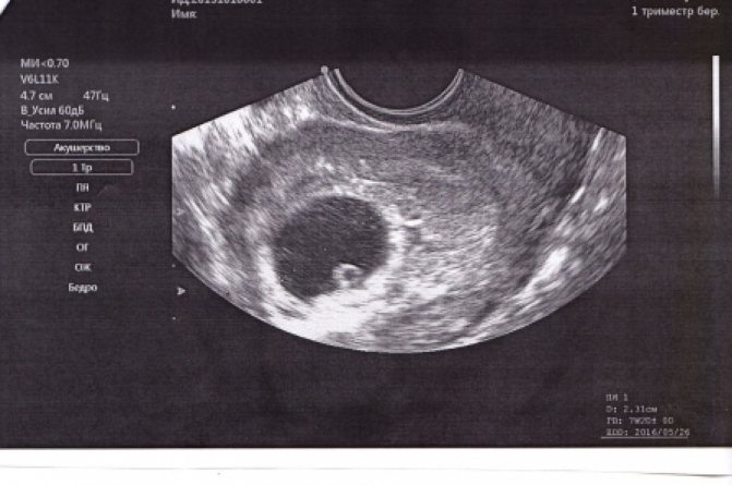
Study in the first trimester
Of particular interest to women is the procedure in the early period. The goal is to confirm the fact of pregnancy. For ultrasound to be informative, it is necessary to take such points into account. Firstly, preference should be given to transvaginal examination. It allows you to detect the fertilized egg several days earlier than the transabdominal method.
Secondly, diagnosis should be carried out no earlier than 4–5 days of missed menstruation. On the 2nd–3rd day of absence of expected menstruation, it is sometimes possible to visualize the fertilized egg using expert-class devices.
To be sure that the ultrasound is informative, you should take a blood test for human chorionic gonadotropin (hCG) the day before. When the level of the substance is above 2–2.5 thousand units, you can safely go for a transvaginal examination. It will be informative.
From 3 to 12–13 weeks, the gestational period is determined by measuring the embryo.
Table 1.
| Gestation period, weeks. | Diameter of fertilized egg, mm |
| 3 | 3 |
| 4 | 6 |
| 5 | 8 |
| 6 | 14 |
| 7 | 20 |
| 8 | 27 |
| 9 | 34 |
| 10 | 40 |
| 11 | 47 |
| 12 | 53 |
| 13 | 60 |
Already starting from weeks 5–6, the importance of clarifying the presented parameter decreases. This is replaced by an assessment of the coccygeal-parietal size (CTP). The diameter of the fetal egg continues to be determined until 8-9 weeks when examined transabdominally. This is due to the fact that detailed visualization and measurement of CTE in this way is difficult.
The latter method has great accuracy in determining the duration of pregnancy, especially at 11–13 weeks.
Photo: how CTE is calculated.
It is during this period that the first screening examination is recommended. Cardiotocography data depending on the week of the “interesting situation” is presented below.
Table 2.
| KTE, cm | Gestation period, weeks/day |
| 0,3 | 4 |
| 0,4 | 5/2 |
| 0,5 | 6/1 |
| 0,6 | 6/3 |
| 0,7 | 6/5 |
| 0,9 | 7 |
| 1,0 | 7/2 |
| 1,2 | 7/5 |
| 1,4 | 8/1 |
| 1,6 | 8/3 |
| 1,8 | 8/5 |
| 2,0 | 9 |
| 2,1 | 9/1 |
| 2,3 | 9/3 |
| 2,5 | 9/5 |
| 2,7 | 10 |
| 3,0 | 10/2 |
| 3,2 | 10/4 |
| 3,4 | 10/6 |
| 3,6 | 11 |
| 3,9 | 11/2 |
| 4,1 | 11/3 |
| 4,4 | 11/5 |
| 4,7 | 12 |
| 5,0 | 12/2 |
| 5,5 | 12/5 |
| 5,9 | 13 |
| 6,3 | 13/2 |
| 6,6 | 13/4 |
| 7,0 | 13/6 |
| 7,3 | 14 |
| 7,6 | 14/2 |
| 8,0 | 14/4 |
| 8,7 | 15 |
Qualities and benefits of ultrasound

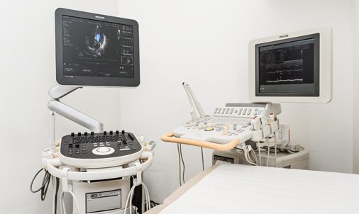
Ultrasound examination is a widespread, painless, very effective method for determining pregnancy and its timing.
The very first ultrasound machines were bulky and unable to produce a clear image on the screen. Therefore, studies often gave inaccurate conclusions. But modern devices are capable of transmitting more detailed information about the state of the human body.
In the modern world, there are various technologies for examining internal organs.
But it is ultrasound that is ideal for determining the timing of conception and the course of pregnancy for the following reasons:
- Determination of conception in the early stages;
- Ultrasonic waves do not harm mother and child;
- Clear visualization of the fetus;
- You can do an ultrasound at any time convenient for the expectant mother;
- No rehabilitation period;
Ultrasound radiation makes it possible to eliminate various types of damage to the skin and mucous membranes. Nowadays, a woman does not have to torment herself with the question “Where to get an ultrasound during pregnancy”?
Because there are a lot of possibilities. An ultrasound scan for pregnancy can be done from private medical institutions to local clinics.
When to do a 3D ultrasound during pregnancy?
Three-dimensional ultrasound is the most modern, improved diagnostic method than the usual 2D. The advantage of 3D ultrasound is that:
- it gives a three-dimensional image of the fetus
- allows you to see the baby’s features and even facial expressions
- The quality of the resulting image on the screen is much higher
Carrying out a three-dimensional study takes longer - about 40-50 minutes.
This procedure is best performed during the second planned ultrasound, when the main features of the fetus have already been formed, and the fetus has grown up and learned to make grimaces. This can make for great keepsake photos. These services are provided by most private medical clinics.
Scheduled diagnostics

After confirmation of successful conception, the expectant mother is registered with a gynecologist. At the antenatal clinic, she learns that during pregnancy, if the pregnancy progresses normally, she will have three scheduled ultrasounds. This procedure does not have any adverse reactions on the fetus or the expectant mother. There are times when, in order to more carefully monitor the course of pregnancy, the diagnosis is carried out 1–2 times more.
The dates for scheduled examinations are distributed by trimester, because each of them differs in the development and growth of the unborn baby:
During the first trimester (period from 10 to 14 weeks), diagnosis may show chromosomal abnormalities that can cause genetic diseases in the fetus. The features of anatomical development are also determined, and the estimated date of birth is calculated;
During the second trimester (period from 20 to 24 weeks), ultrasound shows the structure of the fetal organs, due to which there is a chance to detect deviations from the normal functioning of the heart, kidneys, gastrointestinal tract, liver, as well as malfunctions in the nervous system. At this stage, very often, the sex of the child is determined, you can see the movements of his arms and legs. The length and estimated weight of the fetus are measured, and the placenta is characterized. In the same trimester, a Doppler ultrasound examination is performed to determine the blood flow in the fetal vessels;
The third trimester (the period from 31 to 36 weeks) is considered the final trimester. At this stage, the diagnosis of organ development disorders that could not be identified earlier is carried out. The position of the fetus is determined, which by this time should have taken a position upside down, and the probable entanglement of the umbilical cord around the neck. The child’s parameters are still measured and the date of the upcoming birth is specified. In the third trimester, the doctor gives an opinion - whether the woman will give birth naturally, or will have to resort to a cesarean section.
Along with routine examination procedures, after 12–13 weeks, biochemical screening of the fetus is carried out. A similar method is used to identify pathologies, as well as for early detection of Down syndrome. Ultrasound of the pelvic organs
What does a pelvic ultrasound show? During a pelvic diagnosis, the doctor examines the uterus, appendages, ovaries, as well as the bladder and rectum. For a pregnant woman, this study is carried out to determine the condition of the reproductive organs and, of course, to examine the fetus, amniotic fluid, and placenta.
A feature of a pelvic examination can be considered the ability to detect pathologies such as:
- Ectopic pregnancy;
- Abnormal development of the uterus during pregnancy;
- The process of inflammation of the ovaries, uterus, vagina;
- Malignant and benign tumors on the organs of the reproductive system.
What period does ultrasound examination show?
Ultrasound determines the duration of gestation based on the size of the ovum or fetal parameters. Based on these data, embryonic age is determined. However, ultrasound machines have a program that automatically adds 2 weeks to the data obtained to get the obstetric date.
In what cases do the data differ from the obstetric period?
The most accurate information is obtained using ultrasound in the early stages of pregnancy. During this period, embryos in different women develop approximately the same. After 3 months of gestation, hereditary factors and phenomena accompanying the current pregnancy begin to have an increasing importance on the development of the child.
In this regard, many women note that from the 2nd trimester, the gestational age according to ultrasound differs from the obstetric one. As a rule, an ultrasound shows that it is 2 weeks less than obstetric. The greatest deviations from the obstetric period of pregnancy are observed in the 3rd trimester. Sometimes it is revealed that the ultrasound period is a little longer. The main reasons for the differences between the terms:
- use of different counting methods (real and obstetric);
- influence of genetic factors on fetal development;
- fetal pathologies (developmental delay, congenital abnormalities).
What to do in such a situation?
If you notice a difference in timing, you should not try to draw conclusions on your own. A difference of 2 weeks is the normal limit, and in the 3rd trimester the discrepancy can reach up to 4 weeks.
To determine why deviations were detected and find out the correct period, the doctor will conduct a thorough analysis of other indicators:
- how different are the data from the ultrasound and the obstetrician;
- dates of last menstruation and cycle length;
- correspondence of fetal size to gestational age;
- hCG level;
- the size of the uterus, the height of its fundus;
- volume of a pregnant woman's abdomen;
- degree of placenta development;
- dates of the first fetal movement.
Based on the totality of the data obtained, the gynecologist will make an accurate calculation of the gestational age. If necessary, additional tests will be prescribed (for hormones, infections, Doppler ultrasound). If a delay in fetal development is detected, the woman will be offered treatment. However, in most cases, deviations in ultrasound timing from obstetric ones are explained by the individual characteristics of the unborn child.
Ultrasound of the cervix
Ultrasound examination of the cervix during pregnancy is prescribed as an auxiliary precaution during a routine examination by a gynecologist.
A timely examination makes it possible to detect and exclude various types of pathologies that can harm the health of the fetus and mother.
Throughout pregnancy, the uterine canal tends to change. This is considered the norm, since the physiological process of preparation for the upcoming childbirth is in full swing. Therefore, the slightest deviations in this process may be considered a threat to the normal course of pregnancy, which means you should not postpone visiting a specialist for a long time. The sooner the examination occurs, the greater the chances of a positive birth.
Containment of the fetus is the most important function of the cervix in a pregnant woman. Because, during most of the pregnancy, its closed position protects the unborn child and prevents various types of infections from entering. But after 36 weeks, the cervix becomes deformed, becomes shorter, softer and moves towards the center, thereby starting the process of its opening. Afterwards, a single canal is formed connecting the cervix with the external os. Its full opening reaches dimensions of 12 cm.
If you look at it from an anatomical point of view, the cervix is the entrance to the body of the uterus itself. The organ also includes: internal and external pharynx, which is covered with muscle connective tissue. As a result, a cervical canal is formed, smoothly connecting to the vagina. The muscle holding the unborn child in the form of a ring is located in the internal pharynx.
If the muscle loses tone, the cervix loses the ability to contain the child inside itself.
During pregnancy, the length of the cervix plays an important role. It determines the possible risk of premature birth. It happens that it may have a shortened state, which indicates isthmic-cervical insufficiency (premature pathological dilatation of the cervix due to strong internal pressure of the fetus and amniotic fluid).
For this reason, unfortunately, a miscarriage occurs. But in our time of modern technologies, timely treatment helps prevent premature initiation of the birth process and miscarriage.
Additional Research
It is very difficult to answer the question how many times during the entire period of bearing a child a woman will have to undergo an ultrasound procedure. It all depends on the health and age of the expectant mother, and on whether she had a bad experience in bearing a child. In addition, if a pregnant woman has complaints about her health, the doctor prescribes additional ultrasound examinations.

Each trimester has its own indications for prescribing an unscheduled ultrasound.
In the first three months of pregnancy, it can be caused by spotting and abdominal pain. The doctor sends for an ultrasound to find out the cause and understand whether there is a frozen or ectopic pregnancy.
In the second and third trimesters, ultrasound examination is indicated for multiple pregnancies, detection of abnormalities in the development and size of the fetus, impaired motor activity, the presence of an infectious disease or placenta previa in the mother, leakage of amniotic fluid, etc. In the later stages, a good reason may be the suspicion that the baby is breech. If it is confirmed, doctors will perform a caesarean section to reduce the risk of injury to the baby.
How is the research done and when is it carried out?
A planned anatomical ultrasound examination is prescribed for the period 14–16 weeks of pregnancy. But if in a woman who has previously given birth, with a previous pregnancy, the labor process began earlier than the appointed time, an ultrasound of the cervix is prescribed approximately at the end of the 12th week. After which, observations are repeated at intervals of two weeks.
The following indicators are considered additional reasons for ultrasound of the cervix:
- Miscarriage that occurred in the first pregnancy at a late stage;
- Conception with the presence of two or more fetuses;
- Various reasons for terminating a previous pregnancy;
- Suspicions of isthmic-cervical insufficiency;
Any previous surgical interventions in the field of gynecology.
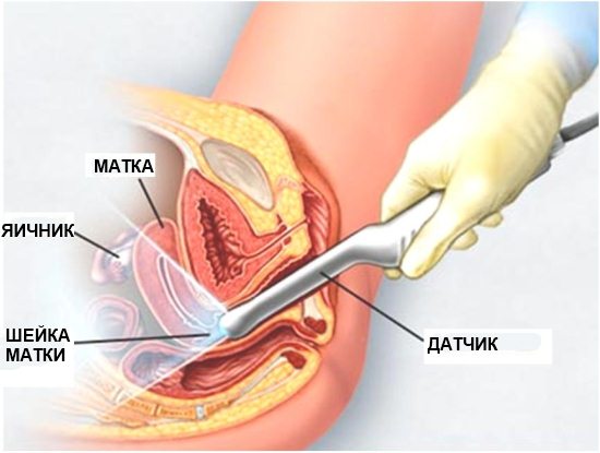
Ultrasound of the cervix, as well as diagnostics to detect pregnancy, is done in two ways, abdominal and vaginal. In the first case, the uterus is visible through the anterior wall of the abdominal cavity. The doctor makes a conclusion about the position, condition, parameters of the uterus, and also gives a description of the external pharynx.
Vaginal ultrasound examination is performed by inserting a special sensor into the vagina. This case is more effective than the previous one, since it becomes possible to measure the length of the cervix, the size of the internal pharynx and the cervical canal.
As a rule, special preparation for an ultrasound is not required; only in the early stages of pregnancy, the doctor may ask you to pre-fill the bladder.
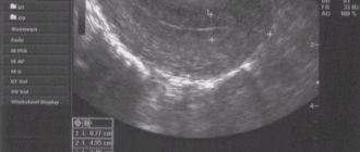
Diagnosis through vaginal and abdominal examination is considered absolutely safe for pregnant women. Visualization is difficult: meaning
When during an ultrasound, your doctor, based on the examination results, says that visualization is difficult, this is not a reason to panic. For personal peace of mind, you should understand what this means.
If the pregnancy proceeds without any problems, a routine ultrasound examination is performed during the 24th week. An ultrasound of the pelvic organs in the first trimester will not provide as much important information as in the second trimester.
Therefore, during this period, future parents will hear the long-awaited and joyful news about the gender of their baby, as well as whether there are any abnormalities or pathologies in the development of the fetus.
The size of the unborn baby in the second trimester is quite large, which allows the doctor to clearly examine all its organs. In addition to growth and assessment of the child’s condition, at 24 weeks, the doctor will examine the placenta, which by this time has become denser in structure than in the first trimester, and will also diagnose the amount of amniotic fluid.
Visualization is difficult during pregnancy; in simple words, this is when the fetus is located in such a way that none of the above is simply visible to the doctor. Of course, this does not mean that you should be upset or consider it a violation in the child’s development, because there will be another third planned ultrasound examination ahead, at 34 weeks.
Survey histories
On the Internet you can now find reviews from forums for pregnant women. The main part of the discussions, of course, concerns the definition of an “interesting position”, its duration and the gender of the child. Most of the stories are banal (they didn’t see the pregnancy at the beginning of the gestational period, the ultrasound weeks do not coincide with the obstetric ones), but there are also unique stories.
More than expected
22.06.2016
Now happy mother Ksenia shares her review that before 32 weeks of pregnancy she and her husband were expecting their first child. The thought that the child was not alone there did not appear at all. True, the stomach was too big, and the hCG level was high in the early stages. But this did not bother the doctors, so the parents were not worried. Ultrasound was performed at the established screening times twice. At 33 weeks, the doctor, upon examination and listening to the heartbeat, had doubts and sent me for an ultrasound examination. As a result, they found the “lost” second child, who was not noticed either at the first or at the subsequent ultrasound.
Twins are enough
06.06.2015
But Anna already had 2 embryos at the first ultrasound screening. The happy parents waited long enough for twins - up to 28 weeks. It was at this stage that the ultrasound doctor said with surprise that there was a third child. Everyone agreed that, having discovered the second fetus, the doctor at the first ultrasound did not continue the search, but focused on a detailed study of these two.
And the fibroid is moving
12.12.2016
Tatiana's ultrasound showed that her menstruation was delayed for 5 days. The hope of a long-awaited pregnancy glimmered. But the ultrasound result was not pleasing, but quite upsetting - fibroids, 6 mm. This is how the doctor explained the cycle disorders and advised me to go to a gynecologist. Tatyana never got to see the doctor: either she didn’t have the strength or the time.
The discovered tumor, although benign, drove me into depression. Like many women, I began to “eat” my stress. This explained her weight gain. And the absence of menstruation, Tatyana consoled herself with the growing tumor. But I still had to go to the doctors, albeit a little later.
After 4 months, the woman felt strange tremors in her stomach. She couldn’t explain them to herself. The therapist already recommended undergoing an ultrasound examination, which confirmed the presence of an “interesting situation” at 20–21 weeks.
That’s how funny it all turned out: the fibroid turned out to be a fertilized egg, and the strange sensations in the stomach turned out to be the baby’s movements.
The climax has arrived
31.01.2016
The fact that menopause occurs is not surprising. Especially if the age has exceeded 40. Many try to slow down the processes, some try to stop them, while others take it for granted and don’t pay attention (“What’s the point of running to doctors in vain?”).
So Nina decided that at 42 years old, the absence of menstruation is an everyday matter. The menopause has arrived. Having read on the Internet that the signs were different, she calmly attributed all the sensations and manifestations to her hormonal problem. After suffering for 3-4 months, with a feeling of despair (“how long will I have to suffer like this?”) I made an appointment with a gynecologist. The purpose of the visit was one - maybe there is a medical solution to the symptoms of menopause.
But the gynecologist, after an examination, gave her verdict: pregnancy 16-17 weeks. The doctor's conclusion was confirmed by the results of an ultrasound examination and a blood test.
“That’s how I had to put off old age,” summed up happy mother Nina
Lost time
20.10.2017
Not all stories end equally well. It happens that pregnancy is not confirmed, and sometimes it is there, but not there.
An unexpected thing happened in Zoya’s life - a delay of 16 days. The baby was not planned: the couple took precautions. But there is nowhere to go. A visit to the doctor did not clear up the situation, nor did an ultrasound. The uterine cavity is clean, there is no fertilized egg, but they describe a corpus luteum near the ovary.
With the assumption of an ectopic pregnancy, Zoya spent two weeks going back and forth between the gynecologist and the ultrasound. I did not donate blood for hCG. There was no talk of hospitalization. At the end of 3 weeks of delay, everything ends with a rupture of the fallopian tube, massive internal bleeding and a long recovery.
Of course, there remains another healthy organ and hope for motherhood in the future. But everything could have been identified and corrected in time.
An unexpected find
01.02.2015
Inna also decided to share her unusual story. When planning a second child, a long-awaited delay appeared. True, the joy was short-lived. On the 20th day of absence of menstruation, red discharge formed: at first scanty, then more. This was not accompanied by pain, so the idea of going to the doctors did not come immediately.
Somewhere on the 5th day of discharge, Inna finally went to the hospital. The gynecologist suggested an ectopic one, but the ultrasound doctor did not confirm the diagnosis. As a result, due to a miscarriage, a cleaning was performed and a course of antibiotics was given.
For some time, this stopped Inna and her husband from trying to have a child. And only 4 years later another pregnancy occurred (almost immediately after the first attempts to conceive a child). It proceeded well, without any peculiarities or problems. True, the birth was difficult, and I had to “caesare” it, and under anesthesia. This is probably what saved Inna.
After the caesarean section, the doctor who performed it came to the ward and told about the accidental discovery. It turns out that after removing the baby, he conducted an examination. The specialist was confused by the strange bend of one fallopian tube. Upon closer examination, it turned out that a frozen ectopic pregnancy had developed here and was clearly not fresh. It was decided to remove the uterine labor, since it could rupture at any moment.
This is how an ectopic pregnancy almost 5 years ago not only did not manifest itself in any way and was not visible on an ultrasound, but also did not prevent me from carrying and giving birth to a healthy baby.