In what cases is ultrasound examination of the blood vessels of the legs prescribed?
Ultrasound examination helps to understand the causes of symptoms such as:
- Pain, heaviness in the legs.
- Numbness, unpleasant sensations: tingling, chilliness, “pins and needles.”
- Change in skin color: pallor, cyanosis, dark spots.
- Swelling on the feet and legs.
- Feeling cold, feet quickly freeze even when warm.
- Signs of atrophy: decreased circumference of the legs, decreased tone, muscle strength.
- Leg cramps.
- Rapid onset of fatigue and pain in the legs while walking: you often have to stop and rest.
- Pain that intensifies when you raise your legs up and subsides when you lower them from the bed.
- Dilated veins under the skin, spider veins.
A woman should undergo an ultrasound examination of the vessels of the lower extremities during pregnancy if she begins to experience pain, swelling in the legs, or dilated veins become visible under the skin.
Ultrasound of leg vessels is prescribed to people who suffer from arterial hypertension, diabetes, atherosclerosis, have had a heart attack, or vascular surgery. It will not be superfluous to undergo a study if you are obese or have a long history of smoking.
What does an ultrasound of the lower extremities show?
Examination of the joints and adjacent soft tissues of the lower extremities with sound waves with a frequency of 2 to 10 MHz allows us to assess the condition of the muscles, tendons, cartilage tissue and articular synovial bursa. Ultrasound quickly and reliably diagnoses tissue rupture, damage to the patella, congenital pathologies, and also helps to identify hematomas and neoplasms. This type of study helps diagnose the presence of effusion in the joint cavity. An ultrasound can determine the amount of fluid and its density, which indicates the degree of inflammation and its duration. Ultrasound of the lower extremities, the cost of which is much lower than MRI or CT, is performed if the development of diseases such as:
- arthrosis;
- arthritis;
- synovitis;
- osteoarthritis;
- bursitis;
- rheumatism;
- tendonitis and others.
Due to its harmlessness, ultrasound of the lower extremities before surgery can be performed many times. The examination is also used to analyze the development of the disease over time, to monitor the effectiveness of the prescribed treatment and the accuracy of the needle entering the joint cavity during intra-articular injections or punctures.
The specialized clinic “Stoparthrosis” offers qualified medical care for joint pain, including ultrasound diagnostics. Studies are carried out using the latest equipment at a time convenient for the patient, without queues. Diagnostics can be done by appointment, as well as urgently for injuries of various types.
Author of the article:
Litvinenko Andrey Sergeevich
Orthopedist
Make an appointment
Recent publications by the author:
- Ultrasound of soft tissues of the abdominal wall
- Sonography of joints
- Ultrasound of a foreign body
- Magnetic resonance imaging
How is ultrasound examination of the blood vessels of the legs performed?
In the ultrasound diagnostic room, you will be asked to remove your clothes from your legs and lie down on the couch. The doctor will apply a small amount of a special gel to your skin and place an ultrasound probe. After the vessels in your legs have been examined while lying down, you will be asked to stand up and the examination will be repeated. In an upright position, it is more difficult for blood to rise through the veins of the legs, since gravity acts in the opposite direction.
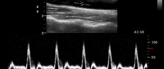
In order to study the speed and other parameters of blood flow, ultrasound is combined with Doppler ultrasound, duplex, triplex scanning. You may hear the machine make a whistling sound as it records the pulse in your legs. Different blood flows on the screen will be colored blue and red. After the study is completed, the ultrasound doctor will give you a form with a protocol. It should be taken to the attending physician - phlebologist or vascular surgeon.
How is a vein ultrasound performed?
Ultrasound examination of the vascular system of the legs means a procedure for examining the anatomy of veins and arteries, assessing their functioning, the condition of the valves and the characteristics of blood flow. The technique makes it possible not only to find out why blood circulation is affected in a particular area of the vessel, but also to identify inflammation or blood clots. Ultrasound of the vessels of the lower extremities is an important procedure for diagnosing existing problems and helps in planning the correct treatment.
Types of sonography
To study the vessels of the lower extremities, a procedure based on the Doppler effect is used. In medicine, it means the reflection of ultrasound radiation from red blood cells flowing through the vessels.
As a result of a Doppler examination, the doctor gets an idea of the characteristics of the passage of the vascular cord, the speed of blood flow and other nuances of the functioning of the veins and arteries of the legs.
So what types of research are there, and what do they show?
USDG
The term “USDG” means “ultrasound Dopplerography”. This is a technique usually used to: – establish the patency of deep vascular collectors; – assessing the condition of superficial veins; – diagnostics of the condition of valves, including valves of typical main components of the venous system, that is, perforating veins.
UZDS
Duplex scanning combines the principles of Doppler and traditional examination methods and performs the following functions: – studies the operation of vein valves in real time; – makes it possible to assess the condition of the vascular walls; – allows you to analyze the patency of deep and superficial veins; – calculates the presence and location of blood clots.
Technologically, the method remains the most popular and accurate way to assess the performance of the venous system in all respects.
Online scanning
Online scanning is a complex that combines ultrasound and ultrasound scanning. It is intended to determine: – the condition of the walls of arteries and veins; – assessment of valve health; – patency of vascular collectors; – the condition of the veins connecting superficial vessels with deep ones; – the presence of blood clots and their characteristics, including size and location; – degree of blockage of the vessel.
Ultrasound with color mapping
The most modern method of studying the veins and arteries of the legs is distinguished by color identification of the speed of blood flow in different areas. For example, shades of red characterize the flow of blood to the sensor, and blue tones characterize the flow of blood directed away from the sensor.
Important! The brighter the color, the higher the speed of blood movement.
In modern diagnostics of arterial-venous pathologies, the popularity of this method is growing due to its high information content and simplicity.
Preparation
When referring veins and arteries of the legs for ultrasound (as well as other soft tissues, as a rule), there is no need to carry out preliminary preparation for the procedure - it is not required.
How do they do it?
How is the deep veins and arteries of the legs diagnosed?
Ultrasound examination of blood vessels looks like this:
The patient enters the ultrasound diagnostic room, provides the doctor with a referral and frees the veins of the lower extremities. That is, he takes off tights or trousers, remaining in his underwear. The diagnostician applies a little conductive gel to the legs one by one, which will ensure better adhesion of the sensor to the surface of the skin.
During the manipulation, the doctor can change the frequency of the sensor for better visualization of deep vessels, but the patient will not feel this in any way.
The difference between an ultrasound scan is that during the examination the doctor will measure the pressure in both the upper and lower extremities. During the procedure, the patient will change his position from sitting to lying down and back.
During the examination of the leg veins, they are first examined with the patient lying down, then he will be asked to stand up. In addition, as part of the examination, special tests are carried out to determine the blood flow between the superficial and deep vessels. The test consists of taking a deep breath, without interrupting it, you need to make a strained effort.
The deep veins of the legs are examined in traditional and color modes. For a better examination, the patient is asked not only to perform stress tests, but also the areas where the veins are located are palpated with different intensities. Such tests (like the ultrasound itself) are performed in different positions: lying on your back or stomach, as well as standing.
Contraindications
In addition to the indications for ultrasound of the blood vessels of the legs, there are also contraindications.
Contraindications to duplex vascular examination are associated with the relatively long duration of the procedure - it takes about 40 minutes.
It may turn out that this examination turns out to be secondary and the study of adjacent tissues or internal organs comes to the fore.
Important! You should not perform an ultrasound in the area of the veins of the lower extremities if the skin is broken or damaged.
The main contraindications to studying vessels using the duplex method are:
– acute infectious processes; – any diseases accompanied by open wounds on the skin at the location of the sensor; – burns; – emergency conditions; – mental illnesses that make it impossible to carry out the procedure.
There are also conditions in which it is not recommended to scan blood vessels due to possible discomfort for the patient during prolonged immobility. These conditions include:
- excessive fullness;
- bloating;
- cystitis and some diseases of the genitourinary system;
- lymphostasis, causing severe swelling of the limbs.
Advantages and disadvantages
The advantages of the ultrasound method of studying limbs are:
- no pain;
- non-invasiveness, that is, the absence of punctures and other damage to the skin;
- economic accessibility of the procedure;
- ease of implementation;
- absence of radiation or ionizing load;
- linking the research picture to real time;
- the ability to perform a biopsy under ultrasound guidance;
- good visibility of all soft tissue features;
- the possibility of repeated repetition (for example, during treatment therapy to monitor its effectiveness).
Minuses:
- there may be insufficient data for a complete diagnosis;
- Adequate assessment of small vascular formations is not always possible;
- with atherosclerotic changes, the patency of the ultrasound wave may be impaired;
- is not a replacement for angiography;
- If carried out on old equipment or with insufficient qualifications of the doctor, the procedure may have low diagnostic value.
How much does it cost, where is the best place to do it?
It is best to contact your attending vascular surgeon, who will recommend a good specialist or tell you where and at what time he himself conducts ultrasound examinations.
In the Department of Vascular Pathologies, ultrasound can be performed free of charge, as prescribed by the surgeon. For a fee, in the absence of a referral, ultrasound examination of the extremities is carried out in phlebological clinics or multidisciplinary medical centers. You can find out the cost of the examination from the administrator by phone or by making a personal appointment. But it is better to refuse to perform vascular ultrasound in the offices of small diagnostic centers where any organs are examined.
Reference! The price of an ultrasound scan will depend on the specifics of the procedure and which vessels the patient needs to examine.
For example, for an ultrasound scan of the legs, the price will be from 1300 to 3500 rubles, and duplex angioscanning will cost 800–5000 rubles, color scanning using the duplex method will cost from 900 to 6500 rubles. On average, the cost of examining the vascular sections of the legs using ultrasound will cost about 2 thousand rubles.
There are also portable ultrasound machines, so ultrasound can be performed at home: this route naturally requires specific knowledge. Still, it is better to entrust this matter to a specialist.
Conclusion
Any patient, if indicated, can safely undergo angioscanning of blood vessels, because this manipulation is safe and painless. It does not require preparation and allows you to know exactly how healthy the vessels are an hour after the start of the examination.
After research
After the examination, you are wiped off the remaining gel from your skin and allowed to get dressed. After the ultrasound examination, no restrictions on normal activities are required. It will be necessary to wait for a written conclusion from a specialist on the results of the study. No complications after ultrasound examination of veins have been noted so far.
An ultrasound diagnostic doctor or phlebologist will analyze the results of the study and draw up a conclusion, which will be the main document on the diagnosis performed. In most cases, ultrasound of the veins is the final diagnostic method for venous diseases, but sometimes additional research methods such as venography are required.
The conclusion on ultrasound of the veins should answer the following questions:
- Are the venous trunks passable?
- What is the diameter of the trunks of the saphenous veins
- Are there thromboses in the superficial and deep veins?
- Do venous valves work and what is the direction of venous blood flow?
- Are there incompetent perforating veins?
Indications:
- Edema.
- Feeling of heaviness.
- Varicose eczema.
- Bursting pains.
- Cramps in the calf muscles.
- Spider veins.
- Acute venous insufficiency.
- Feeling of heat or cold in the foot.
- Darkening of the skin in the ankle area.
- Trophic ulcers and non-healing abscesses.
- Suspicions of congenital pathologies and traumatic injuries.
Ultrasound of the veins of the lower extremities is included in the complex of postoperative monitoring in patients who have undergone operations on the venous vessels, and is recommended to be periodically performed by people in the age group 50+, as well as women who have undergone a difficult pregnancy and difficult childbirth. Smoking, a sedentary lifestyle, being on your feet and in the cold for a long time, uncontrolled and long-term use of hormones and antibiotics - all this also negatively affects the condition of the venous lines. Therefore, patients at risk should also undergo ultrasound diagnostics, which allows many venous diseases to be detected at very early stages.
Detected pathologies
Dopplerography of the vessels of the lower extremities and other types of diagnostics help to identify the following pathologies:
- varicose veins;
- asymptomatic initial vascular lesions;
- the presence of segments of narrowing of the arteries (stenosis);
- impaired blood flow;
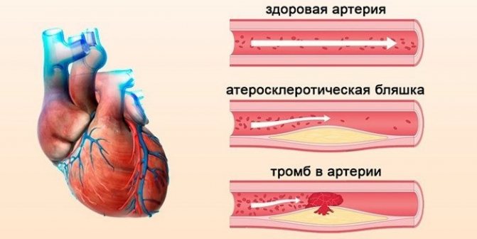
- presence of cholesterol plaques;
- a- and hypoplasia;
- thrombus in blood vessels;
- aneurysms;
- thromboembolism;
- phlebothrombosis;
- Raynaud's syndrome;
- valve disease;
- chronic vascular insufficiency;
- extravasal compression;
- trauma, arteritis; postthrombophlebitic syndrome.
Leg vascular disease is an epidemic of our time!
Pain and constant fatigue in the legs is a pressing problem for modern people. Due to the nature of our work, some of us constantly stand, some of us walk tirelessly, and others sit all day long.
The cause may be problems in the vascular bed, as well as inflammation of the joints and muscles. The movement of blood through the vessels is equivalent to the movement of a car along a busy highway. If there is an obstacle on the way, then a traffic jam will certainly arise, which can lead to a huge and painful “traffic jam”.
The same thing happens when blood circulates through our vessels. When something impedes the movement of blood, a disruption occurs in the blood flow throughout the vessel. Such undesirable changes lead to the emergence of dangerous diseases.
Today, medical ultrasound doctor Vera Aleksandrovna Abramkina will talk about the important secrets of the health of our legs and answer the most pressing questions.
— Vera Alexandrovna, tell me, what pathologies and diseases can be associated with blood vessels? Is this serious?
— Healthy blood vessels support the functioning of the entire circulatory system. Elastic walls allow the vessels to narrow and expand as needed. It is very important. Due to the dilation of blood vessels, blood flow increases, therefore, nutrition and metabolic processes in tissues improve. Due to the narrowing, all processes are slowed down.
Diseased blood vessels can be the result of diseases that manifest themselves in different ways. For example, diabetes, atherosclerosis, autoimmune diseases, vasospasms, heart failure, migraine, hypertension, heart defects, liver cirrhosis, kidney disease, erectile dysfunction, tumors, blood diseases, radiation injuries, injuries, surgeries, infections, varicose veins.
I can name a dozen more diseases. (winks)
- Uff... my head is spinning. Is it really that serious?
- Yes. Everything is more than serious.
— Can a person independently determine that he has problems with blood vessels?
— There are clear signs of problems with blood vessels. True, this is very bad if you already have symptoms of the disease. After all, very often the disease proceeds unnoticed, and the symptoms are already an advanced stage.
Run to the doctor if you have: swelling in the legs, pain and redness, cold feet, pain when walking short distances, night cramps, the presence of saccular formations under the skin, arterial hypertension, tinnitus, headache, heredity aggravated by atherosclerosis, were strokes or heart attacks.
— Recently, people are increasingly talking about the benefits of ultrasound diagnostics. Tell us in more detail how an ultrasound of the lower extremities is performed? How to prepare for the procedure?
— You know, no special training is required for ultrasound diagnostics. You come to the doctor, bare your legs, lie down on the couch and the specialist begins the procedure. He smears your feet with a special gel and runs a special sensor over certain areas. At this moment, you lie and see on the monitor what is happening inside your body. Of course, all these pictures are a “dark forest” for you, but that’s why you need a doctor to figure it all out. To obtain more reliable information about the state of your health, the examination is done comprehensively: lying down and standing. I advise you to eat so as not to fall from hunger or fear (laughs).
— Vera Alexandrovna, you are so positive. Do you joke with your patients too?
- Certainly. Where would our life be without humor?
— Who turns to you most?
“Today, unfortunately, our patients are getting younger. People over 30 are increasingly coming to us for research. This is due to the unfavorable factors that we encounter every day:
- unbalanced diet
- bad habits
-sedentary lifestyle
— Here’s what else we’d like to ask you about... What are the contraindications for ultrasound of the lower extremities?
— The good thing about ultrasound diagnostics is that it has almost no contraindications. It all depends on the severity of the patient’s condition. There are diseases that can only be examined in a hospital.
-Thank you very much for such useful information. How often do you need to do an ultrasound of the lower extremities to be healthy?
— I would like to note that ultrasound does not cure, we identify changes and carry out dynamic monitoring. Ultrasound is not a panacea. Such a study shows the doctor what is inside the patient and allows him to make an accurate diagnosis. When there is a research protocol, the doctor does not act blindly
In general, experts recommend doing an ultrasound as part of a clinical examination. The absence of contraindications allows you to do an ultrasound as many times as the attending physician considers necessary.
— Vera Alexandrovna, what else can you recommend for excellent leg health?
— If we talk about the legs, then sometimes you also need to look at the veins along with the arteries. Since there are many concomitant diseases that negatively affect our health while developing in parallel in the human body. Otherwise, it will turn out that you are treating one thing, but the deeper and more serious problem is in another. Only a doctor can figure out what the specific problem is.
— Thank you, Vera Alexandrovna, for such an interesting conversation.
- Contact me, I will always help in any way I can. Take care of yourself and be healthy!
Contraindications
Ultrasound examination has no permanent contraindications. The patient’s poor health can affect the result of the examination, therefore it is worth rescheduling the diagnosis if there are signs of acute respiratory viral infection, high blood pressure and a significant deterioration in health.
Attention! Ultrasound diagnostics does not affect the patient’s well-being and does not aggravate the disease. The method is used to obtain information about violations.
The device does not provoke adverse reactions and does not emit radiation, therefore the use of the product is permitted by patients of different health groups.