Endoscopic examination of the lower digestive tract
Sigmoidoscopy is a type of endoscopic examination that allows you to study in detail the lower (final) part of the digestive tract, as well as take tissue for biopsy, remove a foreign body and other manipulations.
This endoscopy method is performed by introducing a device into the rectal cavity - a sigmoidoscope, with the help of which the image is transmitted to the monitor screen. The procedure is performed on an outpatient basis after preliminary preparation, including bowel cleansing.
Unlike colonoscopy, this examination concerns a small area of the colon (rectum and part of the sigmoid colon - only up to 25 cm), so it does not take much time, is carried out without anesthesia and is easily tolerated by patients.
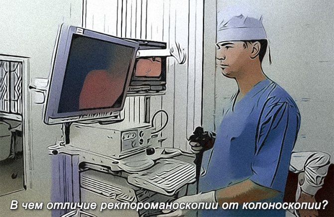
How is sigmoidoscopy different from colonoscopy?
Many patients have heard about another method of diagnosing the intestines from the inside - colonoscopy - and want to know how it differs from sigmoidoscopy of the rectum. Although both procedures are equally valuable as diagnostic tests and can be performed alongside biopsies and medical procedures, they have a number of differences, which are presented in our table below:
| Options | Sigmoidoscopy | Colonoscopy |
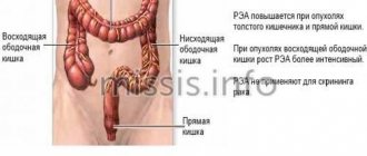
| Field of study | Rectum and part of the sigmoid | All parts of the large intestine |
| Invasiveness | Small | High |
| Painful sensations | Minimal or absent. | Palpable and require anesthesia. |
Indications
For an accurate diagnosis
The sigmoidoscopy procedure is widely used by doctors for primary diagnosis, that is, to determine the diagnosis of a patient who first contacts a proctologist with specific complaints.
A coloproctologist, gastroenterologist or therapist may recommend undergoing endoscopy of the lower digestive tract if you have the following complaints:
- blood, mucus or pus in the stool;
- pain, sensation of a foreign body in the anus;
- changes in the shape of feces (ribbon-shaped feces);
- persistent constipation followed by diarrhea.
Indications for the procedure of sigmoidoscopy of the intestines may appear during a consultative examination of a patient who has applied for an anal fissure or hemorrhoids. Such a study is necessary to exclude a tumor, inflammatory process, foreign body or other pathology.
For clinical observation and screening examinations
Sigmoidoscopy refers to studies that are not associated with radiation exposure to the body, and therefore can be performed as often as desired. At the same time, endoscopy provides maximum information about pathology when it comes to diseases of the mucous membrane.
Therefore, endoscopic examination of the rectum and sigmoid colon is widely used for preventive purposes - for clinical observation and screening examinations.
Preventive endoscopy of the rectum diagnoses cancer at the earliest stage of development, when the patient does not make any complaints, and the surgeon cannot palpate the tumor during a digital examination.
Dispensary monitoring of already diagnosed diseases using endoscopic methods makes it possible to prevent complications of the disease.
For diagnostic and therapeutic procedures
Another advantage of the sigmoidoscopy procedure is the ability to combine endoscopic examination with diagnostic and therapeutic procedures.
An endoscopic examination can be combined with a biopsy - tissue sampling from a suspicious tumor for cytological examination, which is the main diagnostic procedure when diagnosing a malignant tumor.
During sigmoidoscopy, the doctor can perform a number of microsurgical treatment procedures - cauterize a bleeding vessel, remove a foreign body, remove a polyp (benign neoplasm) of the rectum.
It must be said that the vast majority of polyps are dangerous in relation to malignant degeneration, therefore, after removal, doctors recommend regular preventive examinations (every 3-5 years).
What's better?
From the point of view of diagnostic value and information content, primacy is given to colonoscopy. It allows not only to control the disease, but also to detect it in the early stages with great accuracy. At the same time, the method is more unpleasant to carry out and requires the most serious preparation and recovery period. The risk of complications is increased, the list of contraindications is wider.
Rectoscopy is much easier to tolerate, is not accompanied by any particular discomfort or pain, passes quickly, and has a minimum of contraindications. However, the information content is limited to only 35 cm of the lower intestine and there is a risk of missing the progression of the pathology.
Despite all the differences, it is impossible to single out one procedure as a favorite. These diagnostic methods are not opposed to each other; they are used according to indications for a specific situation. The choice is made taking into account maximum benefit, as well as minimal harm to the patient.
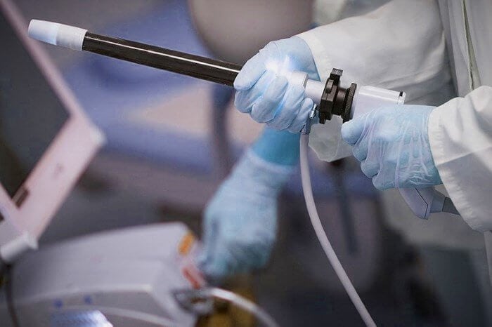
Sigmoidoscope
A cylindrical diagnostic device, which is inserted into the rectum during an endoscopic examination, is called a sigmoidoscope.
The lower part of the digestive tract has no bends. Therefore, for its endoscopic examination, both soft plastic and hard metal sigmoidoscopes are used.
Each sigmoidoscope is equipped with a set of additional devices of a fixed (illuminator) and removable nature. Replacing each other, removable elements allow you to perform additional tasks.
The standard package of the sigmoidoscope includes:
- air supply device;
- optical system, including an eyepiece and a light guide harness;
- forceps;
- biopsy channel;
- obturator
During the examination, the LED fiber of the sigmoidoscope transmits the image to the monitor screen, so that all manipulations are carried out under visual control.
After the study, a record is saved, which is of particular importance for dispensary patients, since it allows you to track the dynamics of the development of the disease.
How is sigmoidoscopy performed?
After the morning bowel cleansing, the patient comes for the procedure. In the office, he will be asked to undress from the waist down and take a position convenient for inserting the sigmoidoscope. There are 2 options:
- “lying on your side” - the patient lies on his right side, bending his knees at a right angle;
- knee-elbow pose - kneeling, the body is tilted forward, leaning on the elbows.
The second option is considered preferable - in this position the peritoneal wall lowers, facilitating the insertion of the device tube into the intestine.
The hardware examination procedure begins only after a digital rectal examination. This allows you to prevent possible damage in the presence of acute anal fissures and other pathologies.
Procedure for RMS:
- A sigmoidoscope tube with an expander, lubricated with petroleum jelly, is inserted through the anus into the rectum to a depth of 5 cm, after which the patient is asked to take a deep breath and strain, as if defecating, to facilitate the passage of the tube through the intestines.
- When the required depth is reached, an optical device is inserted into the tube to visually examine the intestinal walls.
- In the zone of transition of the rectum to the sigmoid colon, air is pumped. This allows the folds to straighten out, but may cause a feeling of painful fullness.
- If fecal residues enter the viewing lumen, cleanse with a tampon inserted through a tube; if there is a large amount of pus, mucus, or bloody discharge, use suction.
- In parallel with the visual examination, tissue biopsy, cauterization of polyps, blood vessels, and microtumors are performed during RRS. For this purpose, a special coagulation loop, biopsy microforceps and brushes are used.
The entire procedure takes 5-10 minutes.
If RRS was carried out in the knee-elbow position, after its completion the patient should lie on his side for several minutes - this avoids orthostatic hypotension - “floaters” and loss of balance when rising. During this time, the doctor measures blood pressure, pulse and assesses the patient’s general well-being. During the first minutes after all manipulations, there is an intense release of air pumped into the intestines during examination.
Attention! After insertion of the endoscopic tube, the patient may feel the urge to defecate. There is no need to be afraid of these sensations - they help the endoscope move through the intestines. The best solution is to relax and breathe slowly and deeply.
In case of increased excitability of patients, the examination is carried out under sedation with sedatives. In special cases, a light form of anesthesia is used.
What the study will show
Endoscopy is an examination of the mucous membrane of the digestive tube. Doctors receive information about the condition of other layers of the intestinal wall indirectly from changes in the pattern of the mucous membrane, abnormal bends, etc.
Sigmoidoscopy of the intestine will provide the maximum amount of information about the following diseases of the lower intestinal tube:
- inflammatory reactions;
- benign neoplasms;
- oncology;
- enlarged hemorrhoids;
- foreign bodies in the intestinal lumen;
- injuries;
- congenital anomalies.
During a sigmoidoscopy examination, the doctor will examine all parts of the rectum (anal canal (4 cm), the following ampullary extension (8-10 cm) and the supramullary part (2-4 cm)), as well as the adjacent part of the sigmoid colon (10-15 cm).
If there is a need to examine the upper parts of the large intestine, then a colonoscopy is prescribed, but if it is advisable to limit the diagnosis to the study of the anal canal, an anoscopy is prescribed.
Sigmoidoscopy and colonoscopy: areas of examination
Sigmoidoscopy of the intestine is a method based on visual inspection of certain parts of the intestine. This is mainly the mucous membrane of the rectum; less often, this method is used for the distal parts of the sigmoid colon (on average, up to a distance of 20-25 cm from the injection site - through the anus). This diagnostic method is considered one of the most informative in modern medicine.
It allows you to visually assess the condition of the intestine in the area it covers. Such diagnostics are often used to check for the presence of various neoplasms, since the technique allows not only to examine the surface of the intestine, but also to immediately perform a biopsy if something suspicious is noticed.
Colonoscopy is an endoscopic examination in which the entire large intestine is examined using a special instrument. That is, the instrument penetrates 120-150 cm of the total length of the intestine. This makes the technique more informative compared to sigmoidoscopy. And this is the main difference between these two methods - in the areas of research coverage.
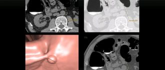
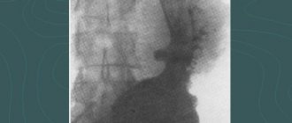
There is another research method - irrigoscopy . What kind of diagnosis is this? This method is an x-ray examination of the colon using a special x-ray contrast agent.
Often the doctor is faced with a choice - sigmoidoscopy or irrigoscopy. Since the second method is considered more gentle (only a substance is injected, and not a rigid instrument), then if there are contraindications to sigmoidoscopy or colonoscopy, this particular X-ray examination is chosen. But since it is more suitable for contour diagnostics and not all changes are visible on it, it can be considered a kind of compromise.
Both sigmoidoscopy and colonoscopy help assess the condition of the intestinal walls according to several basic characteristics, changes in which may indicate the presence of a serious disease - the color of the walls, vascular pattern, elasticity, tone, and, to identify tumors, relief.
Contraindications
There are no absolute contraindications to endoscopy. The exception is pathologies that do not technically allow this absolutely safe study to be carried out (mechanical obstruction of the rectum).
Doctors advise postponing the examination if the patient is in serious condition:
- acute infectious diseases;
- exacerbation of chronic diseases of the pelvic organs;
- decompensated cardiovascular, renal or liver failure;
- condition after a heart attack, stroke, etc.
Difficulties with sigmoidoscopy of the intestines can arise in mentally ill people. In such cases, the issue is resolved individually (examination under anesthesia or replacement with another type of diagnosis).
Colonoscopy technique
The patient is placed on his left side, with his knees brought to his chest. The tip of the colonoscope is lubricated with anesthetic gel and inserted into the rectum - the examination begins. The doctor slowly advances the tube with the camera deep into the intestine, examining it along its entire length. The intestine, as well as sigmoidoscopy, is pre-inflated with air, which is then pumped out. A simple diagnostic examination takes no more than 10 minutes; if additional manipulations are necessary, the time increases.
Preparing for the study
Preparation for sigmoidoscopy involves cleansing the intestines of gases and feces. To prevent increased gas formation, three days before the procedure you should avoid peas, pearl barley, cabbage, beans, fresh vegetables and fruits, juices, sparkling water, kvass, alcohol, rye bread and other foods that provoke flatulence.
The day before the procedure, they switch to a slag-free diet (low-fat dietary meat (chicken, turkey), fermented milk products, eggs). Dinner on the eve of the event should be light and early (no later than 12 hours before the examination).
Immediately before the study in the evening and/or morning, the intestines are cleansed with an enema (to clean water) or with a laxative such as Fortrans, which is taken according to the instructions included with the drug.
Preparation for the procedure
The process includes a special diet for 2-3 days before RMS, as well as a complete bowel cleanse the day before the procedure.
Diet requirements:
- completely eliminate foods that provoke gas formation and fermentation: legumes, cabbage, sweets, flour products, fruits;
- give up alcohol, tonic, sweet and carbonated drinks;
- dietary meats, boiled or stewed fish, water porridge (semolina, rice), boiled vegetables are allowed for consumption;
- For drinks, it is preferable to stick to green teas and clean still water;
- on the day before the procedure, breakfast and lunch should be light, avoid dinner completely;
- On the day of RRS, you are allowed to drink only water or weak green tea.
Attention! If you are taking anticoagulants, be sure to tell your doctor about the risk of bleeding.
Methods for colon cleansing:
- Cleansing with an enema is carried out in the evening before the prescribed RRS and early in the morning on the day of the procedure: each time do 2 approaches, pouring 1-1.5 liters of warm water into the intestines. Ideally, rinsing is carried out until clean water flows.
- The use of laxatives requires additional consultation with a doctor. The choice of drugs depends on the individual characteristics of the patient and can be adjusted. Oral laxatives are taken the evening before the procedure with 3-4 liters of water. If the patient is not able to drink such a large volume, part of the dose is transferred to the early morning - in this case, it is enough to drink 2 liters of water in the evening, and 2 liters with the corresponding volume of medicine will remain for the morning. Rectal medications are also administered in the evening and morning (or as directed in the prescription), but large volumes of water are not required.
How does the procedure work?
Before an endoscopic examination of the lower intestine, you will need to undress from the waist down and remove your underwear. If the device is equipped with a metal sigmoidoscope, the examination is carried out in the knee-elbow position. When using a soft plastic device, the procedure is performed in a lying position on your side with your knees pulled up to your chest.
Immediately before the procedure, the doctor conducts a digital examination to assess the tone of the sphincter, ensure the patency of the anal canal, and identify swelling, soreness or other pathological symptoms.
After a digital examination, the doctor carefully inserts a sigmoidoscope lubricated with medical petroleum jelly, pumps air, straightening the folds of the mucous membrane, then conducts an examination, and performs diagnostic and therapeutic manipulations according to indications.
The entire procedure takes 10-40 minutes, depending on the structural features of the patient’s intestines, the complexity of diagnosis, and the need for additional manipulations.
Carrying out anoscopy
The patient can take any comfortable position: on the gynecological chair, lying on his back, standing in a knee-elbow position on the couch, or lying on his side with his knees raised to his chest. The proctologist lubricates the anoscope for easier passage into the anus with medical lubricant. The device is carefully inserted, while the doctor tells the patient how to behave correctly to eliminate the possibility of pain or discomfort. If the examination is carried out for purely diagnostic purposes, its duration is on average 10-15 minutes. More time may be required if medical or surgical intervention is also performed during anoscopy.
Rules of conduct after the examination
Endoscopic examination does not require hospitalization of the patient. After the procedure, the doctor will suggest you rest for a few minutes on the couch, then you can go home.
Since the procedure is accompanied by the injection of gases into the intestines, their free passage is possible throughout the day. This is fine.
When washing the intestines with Fortrans or an enema, beneficial bacteria are “washed out,” and the procedure itself can have an irritating effect on the mucous membrane. Therefore, for 5-7 days after the procedure you need to adhere to a diet (fermented milk products, dietary meat, eggs).
After a week, if there are no other indications, you can abandon the restrictions.
Patient reviews about the procedure
We analyzed reviews of intestinal sigmoidoscopy. It can be concluded that the vast majority of patients who underwent the procedure characterize the study as unpleasant, but painless.
Many reviews record the impression that waiting for the procedure brings more unpleasant worries than the examination itself. In some patients, the bowel cleansing procedure caused discomfort. To avoid such sensations, it is better to use laxatives like Fortrans instead of microenemas, which can cause irritation of the mucous membrane.
There are relatively few complaints of pain during sigmoidoscopy examination of the intestine. Patients complain of pain and discomfort during the entry of air into the intestine, as well as during the examination procedure.