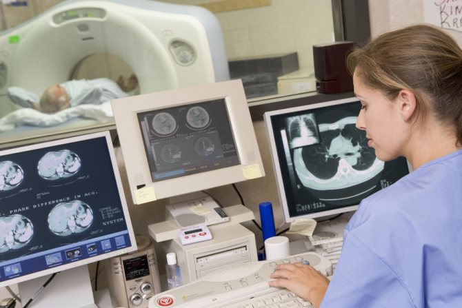
With the advent of x-rays, the task of making a diagnosis for diseases of the gastrointestinal tract has been significantly simplified. Further development of technology served as the basis for the development of new, less harmful to the body, examination methods. Today, more and more often, if it is necessary to examine the gastrointestinal tract, patients turn to magnetic resonance imaging and computed tomography, which allows them to look inside the body without resorting to instrumental intervention. However, the interpretation of MRI and CT results is simpler compared to interpretation; x-ray.
Introduction
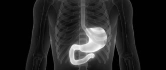
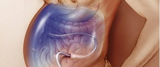
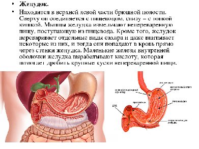
The stomach
is a hollow muscular organ located between the esophagus and the duodenum. It is a pouch-like expansion of the digestive canal in which swallowed food accumulates and is digested. Gastric juice secreted by the glands of the gastric mucosa contains digestive enzymes, hydrochloric acid and other substances, digests proteins, partially fats, and has a bactericidal effect. In addition, mechanical grinding of food occurs in the stomach.
Magnetic resonance imaging of the stomach can be part of a comprehensive diagnostic system of abdominal organs or prescribed on an individual basis if there are signs of a pathological condition of the organ.
|
|
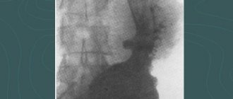
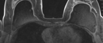
| Anatomical diagram of the structure and location of the stomach | MRI image of the stomach |
Indications
A local internist or gastroenterologist can refer you for an MRI of the stomach, or the patient can independently undergo an examination through the sphere of paid services. The need for the procedure arises when the following symptoms occur systematically:
- Aching, cutting, stabbing pain in the abdomen, appearing with a certain frequency.
- Flatulence.
- Feeling of heaviness and bloating.
- Nausea and vomiting for no apparent reason.
- Lack of appetite.
- Unexpected weight loss.
- Change in urine color.
- Traces of blood in the stool.
- Laboratory tests and/or ultrasound revealed signs of degenerative processes, neoplasms, and other pathologies.
- Helminthic infestation in acute form.
MRI scanning is also necessary in the work of oncologists and surgeons when:
- Determining the stage of the neoplasm.
- Assessing the extent of metastasis.
- Checking the effectiveness of the therapy.
- Preparing for surgery.
- Quality control of surgical intervention.
An MRI examination of the stomach allows you to establish an accurate diagnosis, select treatment, check the feasibility of surgical intervention and increases the chances of successfully combating the disease.
OLYMPUS VIDEOSCOPES: WIDE SELECTION, REASONABLE PRICE, DELIVERY - lkmed.ru/../videoskopy-olympus/
Contraindications for MRI
Contraindications can be divided into two groups: absolute and relative. If there are absolute contraindications, MRI examination is unacceptable. In case of relative contraindications, the study is possible under certain conditions.
Absolute contraindications
- pregnancy, first trimester
- presence of a pacemaker
- presence of middle ear implants
- presence of metal foreign bodies (splinters, shavings, etc.)
- the presence of metal clips/clamps on the vessels of the brain
Relative contraindications
- pregnancy
- joint prostheses
- insulin pump
- artificial heart valve
- vascular stents, vena cava filter
- claustrophobia, panic attacks
- inability to maintain a still position
- grafted electronic devices (neurostimulators)
You can see prices for services
Preparation for MR enterography
| Diet on the day of examination |
| |
| Bowel preparation | not required | |
| Take with you |
| |
| Keep in mind |
| |
What does it show
If the cause of the painful condition lies in the pathology of another organ, then the MRI image will demonstrate the correct location and size, the absence of signs of inflammation, deformations and neoplasms.
In the presence of pathologies, MRI allows us to establish the following conditions with an accuracy of 95%:
- Abnormal structure of the stomach from birth.
- Developmental defects of the organ.
- Inflammatory processes in colitis.
- Deformation of lymph nodes.
- Mechanical damage to blood vessels and internal bleeding.
- The presence of ulcers and erosions.
- Poor circulation in blood vessels (aneurysms, thrombosis).
- Tissue necrosis, peritonitis.
- Oncological diseases.
- Penetrating and intrinsic metastases.
- Cysts and benign neoplasms.
MRI allows you to distinguish fibrous compactions from an acute inflammatory process, determine the shape and size of the tumor, detect pathology and record relapse of the disease.
How to prepare for scanning
The most important thing when preparing for an MRI of the stomach is to stop eating 8 hours before the examination. This is convenient to do if the procedure is scheduled for the morning. You should also stop eating foods that cause gas for a few days.
An hour before the examination in the office, the doctor gives the patient one liter of iron-containing liquid to drink. This is necessary to stretch the organ. This is where the preparation ends. If you have metal jewelry, you must remove it; you also cannot take a mobile phone, watch or other electronic device with you.
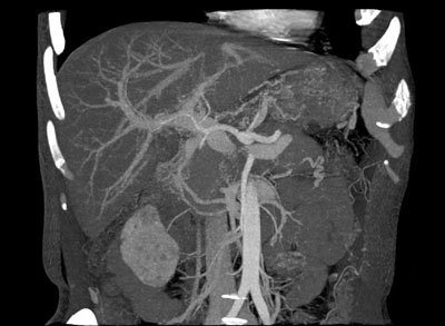
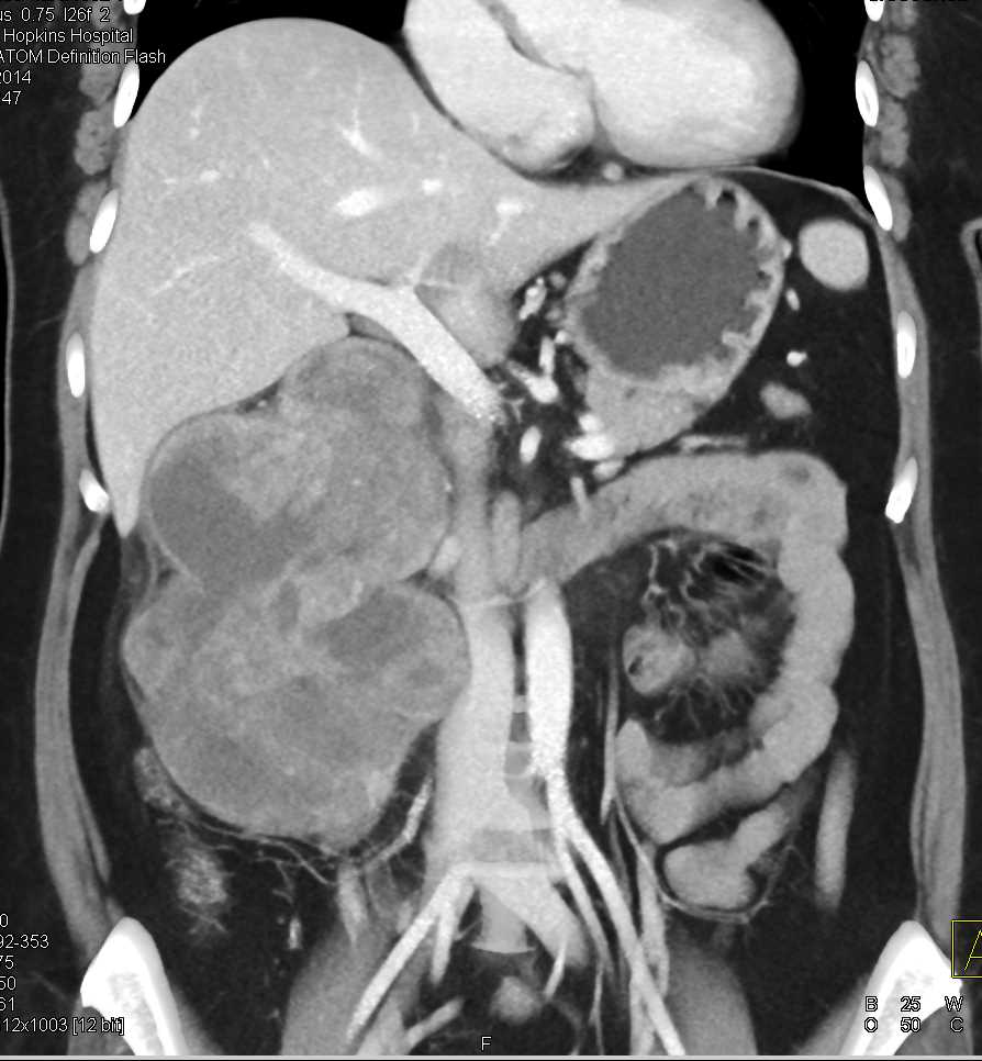
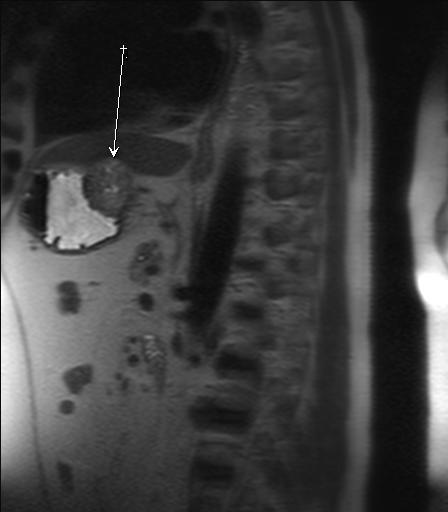
Photo
|
| Gastrointestinal stromal tumor of the stomach |
|
| Gastric leiomyoma on MRI |
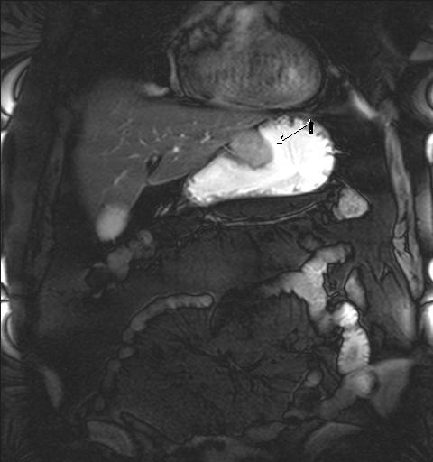
|
| Gastric leiomyoma |
|
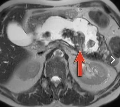
| Abdominal cancer with metastases to the stomach |
|
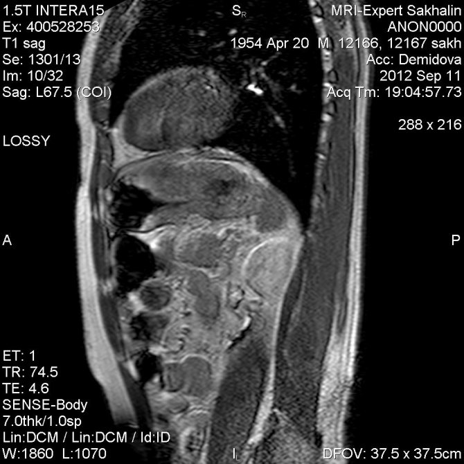
| Enlarged stomach on MRI |
Price
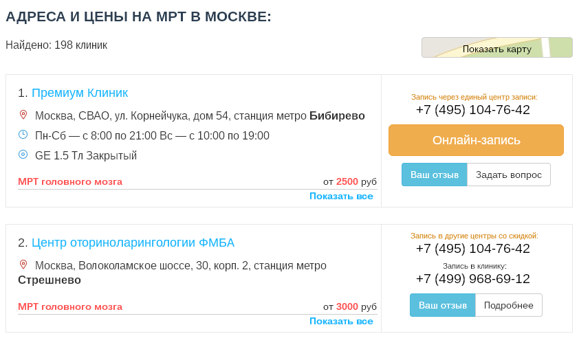
On our website, many clinics list prices for MRI, including MRI of the stomach.
To see prices, you need to go to the list of clinics in your city. To do this, select your city on the “Addresses” page.
On the page for the list of city clinics, in the form for selecting a clinic based on parameters, in the “Research area” field, select “MRI of the stomach.”
The list will be filtered and the clinics will be sorted by ascending price, which will allow you to find out where to get an MRI of the stomach cheaper.
|
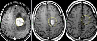
| List of clinics filtered by MRI of the brain using the example of the city of Moscow |
If prices are not indicated on the website, then you need to call all the clinics and find the best price for you yourself.
When talking with the operator, be sure to ask if there are any promotions or discounts. For example, some 24-hour clinics offer discounts on MRIs at night.
You can get an MRI with compulsory medical insurance or VHI insurance. To do this, you need to find out which clinic does MRI under insurance, using the information on the website or calling the clinics yourself if there is no information on the website. In the form for selecting a clinic by parameters, there is a field “MRI under insurance”. Next, you need to find out the list of documents that the clinic operator will tell you, collect them and you will be able to undergo an MRI with insurance, which will allow you to save a lot.
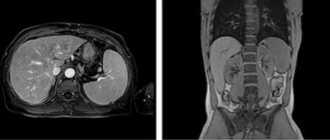
Abdominal MRI
Preparation
MRI of the abdominal cavity with contrast
MRI of the abdominal cavity requires little preparation. This is necessary for image clarity and to eliminate motion interference. 24 hours before the session, you need to pay attention to the menu, from which you should remove foods that cause gas formation: cabbage, beets, peas and beans, carbonated lemonade, kefir. Other items include:
- a carbohydrate-free diet two days before the session;
- availability of medical documents: outpatient card, referrals, extracts, information on the results of previous studies;
- eating light food on the day of the procedure;
- exclusion of fluid 5 hours before an MRI of the abdominal cavity;
- taking an antispasmodic half an hour before the start;
- adoption of activated carbon to reduce gas formation.
An MRI of the abdominal cavity, the price of which depends on the area of study, requires the removal of all metal objects - hairpins, earrings, rings, chains. If you have dentures, crowns, implants or other non-removable items on your body, you must inform your doctor about this. Otherwise, the analysis results will be distorted, and watches, mobile phones and credit cards may be damaged under the influence of the magnetic field.
Studying patients with internal defibrillators, drug dispensers, ear implants, or cerebral aneurysm attachments is prohibited. At the same time, patients with coronary stents and pacemakers are allowed to undergo MRI of the abdominal cavity. Metal objects do not pose a health risk, but will interfere with the correct determination of the cause of the disease.
MRI of the abdomen with contrast
What does MRI of the abdomen with contrast show?
The procedure lasts approximately half an hour to an hour. To improve detail before surgery, after surgery and in difficult cases, the doctor prescribes the introduction of a gadolinium-based contrast agent into the body. This element, unlike iodine-containing drugs, is completely harmless, does not cause allergies or side effects, and also has the ability to accumulate in pathological sites, oncological tumors, and areas with impaired blood circulation.
The procedure using contrast involves the following measures:
- introduction of the drug into the blood once by injection or in doses through a dropper;
- performing a scan.
- monitoring its action through a monitor.
The injection method of administering contrast is carried out when studying the boundaries, structure and nature of neoplasms, before operations, pathologies in the mammary glands, brain, to determine multiple sclerosis, to identify foci of a hidden nature, in differential diagnosis. The second method, called dynamic magnetic resonance imaging, is used to monitor processes in real time, for example, during surgery.
If you would like to have an MRI of the abdominal cavity done in Moscow, the price , address and conditions can be clarified by phone. You can also sign up for the procedure by telephone or by filling out the feedback form on the website.
Price for MRI of the abdominal cavity
The cost of the procedure depends on the type of medical institution, the conditions of the study, for example, a combination of magnetic resonance imaging with cholangiopancreatography, urography, and the use of a contrast agent. The amount is affected by the modification of the device, the specified type of image, the time it takes to decrypt the result, and the presence of digital media - a flash card, a CD.
Reviews
On our website, people leave reviews for clinics about their MRI experience.
To read patient reviews, you need to go to the list of clinics in your city. To do this, select your city on the “Addresses” page.
The list in the block of each clinic will show a button to go to the list of reviews, if there are reviews, or a button to go to write a review, if there are no reviews yet.
|
| This is what review buttons look like, using the example of the city of Moscow |
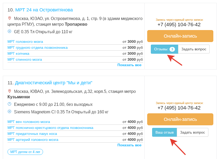
You can also leave a review about your MRI experience so that other people can choose the most suitable clinic.
To leave a review, you need to click the “Your review” button and you will be taken to the review writing form. Or if there are already reviews, then you need to go to the end of the list of reviews and there will be a form for writing a review.
To open the review writing form, click the “Leave a review” button.
Fill out the form and send it for moderation.