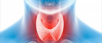
Indications for the procedure
There are a number of indications for magnetic resonance imaging of the stomach that are directly related to diseases and symptoms.
Stomach ulcer
This disease can be prevented if a procedure such as an MRI of the stomach is performed in a timely manner and the right diet is chosen already at the stage of gastritis.
Chronic or acute gastritis
One of the most common stomach diseases - gastritis - after some time can become chronic and develop into an ulcer. This can be avoided if you undergo a timely examination of the stomach and esophagus using magnetic resonance imaging.
Stomach cancer
MRI of the stomach allows you to get clear and clear images, thanks to which you can identify tumors with 100% accuracy.
Esophageal spasm
This rather painful process can occur not only due to a malfunction of the gastrointestinal tract, but also due to the presence of concomitant diseases. An MRI of the esophagus shows what is causing the unpleasant symptoms. Doctors recommend undergoing a tomography examination to be sure of the accuracy of the diagnosis.
Diaphragmatic hernias and more
It is not always possible that a routine examination and questioning by a general practitioner allows one to make the most accurate diagnosis, so one has to resort to various diagnostic methods that can show a clear picture. The same is the case with diaphragmatic hernias. Experts recommend examining the stomach and esophagus using MRI.
Medical Internet conferences
Comparative information content of gastric fluoroscopy and gastroscopy
with hiatal hernia
Scientific supervisor: candidate of medical sciences, associate professor Ilyasova E.B.
Comparative information content of gastric fluoroscopy and gastroscopy
with hiatal hernia
Scientific supervisor: candidate of medical sciences, associate professor Ilyasova E.B.
GBOU VPO Saratov State Medical University named after. IN AND. Razumovsky Ministry of Health of Russia
Department of Radiation Diagnostics and Radiation Therapy named after. NOT. Stern
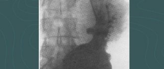
Relevance. Hiatal hernia (HH) occurs in 30% of gastrointestinal diseases in middle-aged and elderly people. To diagnose hiatal hernia, many clinicians consider the use of gastroscopy sufficient and exclude fluoroscopy of the stomach from the examination plan.
The purpose of the study is to comparatively assess the information content of gastric fluoroscopy (RG) and fibrogastroduodenoscopy (FGDS) in the diagnosis of hiatal hernia.
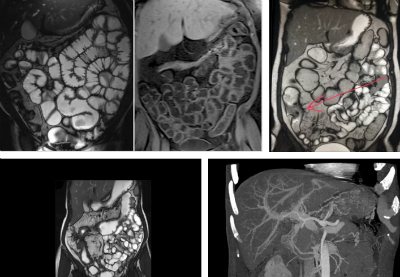
Material and methods. The study material included 22 patients, aged from 38 to 65 years, with suspected hiatal hernia, who were treated at the Clinical Hospital named after. S.R. Peacemakers SSMU. All patients underwent: RSJ using a device with digital technology DH-90 (Apelem, France), FGDS, survey fluoroscopy (ORS) of the abdominal and thoracic cavities.
Results. In 3 (13.5%) patients with ORS of the chest and abdominal cavities, a ring-shaped shadow with thin walls and a horizontal fluid level was detected in the posterior mediastinum; a gas bubble of the stomach was visualized under the diaphragm. This shadow with RSG in a vertical position turned out to be fixed by the hiatal hernia. In the remaining 19 patients (86%), partial displacement of the stomach was determined only in the Trandelenburg position - sliding hiatal hernia. In 15 (68%) cases, gastric prolapse was noted, measuring 2x3 cm, in 4 cases - 3x4 cm, in 2 cases - 4x5 cm, in 1 case - 5x6 cm. In all 22 cases, gastroesophageal reflux (GEFR) was noted ) and signs of esophagitis, of which erosion was suspected in 4 cases. During FGDS, signs of esophagitis and hiatal hernia were detected in all cases, GEFR – in 19 patients (86%) .
The size of the prolapsed area of the stomach and its mobility were not determined. Erosive esophagitis was detected in 9 (40%) patients. In 2 patients (9%) it was possible to identify Saint's triad and Castaing syndrome (partly with the help of RSG)
Conclusions: When diagnosing a hiatal hernia, the use of FGDS or ORS of the thoracic and abdominal cavities is not enough. The advantage of RSG is that it is more accurate than with FGDS in identifying the hiatal hernia, determining its fixation, establishing the volume of the prolapsed stomach and the presence of GEFR. FGDS clarifies the presence of esophagitis and reveals erosions in the esophagus. Consequently, RS of the stomach and FGDS cannot replace each other, but should be used in combination in all cases of suspected hiatal hernia.
How to prepare for an MRI examination?
This procedure does not require special preparation. Your doctor will recommend that you stop eating and drinking immediately before the test. To obtain the most accurate result, it is recommended to limit yourself in the consumption of tea, coffee and flour products a few days before the procedure. The tomograph creates a special magnetized field, as a result of which before the procedure it is necessary to remove jewelry and metal objects, glasses, piercings, watches, belts, remove your mobile phone, flash cards, coins, bank and transport cards from your pockets. Females are advised to limit themselves to minimal makeup, as some products contain metal particles.
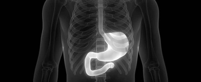
Computed tomography of the stomach
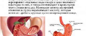
Before the advent of computed tomography, the main diagnostic tests for identifying various gastric diseases were endoscopy and x-rays. An endoscopic examination allows you to examine the mucous membrane of the esophagus, stomach, the beginning of the small intestine (examination of the upper part of the gastrointestinal tract), as well as the large intestine (examination of the lower part of the gastrointestinal tract). Examination of the stomach is carried out using gastroduodenoscopy, the following pathological conditions are detected:
- Gastritis.
- Gastroesophageal reflux disease.
- Portal hypertension.
- Stomach ulcer.
- Polyps.
- Cancer of the esophagus, stomach.
- Gluten intolerance.
- Anemia.
An endoscopic examination allows for therapeutic measures to be taken - to stop bleeding, remove foreign bodies, and also during the examination a biopsy of stomach tissue is performed.
X-ray of the stomach is a dynamic research method that allows you to take a picture of the stomach and select the desired projection of the organ. An X-ray with barium is also performed; the study allows you to observe the passage of contrast through the gastrointestinal tract using images. X-ray examination allows us to identify the following pathological conditions:
- Varicose veins of the esophagus and stomach walls.
- Polyps.
- Stomach ulcer.
- Malabsorption syndrome.
- Gastrointestinal motility disorder.
- Benign and malignant tumors of the gastrointestinal tract.
- Narrowing of the duodenum.
- Hiatal hernia.
Computed tomography is most often performed to detect stomach tumors. Indications for use include suspected ulcers, Crohn's disease, colitis, cancer and many other diseases. Computed tomography allows you to determine the presence of foreign bodies, injuries, polyps, and make a quick decision on whether to perform surgery or prescribe a specific treatment method. Computed tomography can be used to predict the course of the disease. The gastroenterologist refers the patient to a CT scan if unpleasant symptoms appear:
- Indigestion.
- Heartburn.
- Nausea.
- Blood in urine or stool.
- Pain in the stomach after eating.
- Feeling of heaviness in the stomach after eating or having a small snack.
- Loss of appetite, loss of body weight.
- Digestive disorders.
CT with contrast is used to diagnose stomach diseases. Before a computed tomography scan with contrast, preparation is carried out. The patient undergoes tests for allergies to contrast agents, and kidney function is determined. CT scans are performed on an empty stomach; before the study, the patient abstains from eating for 6-7 hours. In order for the contrast agent to quickly leave the body, it is recommended to drink more fluids the day before the examination. To avoid distortion of CT results, you should come to the examination in clothes without metal buttons or jewelry.
A computed tomography scan of the stomach takes an average of 20 minutes and causes virtually no discomfort to the patient. Computed tomography very well allows you to see the walls of the cavity filled with gas or liquid. The contrast agent is administered intravenously or drunk by the patient. The patient is then placed on a rotating CT scanner table, which moves under the scanner. The scanner takes images in slices, then the patient turns over on his right side, which allows scanning of the outlet of the stomach and the upper intestine. The patient must remain completely still during the scan.
The contrast agent is drunk by the patient in several stages, allowing soft tissues and blood vessels to be more clearly visible. Using contrast, tumors of the stomach and intestines are detected in the early stages of development - contrast highlights the smallest malignant and benign tumors in images of the circulatory system. If a reaction to the contrast agent develops during the study, the doctor stops the diagnosis. A reaction to a contrast agent can manifest itself in the form of tachycardia, severe dizziness, and itching.
Computed tomography helps determine the location of the tumor, size, type, circulatory characteristics and other features of the tumor. CT helps to identify the onset of tumor metastasis, the boundaries of the lesion, and the stage of development of the tumor is very accurately determined. The doctor can see signs of perforation of the stomach walls, obtain accurate data on the condition of other abdominal organs, data on the functionality of the organs, and the peculiarities of the functioning of the organs. Computed tomography is a very accurate study, thanks to which the doctor can prescribe effective treatment and timely diagnose a malignant tumor at an early stage of development.
Carrying out the procedure
Before starting the procedure, the doctor explains the entire examination process to the patient, talks about the rules and contraindications. Next, the patient lies down on a special table, which then smoothly moves into the tomograph. To obtain the most accurate results, you should remain calm and still. Often, during the examination, the stomach walls are straightened by slightly stretching with the help of an iron-containing solution, which is prescribed to the patient and relieves pain. Sometimes a specialist administers intravenous contrast, shows the most clearly needed areas, and this does not cause any discomfort.
What can you see on an X-ray of the stomach?
X-ray allows you to visualize bone and soft tissue, as well as the muscle walls of internal organs. Hollow organs, including the stomach and intestines, are poorly visualized in X-rays. However, the use of contrast solutions makes the task easier: hollow organs filled with a solution of barium salt or iodine become visible quite clearly. The resulting images clearly distinguish the muscle wall, the contours of the mucous membrane, and the internal and external outlines of the organ.
Important! The main purpose of using x-rays in examining the stomach is to identify pathological processes inside the muscular layer of the organ, as well as on the mucous membrane.
What diseases can a stomach x-ray help identify:
- congenital anomalies in the structure of the gastric sac, esophagus and sphincters;
- cicatricial changes in the esophagus and stomach after injuries or operations;
- pathological narrowing (stenosis) of the stomach or esophagus;
- defect of the esophageal opening of the diaphragm (esophageal hernia);
- inflammatory processes on the mucous membranes and in the thickness of the muscle layer (esophagitis, gastritis);
- defects of the mucous membrane and muscle layer (stomach ulcer, esophageal diverticula);
- tumor processes (polyps or stomach cancer).
In addition, in the images the doctor can distinguish foreign bodies that have entered the digestive tract and even are located inside their walls.
Note! X-rays are used exclusively to detect stomach diseases. During the examination, the doctor will not be able to correct the situation for the better and alleviate the patient’s condition, as happens when using FGDS.
X-rays of the stomach are practically useless if only the mucous membranes are affected. Although the use of contrast makes it possible to see polyps and hypertrophied areas, this is not enough to make a correct diagnosis.
Contraindications
Many medical diagnostic methods have contraindications, and MRI of the stomach is no exception.
Pregnancy, lactation period
MRI during pregnancy must be agreed upon with your doctor. Pregnancy is not a strict contraindication, but it is not advisable to carry out this diagnosis before 12 weeks, since during this period the organs of the unborn baby begin to form. The lactation period is also not a contraindication for MRI of the stomach. You should not be afraid of the effect of contrast on breast milk, since the time it takes to be removed from the body is no more than 2-3 days.
Presence of foreign metal objects in the body
Patients should definitely inform the specialist about the presence of foreign metal or electronic objects in the body, such as pins, prostheses, implants, etc. They may distort the results or fail. It is possible that such objects may cause injury to the patient during the examination.
What can be seen on an MRI of the stomach and intestines?
Benign tumors are a common anomaly that occurs in the intestinal passages. Since such pathologies do not manifest obvious signs, there is a danger of the disease progressing and the formations transforming into a malignant form. In this case, MRI of the intestine makes it possible to timely detect the disorder and localize it. If the presence of an oncological process is confirmed, a repeat targeted session with contrast is performed. MRI of the stomach often reveals the following abnormalities:
- tumor processes of various etiologies;
- foci of inflammation moving into the erosive stage;
- obstruction of the intestinal canals.
During a hardware examination, radiologists receive a complete picture of the condition of the following departments:
- gastric configuration, relief of the walls;
- rectum;
- middle intestinal tract.
In parallel with the study of the mucous membrane and walls, MRI reveals an increase in lymphatic accumulations and a change in the diameter of the internal lumen of the intestines. The patient does not feel pain or discomfort. There are pathologies that are not accessible to MRI:
- minor changes in the structure of the mucosa;
- scanty foci of inflammation;
- excessive intestinal activity.
In these cases, it is impossible to do without an instrumental colonoscopic method of penetration.
Ultrasound machines for intestinal examination
The intestines are examined using two types of sensors: transabdominal (through the abdominal wall) and endorectal. To study the colon, a 2D device is sufficient, which produces a flat two-dimensional image. Such an examination already provides reliable information about the patient’s health status. The endorectal method is more informative because the sensor is inserted into the anus and examines the organ from the inside.
The doctor decides which sensor to choose depending on the patient’s complaints. In special cases, both methods are used.
- In 15% of cases, the transabdominal sensor “does not see” the rectum, as well as the area of the anal canal. The endorectal method is not possible with stenosis of the terminal gastrointestinal tract (abnormal narrowing).
- The endorectal probe usually examines the distal parts of the rectum. A rectal examination requires preparation.