Ultrasound examination of organs and tissues is carried out using ultrasonic vibrations.
In the process of passing between the boundaries of tissues of different thicknesses, sound reflection does not occur in the same way. The sensor, which performs the function of receiving the reflected echo signal, converts them into graphic symbols, which are subsequently recorded on the device’s monitor and, if desired, printed on special photographic paper.
The huge advantage of this method is that it has absolutely no contraindications; it can be performed as many times as necessary to monitor the course of the disease or diagnose. Even if necessary, it is allowed to do an ultrasound a couple of times a day.
Difficulties in conducting the study or it may not be informative due to the presence of dense scars in the patient after surgery, excess body weight, and flatulence.
History of ultrasound
Ultrasonic vibrations were discovered by Italian scientist Lazzaro Spallanzani back in 1794. While watching the bat, he wondered how it was possible to navigate in space at night while flying. It turned out that this is carried out using high-frequency sound vibrations, inaudible to humans. But animals, particularly bats, perceive them very well. The principles of echolocation are also used by marine animals - killer whales, dolphins, whales, etc. Over time, they became known as ultrasonic waves.
In 1942, for the first time, the German doctor Theodor Dussick and his brother, scientist and physicist Friedrich Dussick, tried to use ultrasound to diagnose a tumor in a patient’s brain. The first medical ultrasound device was created in 1949 by the American scientist Douglas Haury.
Doppler Christian Anders made a great contribution to the development of ultrasound diagnostics. While working on his scientific work “On the collometric characteristics of the study of double stars and some other stars in the sky,” he noticed that the frequency of the signal reflected from an object depends on its speed and direction of movement. This property is called the Doppler effect, which is widely used in Dopplerography to examine blood flow parameters.
What is the thyroid gland?
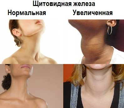
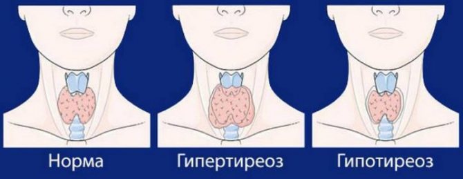
The thyroid gland, or “thyroid” for short, is a gland that is part of the endocrine system (a system for regulating the functioning of internal organs with the help of hormones that secrete endocrine cells into the blood) that stores iodine and secretes hormones containing iodine.
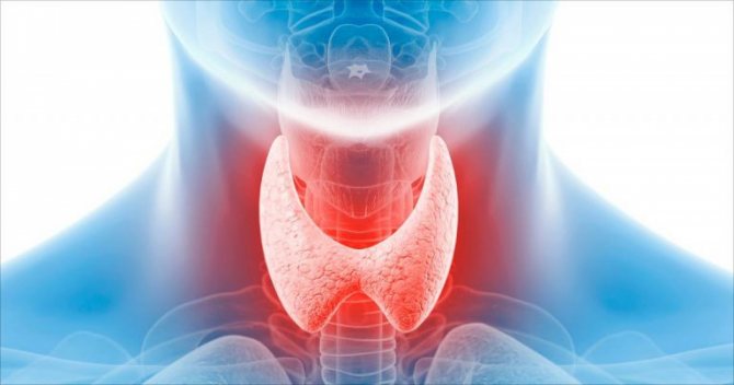
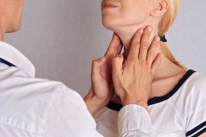
Regulates metabolism, cell growth and the growth of the entire organism in general. The thyroid gland is located in the neck in front of the trachea under the larynx. It looks like a butterfly located on the surface of the cartilage. If there are changes in the functioning of the thyroid gland, the patient will feel unwell (this may indicate an incipient disease), and later various complications may arise. The method of ultrasound examination of the thyroid gland will help to effectively identify incipient diseases.
If the thyroid gland does not function properly, the following pathologies may appear:
- hyperthyroidism – excessive activity of the gland;
- hypothyroidism – low activity of the gland, characterized by a reduced level of hormones produced;
- cancer;
- cyst;
- benign formations;
- thyroiditis is an inflammatory disease of the gland;
We take a test for thyroid hormones (test for thyroid hormones)
An important role for women is to take tests to determine the norm of thyroid hormones. To ensure the result is as accurate as possible, you must adhere to the following rules:
- the last meal should be 8 hours before the test;
- within 8 hours it is allowed to drink only clean water without gas;
- Smoking and drinking alcohol are prohibited at least 24 hours before blood collection;
- the analysis is submitted before 12 o'clock in the morning, in an emotionally calm state;
- It is also necessary to stop taking hormonal medications one day before. If this is not acceptable, then you need to warn the doctor before taking blood.
The next step for women in determining a possible pathology of the thyroid gland will be an ultrasound, which will help determine the norms or abnormalities in the structure of the gland.
Ultrasound of the thyroid gland
, an ultrasound diagnosis of the thyroid gland should be performed . This procedure is invasive, fairly cheap, fast and provides accurate results.
Ultrasound is a study of the body using ultrasonic waves. The echo signals reflected from the internal organs, after certain transformations, create on the monitor screen an image of a section of the gland in different shades of gray.
A specialist can determine the condition of the organ: geometric dimensions, state of borders, lymph nodes, blood vessels. Ultrasound determines the acoustic density of an organ, called echogenicity.
May be:
- normal;
- reduced;
- increased;
- echonegativity.
The condition of the organ being studied is determined by the types of echogenicity.
Reduced (hypoechogenicity) is characteristic of liquid formations (dark gray areas on the screen). Perhaps it is a cyst, liquid formations, vascular formations and cancer in 5% of cases. A doctor cannot always reliably determine the nature of these formations, so other diagnostics, such as a biopsy, will be required.
Increased (hyperechogenicity) indicates a lack of iodine (endemic goiter), damage to the organ by poison (toxic goiter), autoimmune thyroiditis, subacute thyroiditis, oncology, areas of sclerosis. Visualized on the monitor as white spots. Hyperechogenicity may occur in a healthy gland, or it may indicate serious pathologies, therefore, to obtain a reliable diagnosis, it is advisable to conduct additional research.
Echonegativity (anechoicity) is displayed as black areas. Perhaps it is a cyst, pseudocyst, colloid cyst, adenoma. It is also recommended to undergo other examinations to make a more accurate diagnosis.
With normal echogenicity, the silhouette of the gland is smooth and clear. A benign tumor has smooth edges, while a malignant tumor has curved edges.
Since the frequency of ultrasound is not high, there is no harmful effect on the body.
The role of elastography in the diagnosis of the thyroid gland?
Endocrinologists show less interest in elastography than in classical ultrasound, primarily due to the difficulty of obtaining reliable images. Multi-colored stains appearing on the screen are not always clear and understandable. To carry out elastography you need super modern and expensive equipment, which is rarely available.
The sensitivity and specificity of this test for detecting thyroid cancer is about 70-90%, the statistical accuracy parameter is estimated at 90%. Therefore, elastography cannot replace a thyroid biopsy.
The method is only one of the factors determining the endocrinologist's decision to prescribe a biopsy and surgical treatment. The combination of traditional ultrasound and elastography allows you to increase the sensitivity of cancer detection and narrow the group of changes classified as indeterminate. The prognostic value (probability of absence of disease) in this case is 99.8%.
How to prepare for an ultrasound
The method is accessible and simple, does not require special preparatory operations. You can eat absolutely any food product. But there are a number of restrictions:
1. Regardless of the feeling of nausea, older people are not recommended to eat before the procedure. Age-related changes may have an effect.
2. Women are recommended to undergo examination on days 7-9 of the menstrual cycle to avoid inaccurate results.
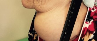
3. Immediately before the procedure, you need to free your neck from elements that interfere: jewelry, collars, etc.
4. Preparation for an ultrasound examination of the thyroid gland in children is no different from the procedure for adults. The child must be psychologically prepared for the procedure so that he is not afraid of either the doctor or what he will do.
Doctors recommend that women undergo testing both during pregnancy and at the planning stage. If it is impossible to get pregnant, gland pathology may be one of the possible causes.
After the child has suffered stress, it is also worth going to the clinic so that an endocrinologist can perform an ultrasound of the thyroid gland.
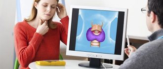
How is the procedure carried out?
The procedure is quite simple.
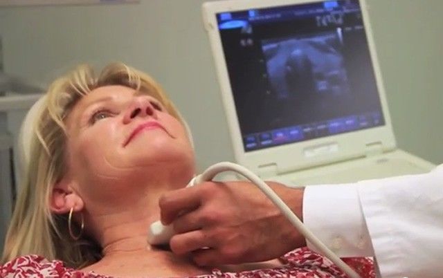
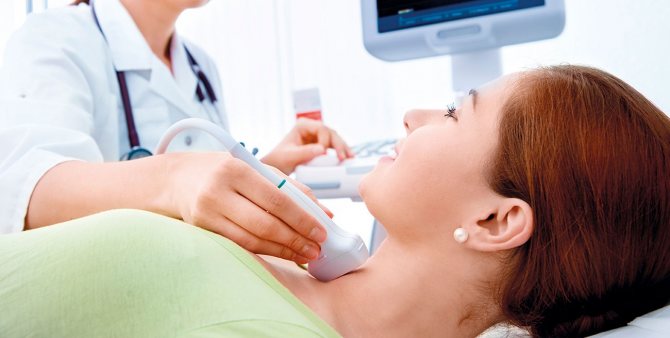
The patient lies down on a special couch with his head thrown back, a soft pillow or cushion is placed at the level of the shoulder girdle. A gel that conducts ultrasound well is applied to the neck in the area of the thyroid gland. An ultrasound transducer is applied to the neck and ultrasonic waves are emitted. The sensor is a transceiver that emits and receives a reflected echo signal. This information is processed by a computer and the result is displayed on the monitor. During the procedure, the patient does not experience any discomfort; sometimes there may be slight discomfort due to incorrect body position.
Interpretation of ultrasound results
The diagnostician describes the results obtained in the study protocol. Typically, the time to prepare a written report does not exceed 15 minutes.
Determination of exact geometric dimensions
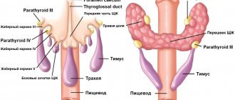
The location of the gland will normally be typical or low, the shape will be classic, the contour will have a clear outline. It consists of two lobes with an isthmus that connects them. Sometimes a pyramidal lobe may be present.
Minor (less than one centimeter) tissue growths may be visible. If during the period of intrauterine growth the gland develops with pathology, the tissue may not be divided into two sides, but may move to one side. This is called aplasia of one lobe.
In the case of a completely undeveloped gland, they speak of complete aplasia. The length of the lobe should be from 4 to 6 cm, width from 1.3 to 1.8 cm, thickness from 1.5 to 1.8 cm; the shares are normally the same; the lintel has a thickness of 4 to 8 cm.
The volume of a healthy gland depends on the patient’s weight and is:
- weight 50 kg, gland volume 15.5 cm³
- weight 50-60 kg, volume 18.7 cm³
- weight 60-70 kg, volume 22 cm³
- weight 70-80 kg, volume 25 cm³
- weight 80-90 kg, volume 28.4 cm³
- 100 kg or more, volume 32 cm³
Non-compliance with standard sizes will indicate possible pathologies.
Definition of structure
The echogenicity of a healthy thyroid gland, without any peculiarities, the structure of the jelly tissue is fine-mesh, homogeneous, echogenic granularity is 1 mm or less. Fibrous and connective tissues are not detected. In inflammatory processes, the structure is heterogeneous.
Focal education. There should be no new growths. If the formation is present, it must be classified. If the size is up to 10 mm, then it is a focal formation, more than 10 mm, then it is a node.
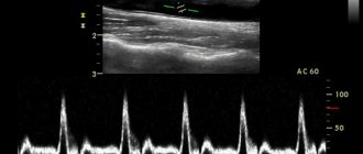
Blood flow analysis. The parameters of blood flow, its nature and density, and parameters of the lymph nodes are determined. Normal nodes have clear boundaries, the width is approximately 2 times less than the length.
Contours. Clear contours are the norm. Fuzzy outlines indicate an inflammatory process or tumor.
Focal formations. Assessed for the presence of nodes, cysts, and calcinitis.
Echogenicity is the grayscale image of the tissue being examined and its tone on a monitor.
Parameters of visible lymph nodes (if present), their structure, size, structure. By their presence, the onset of tumor formation can be diagnosed.
The structure of the salivary gland and the level of response to ultrasound.
The size and structure of the soft tissues of the neck and larynx. Those areas that are located near the thyroid gland are examined.
Women over 35 years of age are recommended to have their thyroid gland examined at least once a year. This category of people is at risk for thyroid diseases.
The human neck is quite complex; it contains many nerve trunks and large vessels, the esophagus, trachea, many lymph nodes, and other glands. Therefore, the specialist performing the ultrasound procedure must be highly qualified.
The doctor conducting the research records the data obtained in a special protocol, which describes the geometric parameters of the lobes and isthmus, calculates the volume of the gland, and writes a conclusion.
The document evaluates the position and contours, tissue structures, analyzes the parathyroid glands and lymph nodes, and the images taken are also attached to the protocol. In case of normal indicators, a corresponding entry is made in the document. This takes no more than 10 minutes.
Frequently asked questions about the normal volume of the thyroid gland on my forum
Mom: Hello. Please tell me, is it necessary to carry out any treatment for elevated TSH (7.8 µIU/l)? T3 and T4, antibodies to TPO are normal. Ultrasound shows a cyst in the left lobe measuring 3.8 mm. Daughter is 21 years old, weight 53 kg. We were prescribed hormone therapy, but I doubt it.
Romanov G.N.: This is a picture of subclinical hypothyroidism. We treat when there are any problems or complaints.
Irina: Hello, doctor! Is there a lower limit for normal thyroid volume in women? What is the norm for a 25-year-old nulliparous girl? The ultrasound doctor, after conducting a study, suggested that the small volume of the thyroid gland is associated with low levels of the hormone; all the main indicators: the thickness of the isthmus, structure and echogenicity are normal.
Romanov G.N.: She’s gone! The main thing is that hormones are normal. And the size of the thyroid gland depends purely on human physiology. In women, the normal volume is up to 18 cm3. Exactly “up to”, which means 1 cm3 is also the norm. The main thing is a hormone analysis. A small gland can work perfectly, but a normal-sized gland may not work at all.
Marina: Hello, I have been doing an ultrasound of the thyroid gland for three years in a row and its volume is decreasing, it was 13, and now it is 9.2 cm3. Is it dangerous? Last year, when she was 11.2, her hormones were normal.
Romanov G.N.: The main thing is not the volume, but the TSH indicator.
Irina: Hello. Do I understand correctly that thyroid hypoplasia has nothing to do with subclinical hypothyroidism?
Romanov G.N.: Any amount of hypothyroidism with normal thyroid volume. I declare as a practicing specialist who constantly encounters this disease. (Hypoplasia is a condition in which disturbances in the structure of an organ and reduced size are observed).
Evgenia: Good afternoon. My daughter is 14 years old and had an ultrasound. Help me decipher: right lobe 14x13x42 mm, volume 3.7 cm3; left lobe 12x13x44 mm, volume 3.3 cm3, isthmus 2.5 mm, total volume 7 cm3. In conclusion, diffuse changes in the thyroid gland.
Romanov G.N.: The conclusion is about nothing. The volume is normal. I'll have to get my hormones tested.