Ultrasound of the prostate gland (prostate) is a non-invasive diagnostic method and allows you to obtain comprehensive information about the health of the male reproductive system and prevent sexual dysfunction. An hour and a half before the ultrasound of the prostate gland, it is necessary to fill the bladder with liquid by drinking up to 2 liters of water. This procedure is prescribed by a urologist, and like other examination procedures using ultrasound, ultrasound of the prostate gland is absolutely painless and safe for the patient’s body. IMPORTANT: Ultrasound of the prostate gland (prostate) must be performed regularly not only by young people, but especially by men over 40 years of age. This prevention will allow you to avoid many complications and diseases of the male reproductive system.
What is a prostate ultrasound and how is the procedure done?
Ultrasound of the scrotum and prostate is a highly accurate diagnosis that allows you to determine the pathological process localized in the organs of the genitourinary system. During diagnostics, ultrasound penetrates tissue structures, reflecting differently from affected and healthy surfaces. The image is transmitted to the display in real time, the information received is analyzed by a specialist, based on which he makes an accurate diagnosis.

Ultrasound of the bladder and prostate gland makes it possible to determine the presence of the following diseases:
- dysfunction of reproductive organs;
- presence of oncological education;
- Ultrasound also shows prostatitis;
- bladder injuries;
- problematic urination;
- the occurrence of stones;
- benign prostatic hyperplasia.
For various indications, ultrasound of the prostate gland is used, the norm of parameters is indicated in special reference books, the indicators directly depend on the age and characteristics of the patient’s body. The expert analyzes the appearance of the prostate, dimensions, density, shape. With the help of a diagnostic measure, you can easily identify adenoma, cancer, prostatitis, glandular growth, the presence of solid formations and bleeding.
Pathologies determined by ultrasound of the prostate
When examining an ultrasound scan of the prostate gland, certain chronic diseases can be identified that can significantly impair the functionality of the body.
Adenoma
Increased size of the gland and tissue density are the primary signs of the development of pathology. In some cases, nodes may appear on the surface of the organ. The diffuse form of the disease is characterized by pronounced heterogeneity, without any inclusions.
Fibrosis
The most common prostate disease. It is believed that the main reasons for the progression of the pathology are an advanced form of prostatitis or consequences after suffering an inflammatory process of the gland. A characteristic sign of the presence of fibrosis on ultrasound is a reduced size of the gland.
Prostatitis
The pathology is characterized by changes occurring in the echogenic background. Depending on the form of the inflammatory process, the prostate tissue becomes denser and the echogenicity decreases. The main features are a blurred contour and similarity of fibrous and glandular tissues. In some cases, abscesses may occur due to uncontrolled changes in echogenicity. An acute form of pathology is recorded when there is a simultaneous enlargement of the gland and a decrease in echogenicity, and vesiculitis is noticeable around the seminal vesicles.
Cysts and stones
Based on tissue structures, all inclusions in an organ can be determined. Often cysts appear as anechoic areas or areas of low echogenicity. Small cysts sometimes appear on a completely healthy organ. The size of such manifestations is no more than 5 mm. The presence of stones of different shapes and sizes is characterized by an increased echo signal.
Neoplasms
The loss of clarity of outlines and contours is determined by the presence of tumors of various types. Benign neoplasms are often located in the central regions of the organ. The presence of transformation and shifts along the edges may signal the formation of cancer cells.
When is it prescribed?
Not everyone knows how to do a prostate ultrasound, so they are afraid of the procedure and delay the date in every possible way, which allows dangerous diseases to progress and enter advanced stages. An ultrasound examination is prescribed by a specialist in a medical institution, this can be a urologist, surgeon, andrologist or therapist. The doctor explains the need for intervention and how an ultrasound of the prostate gland is performed.
Immediately after the appointment, preparations are made for the prostate ultrasound procedure. With the help of an examination, the attending physician can obtain the necessary information about the cause of the pathology, the degree of damage, and the type of illness. Based on the results, complex therapeutic measures are prescribed or additional diagnostics are carried out.
Prostate ultrasound is prescribed in the following cases:
- the presence of traces of blood in biological fluids, that is, in semen or urine;
- problems with urination, including frequent, painful, nighttime emptying of the bladder, accompanied by a feeling of incomplete emptying;
- pain and signs of discomfort in the perineum, scrotum, bladder or rectum, you should first determine how to prepare for a prostate ultrasound in men;
- traces of discharge from the urethra;
- the occurrence of renal colic for unknown reasons;
- the presence of an inflammatory process or injury to the bladder, gland or seminal vesicles;
- infertility;
- rectal ultrasound of the prostate is performed for deterioration of male power;
- the need for diagnostics before or after surgery;
- a preliminary diagnosis related to cancer has been made, in this case, if an ultrasound scan of the prostate is not done, there is a high probability of death.
Screening diagnostics of representatives of the stronger sex over the age of 50 also involves such an examination. Interpretation of prostate ultrasound shows all the problems that are present in the body associated with the organs of the excretory and reproductive systems.
How long does a prostate ultrasound take?
The duration of the examination depends on the method of its conduct.
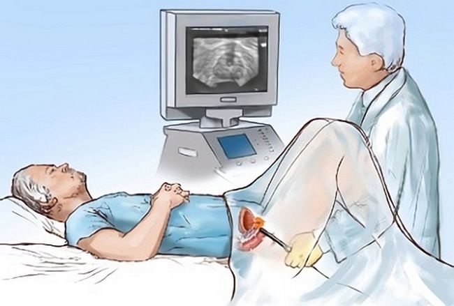
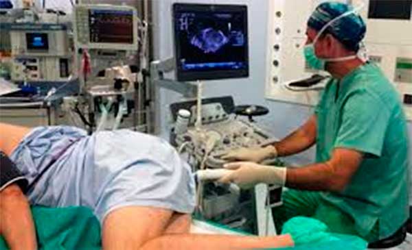
During the rectal method, the patient lies on his side, slightly pulling his knees towards his stomach. The doctor prepares the sensor to reduce the level of discomfort and ensure the best glide. Then the device is inserted into the rectum to a depth of 7 centimeters. This type of prostate ultrasound can last up to 25 minutes, taking into account 10 minutes of preparatory activities and 15 minutes of the diagnosis itself.
An abdominal ultrasound of the prostate is performed using a convex probe to scan tissue up to 25 centimeters deep.
The process looks like this:
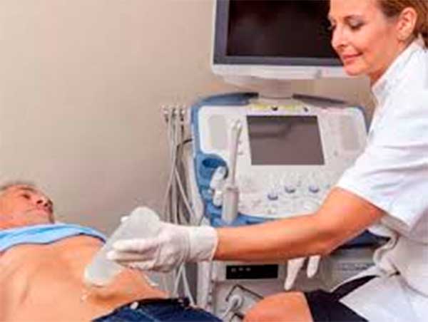
- the patient lies with his back on the couch, having previously exposed his stomach and undressed to the waist;
- the doctor lubricates the surface of the abdomen in the required area with gel to improve the sliding effect and increase the area of contact of the sensor with the skin, which allows for more informative and accurate results;
- then the doctor slowly moves the device over the abdomen;
- the information is transmitted to the screen and subsequently recorded in the study protocol.
With this type of prostate ultrasound, the examination time does not exceed 20 minutes.
How do they do it?
Regardless of the method of carrying out the procedure, preparation is made for an ultrasound of the prostate gland. This process includes several important recommendations, on which the accuracy of the examination will directly depend, so they must be approached as responsibly as possible. There are several methods for conducting ultrasound examinations, for example, TRUS ultrasound of the prostate gland.
For the last few years, only two methods have been practiced in medical institutions: transabdominal and transrectal. The first is included in the category of primary diagnostic measures; it is implemented externally, that is, through the surface of the abdominal cavity. Its disadvantages include significant blurring of the results.
Transabdominal ultrasound of the prostate allows you to determine:
- dimensional characteristics of the gland;
- determine the dimensions of the bladder and compare them with normal values;
- the presence of benign or malignant formations.
Transabdominal ultrasound of the prostate gland is a less informative method than TRUS, so it is used for minor suspicions of a dangerous disease. As a rule, it is supplemented with additional diagnostic methods in order to obtain the clearest picture of the patient’s condition. The transrectal method gives the most accurate results, since the sensor is located close to the prostate wall. These and other methods should be considered in more detail.
Bladder preparation
An equally important step in preparing for such a procedure is preparing your bladder. But how to prepare for TRUS of the prostate gland in this case? To perform this procedure, you need to completely fill your bladder. This can be done in several ways, and the choice will depend on what type of research needs to be performed.

When answering the question of how to prepare for TRUS of the prostate, it should be noted that filling the bladder can be done independently, although most specialists perform this procedure themselves. Typically this is done using a catheter. Here, too, there are two methods for preparing TRUS. Let's look at them separately:
- Method No. 1. If the patient has reduced potency, has infertility, or has an increased level of PSA in the blood, then 1 hour before the test, you need to drink one liter of plain water. Thanks to this, the bladder will fill very quickly, and it cannot be emptied until the TRUS examination is completely completed. Preparation for the study can also be done in another way.
- Method No. 2. If it is necessary to identify the cause of existing difficulties with urination, then a completely different preparation method is used. Such preparation for TRUS of the prostate in men involves taking one and a half liters of still water. After this, it is also forbidden to visit the toilet, as in the previous case. If the patient drinks carbonated water, the specialist will not be able to conduct the study, since such a procedure will have to be postponed for several hours. When the first urge to urinate appears, you must immediately tell a specialist about it, who should begin the TRUS procedure of the prostate.
When the study is completed, the patient can go to the toilet, since urine should not be stored in a bladder under any circumstances. Otherwise, an infection may begin to develop in urine. This is especially true in situations where patients have a severely weakened immune system. Then the bacteria penetrate the human body very quickly, because stagnant urine is an excellent place for the proliferation of various pathogenic microflora.
Those patients who do not know how to prepare for TRUS of the prostate gland will be consulted by a specialist.
Abdominal examination
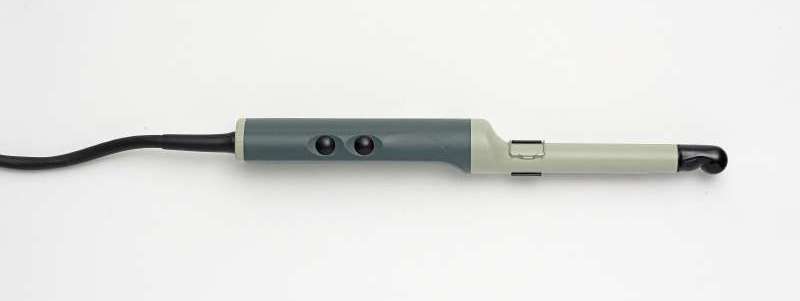
This is a superficial ultrasound method of the prostate; we will discuss how the procedure is done below. Ultrasound waves are directed to the organ being examined using a special sensor. The characteristic differences of this method include the complete absence of contraindications and pain, however, the quality of the results leaves much to be desired.
Before an ultrasound of the prostate is performed, it is necessary to prepare the body. In this study, the preparatory measures are quite gentle. Experts recommend excluding from the diet foods that provoke increased gas formation in the intestines a couple of days before the diagnosis. Before the examination, you should drink 1 liter of clean water to fill your bladder.
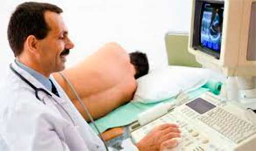
Ultrasound of the prostate gland in men of the abdominal type resembles a banal examination of the abdominal cavity, the main difference is the location of the ultrasonic wave indicator. The patient lies down on the couch, a special gel is applied to the lower abdomen, after which the specialist diagnoses the prostate gland.
The prostate, due to its location, is not scanned accurately enough. Moreover, it won’t even help if a man knows everything about prostate ultrasound, and the preparation for it took place taking into account all the recommendations. Therefore, this method is suitable exclusively for traditional examination without clarifying the exact diagnosis. It should be noted that excessive weight may be a contraindication, since the accuracy of the results will be minimal.
Ultrasound of the prostate at home
It is not always possible to visit a clinic in person. In this case, the modern method of prostate ultrasound at home will allow you to conduct the study in the comfort of your home and significantly save time.
This procedure is recommended for the following categories of patients:
- older people who have difficulty getting around using public transport;
- patients with serious limitations in mobility, which could be caused by injuries of various types or surgical intervention during an operation;
- people with limited time or simply those who do not like to be in medical institutions.
Most often, men experiencing intimate ailments hide it and do not want to visit the clinic. There may be personal reasons or lack of time, due to which a visit to the doctor is constantly postponed. This behavior has a detrimental effect on the general condition of the body subsequently, when the pathology becomes chronic.
When performing an ultrasound at home, equipment is used that is similar to stationary units in the clinic, but smaller in size. The process itself is no different from outpatient diagnostics and is carried out according to the same principles in several stages:
- the patient lies on a flat surface in a horizontal position with his stomach up;
- A special gel is applied to the skin of the study area for better and smoother glide of the scanner over the surface;
- The diagnostic process itself takes about 15-20 minutes;
- after completion, the remaining gel is removed with a damp sterile cloth;
- a patient card is filled out with a detailed description of all the data obtained.
Based on the results of a prostate ultrasound, the doctor makes a diagnosis, after which an effective course of treatment is developed, with reference to the individual characteristics of the patient.
Transrectal diagnostics (TRUS)
TRUS of the prostate gland is considered a more informative diagnostic measure than the previous version of the study. This is due to fundamental differences in the procedure. The sound wave is delivered directly through the surface of the rectum, so the results will be more accurate, they will allow you to determine the location of the organ, diagnose the inflammatory process, the presence of cancer lesions and other problems. However, you should first determine the features of TRUS of the prostate gland, as we will discuss how the procedure is done below.
Accuracy directly depends on proper preparation. Strict adherence to preparatory measures will allow the specialist to obtain the most appropriate data, which will subsequently affect the overall course of treatment. That is why you should find out how to prepare for a prostate ultrasound and what rules need to be followed.
ORDER
Key recommendations include:
- a couple of days before the examination date, you must stop eating cabbage, beans and other foods that can lead to increased flatulence;
- it is important not to drink alcohol;
- before going to bed in the evening, you should cleanse the intestines with an enema, and then repeat the procedure immediately before the test;
- Preparation for TRUS of the prostate involves drinking 1 liter of clean water 1-2 hours before the diagnosis.
Depending on the patient’s condition and the results of previous studies, the doctor can make a number of additional recommendations and advise on how to prepare for a prostate ultrasound correctly. It is strictly forbidden to ignore advice. If you have problems with incontinence, it is better to drink water in the clinic than at home.
Now we should consider how a transrectal ultrasound of the prostate is performed. The indicator is inserted into the anus, which causes catastrophic fear and reluctance in many men to carry out the examination. It is recommended to fully follow the recommendations of the medical professional, relax, remain calm and not worry.
The event is divided into several stages and is performed in the following sequence:
- The patient undresses and puts on a robe.
- Take a position lying on your side on the couch, with your legs bent at the knees and moved closer to your stomach.
- The doctor puts a condom on the sensor to prevent the occurrence of an infectious disease, then lubricates the indicator and the anus with Vaseline.
- The sensor is inserted only 7 cm, so the patient does not experience any particular discomfort.
- Depending on the design of the device and the scanning method, the uzist makes inclined, translational, and rotational movements with the device.
The picture is broadcast on the screen; if necessary, the image is printed. The diameter of the indicator is about 2 cm, so the patient does not feel pain. Sometimes there may be minor discomfort or a urge to defecate, which you do not need to pay attention to. After 10-15 minutes, these sensations disappear without a trace. So we managed to determine what TRUS of the prostate gland is and how it is performed.
Diagnostics allows a specialist to accurately determine even a slight enlargement of organs, which is very important for making a diagnosis in the early stages of the development of adenoma or prostatitis. Thanks to the penetration of the sensor into the rectum, the uzologist can examine the neck of the bladder for the presence of an inflammatory process or dangerous formations.
Contraindications to prostate ultrasound
The presented research method actually has no significant contraindications.
Diagnostics through ultrasound is a completely safe and painless method of obtaining information.
However, for some categories of patients, there are certain contraindications to undergoing the examination:
- the presence of third-party inflammatory processes;
- exacerbation of hemorrhoids;
- tumor of the ampulla in the rectum;
- complications after surgery associated with narrowing of the anal canal;
- anal fissure;
- increased sensitivity of the anorectal area.
Most of the above contraindications will not harm your health when undergoing an ultrasound scan, but will only reduce the accuracy and information content of the results.
Prostate ultrasound results
Interpretation of an ultrasound of the prostate gland allows the specialist to accurately assess the patient’s condition. Based on the results of ultrasound diagnostics, a treatment plan is drawn up, which may consist of a whole range of therapeutic procedures. If the data obtained is incorrectly interpreted, then the treatment will not give the desired results and the disease will be able to develop unhindered.
A clinician can decipher the readings based on the following characteristic parameters:
- localization of the main organs of the genitourinary system;
- boundaries and structure of the prostate gland;
- degree of echogenicity;
- dimensions and shape of structural elements.
Each of these characteristics fits into a special form; all the data shown by ultrasound of the prostate are indicated there. Using this information, the attending physician makes an accurate diagnosis; the patient’s complaints and diseases that the man previously suffered from are taken into account.
Which doctor does prostate ultrasound?
The condition of the prostate gland can be affected by any negative impact, from infectious inflammatory processes to hypothermia. In cases where the patient experiences characteristic symptoms of the development of prostate pathology, it is necessary to consult a urologist.
At your appointment, you will be given a referral for a prostate ultrasound to more accurately diagnose the problem with subsequent treatment.
An ultrasound of the prostate is performed by a doctor specializing in ultrasound diagnostics and having experience in various branches of urology with relevant diplomas. The procedure will be as comfortable as possible for the patient, thanks to modern equipment on which all diagnostic procedures are performed and the high level of training of doctors.
Publication date 2019-10-24
Normal indicators
The normal size of the prostate gland in adults depends on the age and weight of the subject. In a normal state, the prostate corresponds to the established size and shape, with no neoplasms, cysts or tumors.
The normal size of the prostate gland according to ultrasound corresponds to the following indicators:
- 2.4 – 4.1 cm – upper anterior;
- 2.7 – 4.3 cm – transverse;
- 1.6 – 2.3 cm – anteroposterior.
Normal prostate volume according to ultrasound varies from 24 to 30 cm3. In cross section, the seminal vesicles should be within 8-10 mm. Be sure to examine the appearance and indicators of the bladder, also taking into account the shape and dimensions. The wall thickness should be close to 3 - 5 mm. Normal prostate sizes according to ultrasound are usually accompanied by the absence of stones and other pathological formations. After urination, there should be no urine in the bladder; fluid should be efficiently transported into the bladder from the ureters.
It should be noted that the accuracy of the survey is determined by various factors. The normal size of the prostate according to ultrasound is distorted if there is an excessive amount of gas or feces in the intestines. The patient should take the correct position and relax. The fat layer or damage in the abdominal area leads to a significant error, which must be taken into account by a specialist. An enema must be given before the prostate ultrasound.
How is TRUS performed?
During the procedure, the patient lies on his side with his knees brought to his chest. It is advisable to relax and breathe regularly at this time. The sensor is inserted during exhalation to a depth of 5-6 cm and pressed against the anterior wall of the intestine.
The prostate is scanned in several planes, and the doctor evaluates the following parameters:
- The size of the gland, its shape and volume.
- Clarity of contours.
- Features of the echogenic structure.
- The presence of altered areas, their echogenicity, features of the boundaries and relationship with surrounding tissues.
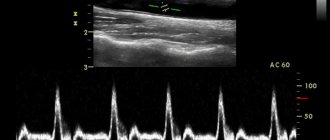
Modernization and development of medical technologies leads to the emergence of new functions that make it possible to increase the sensitivity, specificity and information content of research. In particular, modern ultrasound scanners make it possible to perform color Doppler measurements, obtain three-dimensional images, perform elastography and other useful functions.
For example, three-dimensional ultrasound allows you to accurately calculate the volume of the prostate, allowing for better planning of surgical procedures and radiation therapy. Dopplerography allows one to evaluate the characteristics of vascularization of organ tissues and increase the detection of malignant neoplasms, in which the tissues are hypervascularized and have a more abundant blood network.
Histoscanning technology has great prospects. This system allows non-invasive assessment of the pathomorphological features of tissues, their localization and the distribution of individual types. This study can reduce the number of prostate biopsies.
Another important technology in cancer differentiation is elastography. It is based on the different elasticity of benign and malignant tissues. Cancerous tissues are “harder”, their elasticity is 28 times higher than normal.
What does the diagnosis show?
First of all, ultrasound can be used to determine the size of the prostate; the norm during ultrasound is compared with the obtained values. If serious deviations are observed, then the problem may be hidden in a dangerous pathological process. At the next stages, the physician must check other pelvic organs, which will allow them to assess the condition and diagnose the causes of negative consequences. It is advisable to contact a qualified specialist so that he knows exactly how an ultrasound of the prostate gland is performed and can correctly interpret the results of the study.
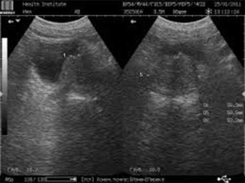
The bladder is located directly behind the bony pubic joint; during the filling process, the organ rises above this element of the genitourinary system, its upper part is adjacent to the abdominal cavity. A correct ultrasound of the prostate and bladder shows the bladder as an oval structural formation with a demarcated edge, and the distant area is visible much better than the near one.
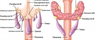
The seminal vesicles are located on the back side of the bladder on both sides, slightly higher than the prostate gland. Ultrasonic waves make it possible to identify them as interconnected formations in the form of bubbles. The prostate gland during ultrasound is visible as a glandular-muscular organ with a dense structure.
It should be noted that this is an important element, especially for a young man, since the quality and functionality of seminal fluid depends on it. The condition of the gland directly determines the reproductive capabilities of the stronger sex. On the screen you can see that the gland is located on top of the bladder. The normal volume of the prostate gland according to ultrasound does not exceed 30 cm3; the younger the man, the smaller this figure. The contours of the prostate are smooth and precise.
Connective tissue is responsible for the localization of organs; it acts as a binder. Each organ is located close to each other, that is, the elements are interconnected. That is why the failure of one organ entails the failure of another. To obtain an accurate assessment result, you should consider how you prepare for a prostate ultrasound.
Price
TRUS of the prostate gland is performed in various medical institutions, reviews are of great importance when choosing a hospital, medical center, clinic dedicated to men's health or dealing with family planning issues. The cost of TRUS of the prostate depends on various factors, including the credibility of the institution, the list of equipment and the qualifications of medical workers. The cost of transabdominal and transrectal ultrasound of the prostate varies widely, but, as a rule, ranges from 550 to 4500 rubles.
Experts recommend choosing a clinic with an average price, since sometimes the accuracy of the data obtained, and, accordingly, the effectiveness of further therapeutic measures depends on this. In most cases, the diagnosis does not end there; in addition, the doctor uses the palpation method, collects anamnesis, and prescribes laboratory tests of biological fluids.
An ultrasound examination of the prostate gland, bladder, and testicles is done to determine the condition of the genitourinary system. As a rule, this method is used when the patient has complaints or other examination methods have yielded appropriate results. Before holding an event, you should adhere to strict preparation rules, because the quality of the results depends on this. The TRUS method is the most informative; it allows you to record the pathological process in the early stages of development.
Where to do a prostate ultrasound
The “Your Doctor” clinic employs doctors who have many years of experience in various fields of urology. The choice of our medical center if there is a need for an ultrasound of the prostate is due to modern equipment, which carries out all specialized studies and subsequent diagnostic procedures:
- for ultrasound examinations of the prostate gland, separate rooms are allocated, equipped with all the necessary instruments for a comfortable position of the patient and obtaining the most accurate and informative results;
- doctors have all the modern technical components for effective work and safe interaction with the patient;
- It is possible to perform an ultrasound of the prostate at home, with a doctor visiting on the day of treatment.
In case of a personal visit, the urologist’s office is equipped with specialized furniture for comfortable placement of the patient. If additional research is necessary, the doctor has a wide range of tools that allows him to achieve the desired results.