Heel spur is a disease characterized by inflammation of the plantar fascia, resulting in the accumulation of osteophytes in the area of the heel tubercle. According to statistics, recently there has been an increase in the diagnosis of heel spurs among the population. Most often, women over 40 years of age are affected by the disease. This is due to prolonged wearing of uncomfortable high-heeled shoes. In addition, there are a number of predisposing factors, the presence of which increases the risk of developing a heel spur. To diagnose the pathology, it is necessary to perform an x-ray of the foot. As a rule, heel spurs are a unilateral disease. To treat growths in the heel area, a set of measures is used, including conservative therapy and surgical intervention.
The Moscow Yusupov Hospital has been diagnosing and treating heel spurs for many years. The latest equipment and experienced staff make it possible to identify the disease in the shortest possible time and begin timely effective treatment.
What is a heel spur?
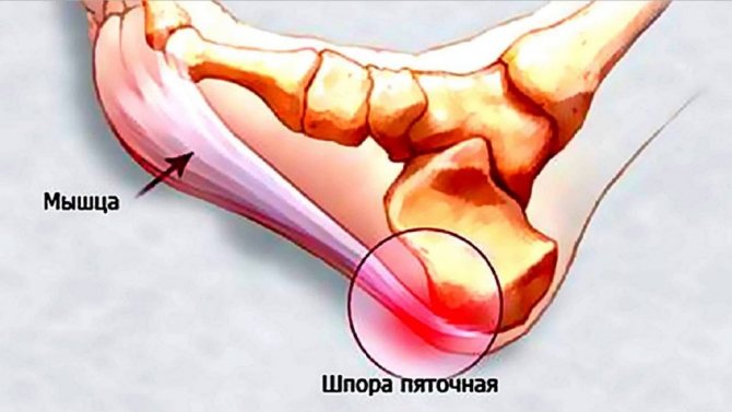
A heel spur is a disease that affects the ligaments, muscles and tendons of the heel area. Due to trauma to the plantar fascia, the center of gravity is redistributed to the foot. A rupture of the ligament activates the inflammatory process, in response to which calcium salts begin to be deposited in the area of the heel tubercle. In this way, a heel spur is formed, the minimum size of which can cause an acute attack of pain. The main clinical sign of a heel spur is pain, which intensifies in the morning and evening. Diagnosis of the disease is not difficult. To do this, it is enough to perform an X-ray examination of the foot. Treatment for heel spurs includes conservative therapy and surgery. Surgery to excise the heel spike is performed in 5% of cases of treatment of the disease. This is due to the sufficient effect of medication and physiotherapy.
In the international classification of diseases ICD-10, heel spur has code M77.3.
Expert opinion
Author:
Alexander Borisovich Tarasov
Doctor specializing in orthopedic rehabilitation
Plantar fasciitis (popularly called a heel spur) is caused by inflammatory and degenerative changes in the plantar fascia, which contributes to the proliferation of bone tissue, the formation of a characteristic growth.
The disease occurs without external signs, although the bone growth can be palpated. Patients complain of pain when walking, physical activity, or transferring body weight to one leg. But traditional methods of treatment are ineffective, and people are in no hurry to seek qualified help. There is no need to endure pain for years, because treatment of spurs in the Yusupov Hospital is carried out in one session.
Diagnosis is carried out through an objective examination and taking an x-ray. After this, the patient has a wide choice of treatment methods, including not only invasive ones. Drug treatment, laser exposure, and shock wave therapy are possible. If there are medical indications, then surgeons at the Yusupov Hospital perform minimally invasive heel spur removal using the endoscopic method or a microscalpel, painlessly.
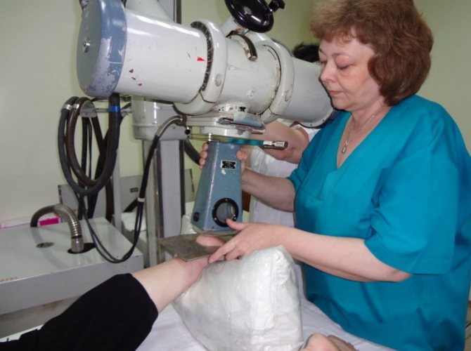
How is an x-ray of a heel spur taken?
X-rays are able to penetrate the human body, resulting in images of bone formations. The calcaneal spine consists of osteophyte cells, so it is clearly visible in the picture.
The principle of displaying bone tissue is based on the calcium content in the bones, which is absent in the surrounding tissues. Passing through calcium deposits, gamma radiation is slowed down and scattered.
The film produces a summation image of bone tissue and osteophytes.
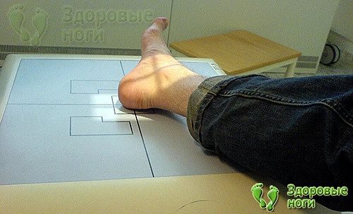
If a diagnosis of heel spur is suspected, an x-ray is taken in one or two projections: longitudinal and transverse. No special preparation of the patient is required before the study.
Advantages of radiography:
- availability of this diagnostic method and ease of the procedure;
- helps differentiate fasciitis from other bone diseases;
- Along the way, other foot pathologies that require treatment may be identified.
The disadvantage of X-rays is the effect of ionizing radiation. The procedure is considered safe, but it is not prescribed to children under 16 years of age, pregnant women and patients with cancer undergoing radiotherapy treatment.
Causes and mechanisms of development of heel spurs
The number of detected heel spurs is growing every year. Most often, women over 40 years old face this problem. Among the main reasons for the development of pathology are:
- Flat feet. It is considered the most common cause of heel spurs. Due to improper distribution of the load on the foot, the center of gravity shifts to the foot.
- Injuries. Any injury to the bones of the heel increases the risk of developing a heel spur.
- Concomitant pathology of the musculoskeletal system. The formation of a heel spur is preceded by certain diseases of the joints and bones. These include:
- Rheumatoid arthritis. Autoimmune diseases trigger the body’s process of aggression against its own connective tissue. In this case, the joints are destroyed and osteophytes are formed.
- Psoriatic arthritis.
- Osteoarthritis. Inflammation in the joint area provokes a redistribution of the load when walking on the heels. This increases the risk of developing heel spurs.
- Ankylosing spondylitis.
- Gout. Deposition of uric acid salts occurs in the area of the heel tubercle, causing destruction of the plantar ligament.
The mechanism of development of heel spurs is associated with constant trauma to the plantar fascia, which supports the arch of the foot. The greatest tension of the fascia occurs at the site of its attachment to the tubercle of the calcaneus. Due to injury to the ligament, the inflammatory process is activated. In response to it, calcium salts are deposited, forming bone growths. A heel spur develops in a similar way.
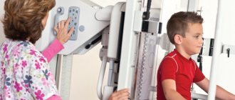
X-ray of the calcaneus at the Kutuzovsky Children's Hospital
The price of an X-ray of the heel bone in a diagnostic and treatment setting starts from 2,100 rubles. X-ray is a simple, fast, painless and safe way to solve the diagnostic and treatment problem facing the doctor and the patient.
X-rays of the heel bone in our clinic are performed using a modern digital device Brivo XR575 Premium, which has a wide range of functions and allows for examination of even people with limited mobility. The X-ray examination is carried out in compliance with all safety rules in order to eliminate any risks of additional radiation exposure for the patient.
The diagnostic and treatment center offers a wide range of medical services - from the simplest laboratory tests to consultations with highly qualified specialists. To make an appointment, you can use the “Online booking” form or call us at our number.
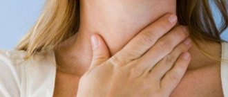
Symptoms and signs of heel spurs
The main and only symptom of a heel spur is pain. In the initial stage, the disease may be asymptomatic. As the osteophytes grow, a sharp, acute pain appears in the heel area. The pain syndrome bothers me in the morning, after rest. Therefore, this syndrome is called “starting pain.” During the first steps after waking up, the pain returns. During the day, the severity of the symptom decreases, but in the evening the acute pain appears again. There were no visible changes indicating the presence of a heel spur. In rare cases, skin hyperemia in the heel area may occur due to the development of the inflammatory process.
The localization of the main symptom of a heel spur can be local or spread throughout the entire heel area. Pain while walking causes lameness and changes in gait. A person tries to step on the toe or outer part of the foot, avoiding the load on the heel.
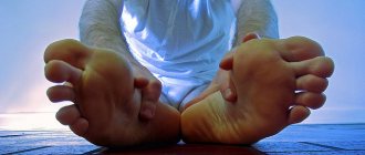
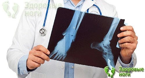
Heel spur: photo
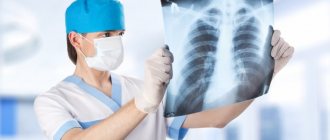
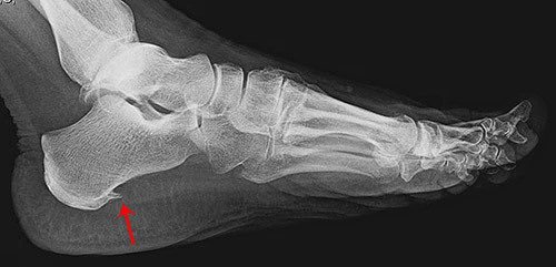
X-ray of the foot. A spike is identified in the area of the heel tubercle.
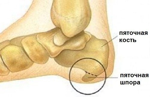
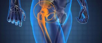
Schematic illustration of a heel spur.

X-ray of a heel spur
Diagnostics
To identify heel spurs, an integrated approach to diagnosing the disease is necessary. To achieve this, the following activities are carried out:
- Collection of complaints and medical history. The main complaints and the time of their appearance are determined. In addition, the presence of predisposing factors is clarified.
- General blood and urine analysis. Thanks to laboratory diagnostics, the presence of an inflammatory process in the body is established. At the same time, the number of leukocytes and ESR increases.
- Blood chemistry. This study allows you to identify or exclude rheumatological diseases. To do this, blood is donated for rheumatoid factor, uric acid and C-reactive protein.
- Radiography. It is the main method for diagnosing heel spurs. An x-ray of the heel reveals a spike in the area of the calcaneal tubercle. Its size depends on how long ago it was formed. However, even the smallest size of a thorn can provoke severe pain.
Yusupov Hospital has a full range of diagnostic services necessary to identify heel spurs. Modern equipment and highly qualified staff are the undoubted advantage of the Yusupov Hospital. Thanks to many years of experience in diagnosing and treating heel spurs, the clinic’s specialists quickly identify the disease of the heel tubercle and prescribe the correct individual treatment.
Heel spur: treatment
Heel spurs are treated with medication, physical therapy, and surgery. The choice of method depends on the severity of symptoms and stage of the disease.
Drug therapy
Conservative therapy for heel spurs includes prescribing medication. The main objectives of this method of therapy are to reduce inflammation and relieve pain. For this purpose, the following groups of medications are used:
- Non-steroidal anti-inflammatory drugs. Due to their action, they reduce the permeability of the vascular wall, relieving edema, and also blocking inflammatory mediators. In this way, an analgesic and anti-inflammatory effect is achieved. Drugs of the NSAID group improve blood circulation in the pathological focus and accelerate trophic processes. The main representatives of non-steroidal anti-inflammatory drugs include:
- Ibuprofen. It has an analgesic and anti-inflammatory effect in the shortest possible time. It is considered the safest drug in the NSAID group.
- Diclofenac. A common drug for relieving acute pain and reducing inflammation. The disadvantage of diclofenac is the side effects. The drug has a strong effect on the gastrointestinal tract. Uncontrolled use of diclofenac provokes internal bleeding.
- Nizilat. The drug has an analgesic and anti-inflammatory effect, relieves edema. The disadvantage of nysilate is its strong effect on the gastrointestinal tract.
- Ketorolac (Ketorol). This drug is prescribed strictly by the attending physician. Ketorol is a potent pain reliever.
The choice of drug treatment is made by the doctor depending on the condition, stage of the disease and the presence of concomitant pathology.
Physiotherapy
Physiotherapy methods are effective in relieving pain and reducing inflammation. The complex of treatment for heel spurs includes the following physiotherapeutic procedures:
- Phonophoresis. The procedure involves introducing hydrocortisone ointment into the body using ultrasound. Thanks to this, there is an anti-inflammatory effect, microcirculation, trophism and lymph outflow in the affected area improves.
- Laser therapy. A laser beam is used to target the inflammatory focus. The effect of this method of physiotherapeutic treatment is based on improving blood circulation, reducing edema, relieving pain and reducing the inflammatory process. The effect accelerates the healing and restoration of the injured plantar ligament. Laser therapy is a safe and painless treatment method. Contraindications for the procedure are pregnancy, tuberculosis, hematological diseases, acute liver and kidney failure.
- Shock wave therapy. The procedure involves applying an ultrasonic wave to the area of bone growths. As a result, pieces of osteophytes break off with their subsequent resorption. Ultrasound treatment improves blood circulation and nutrition in the affected area, relieving pain and inflammation. As a result of therapy, the plantar ligament becomes elastic, which reduces the possibility of re-injury. Shock wave therapy is indicated in the presence of diseases of the musculoskeletal system, flat feet, heel spurs. Contraindications for this method of treatment are pregnancy, cancer, bleeding disorders, acute pathologies accompanied by purulent inflammation.
- X-ray therapy. To relieve pain, nerve endings are blocked using a low dose of radiation.
- Magnetic resonance therapy. This method of physiotherapeutic treatment is used to reduce pain, relieve inflammation and improve blood circulation in the heel area.
To obtain the full effect of physiotherapeutic treatment, it is necessary to complete a course of therapy. On average it is 10-15 procedures. The advantage of this method is the absolute painlessness of the sessions.
Description and treatment of pathology according to the data from the image
X-ray therapy of heel spurs
In case of heel spurs, an assessment of the proliferation of osteophytes is carried out by a radiologist, recording the data in a medical report. Treatment is also prescribed by a doctor and includes:
- Nonsteroidal anti-inflammatory drugs (NSAIDs).
- Physiotherapy, in particular, one of the methods of radiotherapy is short-focus therapy.
- To reduce the intensity of the inflammatory process, corticosteroid ointments are prescribed.
- Physiotherapy (electrophoresis, magnetic therapy).
- Minimizing the load on the injured limb, up to complete physical rest.
- With positive dynamics and a decrease in pain, exercise therapy classes are indicated.
To reduce stress and stimulate tissue restoration, it is recommended to use special insoles, orthopedic massagers, and purchase a silicone heel pad.
Surgery
Surgical treatment is indicated if conservative therapy is ineffective within 6 months. Also, an indication for surgical intervention is a decrease in performance due to pain. Surgical treatment of heel spurs is resorted to in 5% of cases.
There are several ways to surgically remove a thorn in the area of the heel tubercle:
- Traditional. It is carried out in any hospital setting. It is considered the most common and traumatic surgical technique. The recovery period after open surgery is up to 2 months.
- Endoscopic. The operation is performed using endoscopic instruments that are inserted into the heel area through a minimal incision. Endoscopic surgery is the technique of choice due to its low morbidity and rapid recovery period in the postoperative period.
- Minimally invasive. The newest method for treating heel spurs. The operation is performed under X-ray control. Through an incision, a thin tube is inserted into the heel area, at the end of which a microscalpel is located. With its help the thorn is cut off. Minimally invasive surgery is the least traumatic of all available techniques.
An integrated approach to the treatment of heel spurs is the key to getting rid of the disease as quickly as possible. The choice of therapy method is determined by the attending physician based on preliminary diagnosis of bone formation.
In order to relieve pain when walking, experts recommend wearing orthopedic insoles or heel pads during the treatment of heel spurs. The purposes of their use are:
- Relieves stress on the heel.
- Prevention and treatment of flat feet.
- Improving blood circulation and trophism in the affected area.
- Strengthening the ligamentous apparatus of the foot.
- Fixation of muscles and ligaments of the foot.
The selection of the necessary orthopedic products is carried out by an orthopedic doctor based on individual characteristics. The weight and stage of development of the heel spur are taken into account.
The Yusupov Hospital treats heel spurs and other diseases of the musculoskeletal system in Moscow. In a clinical setting, it is possible to carry out a complete diagnosis and appropriate treatment of heel spurs. The clinic's highly qualified specialists have many years of experience in treating such diseases. Doctors at the Yusupov Hospital provide conservative treatment and surgical excision of the spine in the area of the heel tubercle.
| Drug therapy | Physiotherapy | Surgery | |
| Treatment methods | To treat heel spurs, the following groups of medications are used: 1. Non-steroidal anti-inflammatory drugs. Due to their action, they reduce the permeability of the vascular wall, relieving edema, and also blocking inflammatory mediators. In this way, an analgesic and anti-inflammatory effect is achieved. Drugs of the NSAID group improve blood circulation in the pathological focus and accelerate trophic processes. 2.Chondroprotectors. Medicines in this group restore the tissues that make up the vertebrae. This effect improves blood circulation and nutrition in the affected area. Chondroprotectors help restore the plantar ligament, increasing its elasticity and preventing re-injury | The treatment complex for heel spurs includes the following physiotherapeutic procedures: 1. Phonophoresis. The procedure involves introducing hydrocortisone ointment into the body using ultrasound. Thanks to this, there is an anti-inflammatory effect, microcirculation, trophism and lymph outflow in the affected area improves. 2.Laser therapy. A laser beam is used to target the inflammatory focus. The effect of this method of physiotherapeutic treatment is based on improving blood circulation, reducing edema, relieving pain and reducing the inflammatory process. The effect accelerates the healing and restoration of the injured plantar ligament. Laser therapy is a safe and painless treatment method. Contraindications for the procedure are pregnancy, tuberculosis, hematological diseases, acute liver and kidney failure. 3. Shock wave therapy. The procedure involves applying an ultrasonic wave to the area of bone growths. As a result, pieces of osteophytes break off with their subsequent resorption. Ultrasound treatment improves blood circulation and nutrition in the affected area, relieving pain and inflammation. As a result of therapy, the plantar ligament becomes elastic, which reduces the possibility of re-injury. Shock wave therapy is indicated in the presence of diseases of the musculoskeletal system, flat feet, heel spurs. Contraindications for this method of treatment are pregnancy, cancer, bleeding disorders, acute pathologies accompanied by purulent inflammation. 4. X-ray therapy. To relieve pain, nerve endings are blocked using a low dose of radiation. 5.Magnetic resonance therapy. This method of physiotherapeutic treatment is used to reduce pain, relieve inflammation and improve blood circulation in the heel area. | There are several ways to surgically remove a thorn in the area of the heel tubercle: 1. Traditional. It is carried out in any hospital setting. It is considered the most common and traumatic surgical technique. The recovery period after open surgery is up to 2 months. 2.Endoscopic. The operation is performed using endoscopic instruments that are inserted into the heel area through a minimal incision. Endoscopic surgery is the technique of choice due to its low morbidity and rapid recovery period in the postoperative period. 3. Minimally invasive. The newest method for treating heel spurs. The operation is performed under X-ray control. Through an incision, a thin tube is inserted into the heel area, at the end of which a microscalpel is located. With its help the thorn is cut off. Minimally invasive surgery is the least traumatic of all available techniques. |
| Indications | Drug therapy is part of the complex treatment of diseases of the musculoskeletal system, accompanied by pain and inflammatory syndrome. | Physiotherapy is indicated for diseases of the musculoskeletal system, flat feet, and heel spurs. | Surgical treatment is indicated if conservative therapy is ineffective within 6 months. Also, an indication for surgical intervention is a decrease in performance due to pain. |
| Contraindications | Limitations for drug therapy are acute febrile conditions, gastrointestinal bleeding, and individual intolerance to the components of the drug. | Contraindications for physiotherapeutic procedures are pregnancy, tuberculosis, hematological diseases, acute liver and kidney failure. | Limitations for surgical intervention are: § Acute conditions accompanied by an increase in body temperature. § Purulent inflammation of any localization. § Severe concomitant pathology. |
Treatment of heel spurs in Moscow
The Moscow Yusupov Hospital diagnoses and treats heel spurs and other diseases of the musculoskeletal system. Modern equipment, highly qualified doctors and attentive attitude towards each patient are the advantages of the clinic. Based on diagnostic data, hospital specialists provide individual treatment in accordance with the stage of development of the heel spur and concomitant pathology. Before carrying out treatment, you should consult your doctor.
When is a heel spur scan needed?
When you contact an orthopedic doctor or surgeon, after a visual examination, palpation and history taking, the specialist will definitely prescribe an x-ray.
This type of study is prescribed to all patients with complaints of heel pain. X-rays will help quickly establish a diagnosis and begin treatment.
In addition to its diagnostic value, x-rays may be prescribed in the following cases:
- Before surgery. In this case, using an image, the doctor specifies the location, shape and size of the growth.
- To evaluate ongoing treatment. For example, after crushing a heel spur with a laser or shock waves, when it is necessary to evaluate the effectiveness of therapy and make a decision on the further method of treatment.
In some cases, a small heel spur resolves with the use of special ointments and creams. Cream "Pyatkospor Strengthened" with regular use helps to reduce the bone spine, which is also visible on an x-ray.
In addition, if symptoms of a heel spur appear, the doctor may refer the patient for an ultrasound of the foot or an MRI. These studies are additional and are carried out when x-rays do not reflect the entire clinical picture of fasciitis.