The head contains vital organs that are difficult to examine from the outside. Using invasive techniques is dangerous - the slightest damage to anatomical structures can lead to fatal consequences. MRI allows you to examine the internal structure of the head and the organs located in it completely painlessly, without endangering the patient’s health. The high quality of the resulting images makes it possible to accurately diagnose diseases.
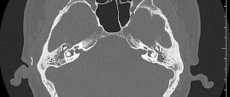
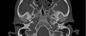
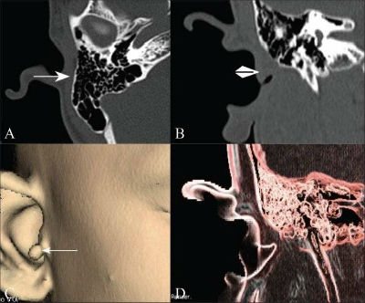
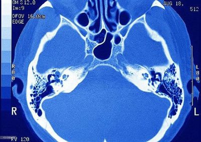
All structures of the ear can be imaged by performing an MRI of the head. Since the tomograph scans along a spiral trajectory, the output is images of cross sections of the head, which includes not only the ear, but also the brain, adjacent vessels and nerves, and eyes. At the same time, the doctor observes everything in its entirety, which expands his diagnostic capabilities.
The human ear consists of three sections:
- external - the auricle and the external auditory canal, ending with the eardrum;
- middle - tympanic cavity with auditory ossicles, auditory tube (opens into the nasopharynx);
- internal - bony and membranous labyrinths in the thickness of the temporal bone, responsible for hearing and a sense of balance.
MRI of the ear allows us to note the pathology of the structure of the internal skeleton of the ear, inflammatory processes, neoplasms, injuries, as well as the condition of the blood vessels.
For the most accurate assessment of vascular disorders or a detailed study of the tumor process, angiography is performed: a contrast agent is injected intravenously, after which a tomograph is scanned. The images are clearer, especially in pathological areas.
X-ray of the temporal bones, what is it?
An X-ray examination is a radiation diagnostic procedure, the result of which is an image of internal organs and tissues, for example, on photographic film.
If a digital X-ray machine is used, the images are displayed on the monitor; they can be recorded on any digital media. X-rays can be used to assess the condition of bone tissue and partially soft tissue quite well. The temporal bone has a complex anatomical structure. It houses the human hearing and balance organs. It is connected to the lower jaw and serves as a support for the masticatory apparatus.
X-ray examination allows us to evaluate the anatomical structure and integrity of the temporal bone. The images show the bony walls of the internal and external auditory canals, the middle ear and the mastoid process.
Since the temporal bone is quite modest in size, it is preferable to choose a digital X-ray method, as it allows you to enlarge images and examine their details.
A simple x-ray of the temporal bone has the following advantages:
- This is an extensive study, the result of which is a visual, quite informative snapshot.
- Speed of execution - the procedure, including preparation and decoding, lasts 10-15 minutes.
- Availability. Almost every medical institution has an X-ray machine that will allow you to remove the temporal bone in various projections.
- Simple technique.
Why CELT?
Multislice computed tomography of the middle ear in the multidisciplinary CELT clinic is performed by highly qualified specialists with extensive experience in performing diagnostic studies. Our specialists have a modern 64-spiral computed tomograph manufactured by General Electric. It has a high scanning speed and provides volumetric images of the highest quality. In the multidisciplinary CELT clinic, after the examination, you can get advice and undergo treatment from a specialized specialist (otorhinolaryngologist).
When is the examination scheduled?
X-rays of the temporal bones may be prescribed if the following indications exist:
- bruises, blows, injuries, temporal bone fractures;
- ear diseases: acute otitis with complications, chronic inflammation of the middle ear, cholesteatoma and others;
- suspicion of tumor growths in the temple area.
Today, CT and MRI are more effective and safe techniques than x-rays.
This is why there is such a narrow range of indications for x-rays. However, this study helps to reliably identify the presence of pneumatization in a patient (in other words, cavities filled with air), as well as to study the position of intra-ear implants. X-rays are also prescribed for other indications, when CT and MRI machines are not available in the medical institution.
Indications for use

If complicated acute or chronic otitis is suspected, radiography of the temporal bones may be recommended to the patient in order to clarify the diagnosis.
X-rays of the temporal bones can be performed to diagnose the following pathological conditions:
- complicated acute otitis;
- chronic inflammation of the middle ear;
- inflammatory process in the cells of the mastoid process;
- cholesteatoma;
- traumatic damage to the structures of the hearing organ;
- tumor.
Also, an indication for this diagnostic procedure may be the need to assess pneumatization of the mastoid process at the end of treatment or to study the position of the cochlear implant after surgery.
If it is possible to perform a CT or MRI, then conventional X-ray examination is not advisable. After all, these methods have significant advantages and will be more useful in terms of diagnosis.
Pregnancy is a contraindication to x-rays.
Methodology
Extraoral, or also called “extraoral,” X-rays of the facial bone can be taken with dental or stationary devices. Using an extraoral technique, they diagnose the bones of the facial skeleton: zygomatic
, temporal - as well as the upper and
lower jaws
. In practice, three types of radiography techniques are most often used.
Schuller projection
X-ray photography to obtain an image in the lateral Schüller projection is performed with the patient lying on his side. Projects the area of the mastoid process, helps to clearly display in the image the cavity of the middle ear, the bulb of the jugular vein, and the tympanic part of the pyramid of the temporal bone. Tumor, inflammatory, and bone-destroying processes are identified by Schuller placement.
Mayer projection
Using the Mayer projection, an axial view of the temporal bone is obtained on the image. Pathologies of the tympanic cavity, the entrance area to the antrum and adjacent structures can be identified. Also, such an examination helps to diagnose purulent-inflammatory diseases and identify foci of destruction in the area of the temporal bone.
Stenvers projection
The Stenvers projection is an overview method of examination, performed in a transverse projection. The X-ray image clearly shows the upper part of the pyramid of the temporal bone. The internal auditory canal, the structural units of the inner ear, is identified.
This research method makes it possible to detect purulent-inflammatory diseases and destructive changes in the hearing organs.
In addition to the above methods of laying and shooting, they also use clarifying ones:
- Two-moment isolated or tangential shot. It visualizes the mastoid process (essentially, this is a modified oblique Stenvers projection).
- Two-stage lateral image with a downward displacement of the labyrinth - Lysholm placement.
- Lateral image with anterior displacement of the labyrinth – Lange-Sonnenkalb method.
Tinnitus: causes of pathology
This phenomenon is otherwise called tinnitus. The cause may be damage to the inner ear or vestibular system by disease or injury, vascular disorders, or neurological disorders. There is no external source of such noise; ringing, humming, hissing, and crackling are subjectively felt.
The cause of tinnitus may be diseases of the hearing organ:
- hearing loss due to sensorineural hearing loss, including in workers in noisy industries;
- tumor of the auditory nerve, tympanic cavity;
- inflammatory diseases of the ear;
- accumulations of earwax in the form of a plug, foreign objects in the ear;
- Meniere's disease;
- sudden impact on the eardrum of a loud sound or external pressure;
- otosclerosis is a pathological growth of the auditory ossicles.
Ringing in the ears also accompanies vascular disorders that occur with arterial hypertension, diabetes mellitus, atherosclerosis, aneurysms, and stenoses. The phenomenon occurs in pathology of the temporomandibular joint, osteochondrosis, lesions of the spine in the cervical region, psychoneurological diseases (schizophrenia, depression, Alzheimer's disease), overwork, intoxication with certain drugs or poisons (benzene, methyl alcohol, lead, some antibiotics, diuretics, antitumor drugs ).
Possible complications
Radiography has no complications, since it does not require making incisions on the patient’s body, introducing contrast, or performing other traumatic manipulations.
Contraindications for
X-ray examination of the ear is not recommended if:
- The patient is a pregnant woman (regardless of gestational age).
- The patient is a child under 3 years old. The radiation effect on a child's body is much stronger than on an adult. X-rays can theoretically cause a malignant change in the cells of any of the child’s organs.
- Excess body weight – over 160 kg.
- The patient’s severe general condition or his inappropriate behavior associated with a mental disorder, taking narcotic drugs, etc.
Purpose of the diagnostic method: CT or MRI
The choice of method depends on the expected diagnosis and is carried out individually after consultation with a doctor.
Computed tomography will show in detail deformations and damage to bone structures, and magnetic resonance imaging takes a leading position in the visualization of soft tissues, including nerves and blood vessels.
It should be remembered that CT of the inner ear is based on exposure to X-rays on the head area, which poses a potential carcinogenic hazard and is undesirable for children and pregnant women, as well as people with thyroid diseases.
Magnetic resonance imaging is safe for human health, but there are contraindications that make it impossible to perform.
When not to do magnetic resonance imaging:
- presence of internal implants (pacemaker, cochlear implant, pacemaker, insulin pump);
- metal objects implanted into the body (knitting needles, prostheses, vascular clips, fragments);
- relative contraindications - mental disorders, epilepsy, convulsions, claustrophobia, severe general condition.
Due to its safety, magnetic resonance imaging can be repeated without restrictions, which makes the method convenient for assessing the dynamics of the condition and the effectiveness of treatment, including in the postoperative period and for cancer.
Decoding the results
Based on x-rays taken in various projections, the following indicators are assessed:
- integrity of the temporal bone;
- symmetry of the temporal joint;
- size of the interarticular space;
- size, shape and structure of the articular surfaces of the temporal bone and mandible;
- presence of formations.
Most temporal bone pathologies have certain X-ray symptoms:
- Ankylosis (fibrous/osseous). The image shows how the articular process of the lower jaw is connected to the temporal bone without an area of reduced density (disc).
- Flattening. The image will show a loss of convexity and congruence (spatial correspondence) of the articular surfaces. Such a local defect can develop due to thinning of the compact layer of bone tissue. Flattening is quite often the first sign of degenerative diseases.
- Osteophytes. The photographs show pathological growth of bone tissue due to degeneration of articular cartilage.
- Subchondral cyst. With such a pathology, the images will show a cavity formation of osteolysis (dissolution of bone tissue) of a regular round shape localized under the articular surface.
To obtain the most accurate and complete information about the condition of the temporal joint, in addition to X-ray examination, previously performed CT and MRI can be taken into account. Other specialists, for example, an otolaryngologist and a surgeon, may take part in the interpretation along with the radiologist.