Reasons for the development of the disease
In most cases, the nature of bronchitis is viral; this disease accompanies adenovirus infection, pneumonia, and influenza. However, in some cases, the development of bronchitis can be caused by bacteria: staphylococci, streptococci and others. They often accompany viral diseases. Smoking and prolonged inhalation of dust and pollutants can contribute to the development of bronchitis. Less common are cases of bronchitis of a fungal nature, as well as due to anomalies in the structure of the bronchopulmonary system. Bronchitis is more common in adults than in children.
Treatment of the disease
If bronchitis is detected on a FLG image or X-ray of the lungs, confirmed by laboratory tests, the doctor will immediately prescribe an effective course of treatment with medications.
Drugs are prescribed based on the main causes of the disease.
Bacterial bronchitis is treated by prescribing antibacterial agents. These drugs are: Azithromycin, Ceftriaxone, Augmentin. For viral cases, antiviral drugs are used: Grippferon, Kipferon, Arbidol. At high body temperatures, antipyretics are prescribed: Nurofen, Panadol, Paracetamol. In case of a strong wet cough, expectorants are used to facilitate the passage of bronchial secretions: ACC, Ambroxol, Ascoril. Inhalation with saline solution will help alleviate the condition of a severe dry cough. It is important to understand that inhalations are allowed only in the absence of elevated body temperature.

Types of bronchitis
There are acute and chronic forms of bronchitis. Acute is characterized by the sudden onset of mild, moderate or severe symptoms. Acute obstructive bronchitis is divided into catarrhal, purulent, catarrhal-purulent and atrophic, each of which has corresponding symptoms and is an indication for x-rays. Non-obstructive bronchitis is characterized by the appearance of sputum in the bronchi and, with further development of the disease, its entry into the lungs. The chronic form of bronchitis is usually observed in heavy smokers and representatives of “harmful” professions. To identify any type of bronchitis, a comprehensive study is required, including an x-ray, which allows you to identify indirect signs of bronchitis.
| To main |
| Materials of the scientific-practical conference Current issues in radiation diagnostics Minsk, November 5, 2001 |
X-ray criteria for acute forms of bronchitis and bronchopneumonia.
Lazyuk I.I., Sergeeva A.A.
BelMAPO (Conference materials 2001: 42-43)
The clinical manifestations of bronchitis, bronchiolitis and focal pneumonia are identical, especially in the acute period.
There are no direct radiological signs of acute bronchitis and bronchiolitis; the diagnosis is made by indirect signs based on impaired bronchial obstruction. The latter is manifested by swelling of a group of lobules due to endobronchial edema and excess mucus in the bronchi. With complete obstruction of the bronchus, atelectasis may develop.
In older children, atelectasis, caused by bronchitis, is rare.
In young children, due to the anatomical features of the bronchial tree (narrow and short terminal bronchi), edema and excessive secretion of the mucous membrane earlier lead to impaired bronchial ventilation, and the easily changing tone of small bronchi and bronchioles can contribute to the development of bronchospasm. Those. X-ray signs of ventilatory obstruction of the bronchi appear: swelling of part of the entire lung, high-standing apexes, the clarity of the pulmonary pattern in the mantle part is lost and the vascular and root pattern is enhanced.
If the obstruction of bronchial patency is not of the same type, then pulmonary pneumatization is of a variegated nature.
With complete obstruction of the bronchi, atelectasis is formed. More often in the upper lobes and mainly in the form of linear shadows, or segmental, subsegmental.
All these changes become important when taking into account the clinical manifestations of the disease.
The described signs of bronchitis and bronchiolitis may also occur in borderline conditions. To clarify the diagnosis, it is recommended to take chest x-rays with the child in an upright position at intervals of 2-3 days after the first x-rays. The absence of direct signs of pneumonia on the second radiograph allows us to exclude it.
With bronchopneumonia, as a rule, both lungs are involved in the process, with focal shadows localized more often in the basal segments. The vascular pattern is enhanced in the segments where the inflammatory process is localized. The root on the affected side enlarges, its structure is “smeared”. Differential diagnosis, both clinical and radiological, of bacterial pneumonia complicating bronchitis is extremely difficult. In the lungs, the lesions vary in size, their outlines are unclear, they are located against the background of an enhanced vascular pattern, and often the lesions are confluent in nature.
With a favorable course, the lesions disappear after 5-7 days.
It is possible that pneumonia can be combined or layered with acute bronchitis or bronchitis can be added to pneumonia, and then the radiological data will consist of the manifestations of both diseases.
How are x-rays taken to diagnose bronchitis?
X-ray is a procedure that requires the right approach, since the permissible rate of radiation is different for each person. Therefore, if signs of bronchitis are suspected, a doctor will prescribe an x-ray. Before the examination, the patient removes clothing and metal objects that distort the image. The doctor places the patient in front of the device tube in the correct position. Immediately before the scan, the patient will need to breathe into their chest and not breathe or move for several seconds.
Is bronchitis visible on an x-ray of the lungs
? The answer is clear: only its indirect signs are visible, which the lungs perfectly reflect. An additional method of X-ray examination for bronchitis is bronchography, performed with contrast and simultaneously combined with endoscopic examination. However, this diagnostic method is used only if there are clear indications for the study, since it is very unpleasant for the patient.
X-ray and fluorography for bronchitis
Fluorography, like x-ray examination, shows the condition of lung tissue, helps to distinguish fibrosis or the presence of foreign bodies, and see signs of pathological changes.

These diagnostic methods are similar, but if signs of certain pathologies are detected, the doctor will prescribe an x-ray, since this method will give a more accurate result, make it possible to make the correct diagnosis and prescribe adequate treatment.
Bronchitis on fluorography can be suspected by the following changes:
- the pattern of the lungs is enhanced - this is a sign of acute inflammation of both the lungs and bronchi;
- the roots of the bronchial tree are deformed - evidence of probable tissue destruction and inflammation of the bronchi;
- stringy pattern of roots - usually observed in long-term smokers who are at risk of developing chronic bronchitis.
However, the doctor cannot say whether bronchitis is visible on fluorography. A fluorographic image alone cannot make an accurate diagnosis; this study does not provide a complete picture of the disease.
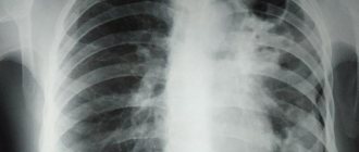
X-ray is a more informative and effective examination in terms of making a correct diagnosis. It will show in detail and accurately what cannot be determined with fluorography. On an x-ray with bronchitis you can see:
- small foci of infiltration, representing an accumulation of fluid in the lung tissues;
- increased thickness of bronchial tissue;
- due to inflammation, small vessels are not visible in the image, the contours of the lungs themselves are blurred in the image;
- focal and lamellar atelectasis - deformations in the form of collapse of part of the lung;
- the roots of the lungs spread and enlarge.
When the disease is advanced, signs of emphysema appear: the pattern of the lungs is significantly distorted due to the accumulation of a large volume of air.

What does bronchitis look like on an x-ray?
Whether bronchitis is visible on an x-ray depends on how advanced the disease is. In any case, the signs of bronchitis on X-rays make themselves felt by the heterogeneous structure of the lung; its changes are accompanied by inflammation.
Acute bronchitis
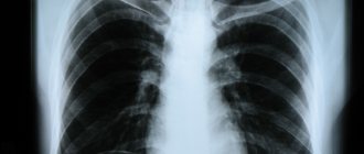
Acute bronchitis on x-ray manifests itself as deformation of the root of the lung, thickening of the walls of the bronchi, unclear contours of the lungs and the disappearance of small vessels from the pulmonary pattern. Advanced obstructive bronchitis, which threatens to develop into emphysema, has the following signs: the presence of abnormal air cavities in the lungs, severe disruption of the pulmonary pattern and changes in the outline of the lung.
Chronical bronchitis
Chronic bronchitis is detected quite easily on X-rays, since the disease usually goes unnoticed for a long time. The main signs of chronic bronchitis on x-ray are: transparency of the lung tissue, vertical position of the heart and thickening of the diaphragm, which means advanced disease. If blood supply problems occur as a result, lung parenchyma is possible, which is visible on x-ray.
What is the difference between acute and chronic form?
Whether an x-ray shows bronchitis depends on the form of the disease. It can be acute and chronic. Acute is characterized by a sharp increase in temperature to high levels, dry barking cough, difficult to separate sputum, chest pain.
But it is difficult to see the sharp shape in the picture. The changes in the organs are still insignificant and not sufficiently manifested. To make a diagnosis, doctors rely not only on external signs, but also on the results of urine and blood tests. Other methods are also used: bronchoscopy, MRI.
The chronic form of the disease develops if the disease lasts more than 3 weeks. It is characterized by an increase in temperature to 37.7 degrees, with fluctuations possible. The cough is dry, but occurs regularly.
In the chronic form of the pathology, changes in the bronchi are bright and visible on an x-ray:
- The gaps in the pulmonary roots resemble tram rails in a pattern. They have thin dark stripes. The roots of the lungs thicken.
- If tissue fibrosis is present, the pulmonary pattern looks like a fine mesh.
- Due to an increase in the volume of air in the alveoli, the lung tissue becomes transparent.
- Stagnation occurs in the small circle and blood circulation is disrupted.
- When examining the bronchi in cross section, ring-shaped threads with uneven edges are noticeable.
If you are wondering whether bronchitis can be seen on an x-ray yourself, the answer is no. It is difficult for a person not involved in medicine to see changes in the image. For diagnosis, be sure to consult a pediatrician and pulmonologist.

Contraindications to X-rays
An absolute contraindication for X-rays for bronchitis is pregnancy and age under 15 years. Otherwise, the radiation dose received during an X-ray of the lungs ranges from 0.15 to 0.40 m3v per session. While the permissible figure for a year is 20 m3v, you will not get it even with regular diagnostic studies of various organs. All this is thanks to the development of X-ray technologies and the protective measures provided (special lead-lined rubber sheets and aprons), which greatly reduce the amount of radiation received. However, despite this, doctors prescribe radiation examination only when justifiably necessary. Therefore, X-ray for signs of bronchitis is one of the most effective and safe diagnostic methods.
Preventive measures
For prevention, it is recommended to do an X-ray of the lungs once a year.
It is better to take care of the health of the pulmonary system in advance than to experience discomfort and spend money on medications later. Maintaining the body's immune system is not as difficult as it seems at first glance.
- It is required to give up bad habits such as smoking, alcohol, and drug use. All of them destroy the human immune system, and smoking irritates the bronchial mucosa, which significantly increases the risk of developing pathology.
- The air in any room should be cool and humid; dust negatively affects the condition of the mucous membranes.
- Playing sports strengthens the human immune system, but all loads should be within normal limits.
- If you suspect the presence of a pathology, you must immediately contact a medical institution for a diagnostic test.
Fluorography allows timely diagnosis of a number of diseases, including: inflammation of the bronchial mucosa, tuberculosis, pneumonia, bronchial asthma. If you take an X-ray once a year, your lungs will not be affected.
Methods for treating bronchitis
Regardless of whether bronchitis can be seen on an x-ray, treatment is prescribed by a doctor and depends on the type of pathogen. For bronchitis of bacterial origin, antibiotics are prescribed, for viral bronchitis, antiviral agents, and for fungal bronchitis, antifungal agents. Symptomatic treatments are used to relieve sore throat and nasal congestion, if necessary. In addition to basic medications, massage and exercise therapy are prescribed to facilitate and accelerate the release of mucus from the bronchi.
With chronic bronchitis, it is very important to eliminate the factors that provoke it. Otherwise, no treatment will help, and there is no doubt whether an x-ray will show bronchitis at the next examination.
Clinical picture of bronchitis
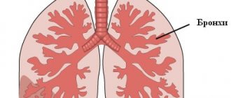
Bronchitis is an inflammatory process in the organs of the respiratory system that affects the lobar, segmental, interlobular, lobular and intralobular bronchi, and bronchioles. It can be observed as an independent process or as a complication against the background of viral or infectious colds.
Bronchitis is characterized by inflammation of the mucous tissues of the bronchial tree. In this case, the onset of the inflammatory process may be accompanied by swelling of the tissues, their compaction, and all this together leads to a narrowing of the respiratory lumen.
The course of bronchial disease can take different forms:
- acute inflammation – has obvious signs of a sharply onset disease, the total duration does not exceed 14 days. The disease manifests itself as a dry, irritating cough. After a couple of days, difficult to expectorate sputum appears. There is an increase in the patient's body temperature to 39 degrees;
- chronic inflammation - mild symptoms appear over a long period, which can last three weeks or more. The appearance of a wet cough with sputum is typical in the morning (immediately after waking up). The body temperature remains within the subfebrile range - no higher than 37.5 degrees;
- obstructive inflammation - a severe course of the disease is complicated by severe attacks of coughing and lack of oxygen (suffocation). The presence of obstruction can be determined by wheezing and changes in respiratory movements. Additionally, the muscles of the neck and abdominal muscles are involved.