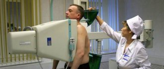
Using an image of the lungs obtained as a result of fluorography, doctors can promptly identify dangerous diseases of the respiratory system. This is the most popular type of screening, which requires a minimum of preparation from the person being examined. Fluorography is carried out quickly, it is relatively inexpensive and allows you to examine large groups of people.
Fluorography has gained the greatest popularity among specialists for monitoring the effectiveness of treatment of patients with tuberculosis and conducting preventive examinations of the population for the timely detection of this dangerous disease in humans.
This technique is used not only as part of the examination of a specific patient complaining of malaise, but also as an effective method of prevention during medical examinations. There are a large number of positions that require a medical certificate of health when applying for jobs. Fluorographic examination is mandatory for all professions whose representatives have to communicate with people en masse due to their work duties.
Fluorography is carried out not only in work groups to prevent tuberculosis, but also in educational institutions among children aged 15 years and older.
The technique is beneficial for mass surveys of the population also because it has a low cost. Unlike an x-ray, a fluorogram is an image reduced several times. If the study is carried out on digital equipment, the image is also projected in a reduced size, but on a workstation monitor. The image is reflected from the fluorescent screen after hitting it with a beam of X-rays passed through the tissue being assessed (the sternum area).
In the context of the prevalence of the procedure, it is important to know whether preliminary preparation for fluorography should be carried out, what can be done, and what is strictly prohibited.
There is a prejudice against such a medical technique, since at the everyday level there is a widespread belief that frequently undergoing examinations that use ionizing radiation is harmful. The article will clear you of misconceptions. It describes why regular fluorographic screening for tuberculosis is so important, to whom it is indicated and to whom it is not, and whether it is necessary to prepare for it in advance.
When is preparation for fluorography necessary?
Thanks to the maximum safety of the technique, almost all adults and children over a certain age can undergo fluorography, and preparation for the procedure does not take much time.
No special preparation is needed before such screening. A patient sent for examination as part of a mass medical examination or to undergo a medical examination when applying for a job only needs to know if he has absolute and relative contraindications to such a procedure.
Thanks to the ease of implementation and maximum safety of this hardware diagnostic, it is possible to obtain an accurate image of the bronchopulmonary system and promptly identify the initial foci of tuberculosis - even before the appearance of characteristic symptoms of the disease.
It is thanks to the use of this diagnostic technique, which allows for rapid examination of large groups of people, that the number of tuberculosis patients has significantly decreased. At an early stage, it is possible to quickly cope with such a dangerous disease, avoiding its chronic course.
In addition to tuberculosis, X-ray screening allows us to identify other diseases of the pulmonary system, blood vessels and heart:
- pneumonia;
- oncology;
- inflammation of the bronchi and lungs;
- vascular pathology;
- heart diseases.
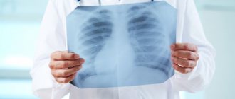
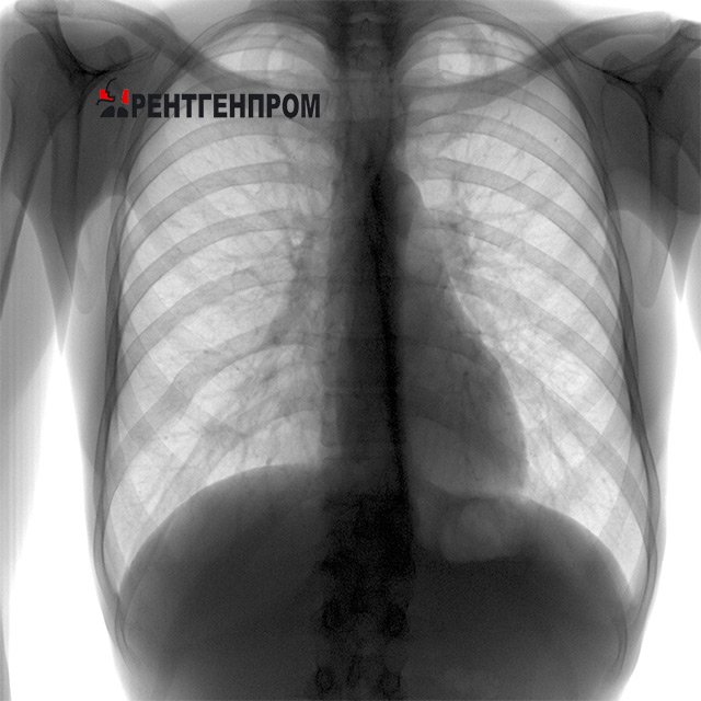
A beam of X-rays directed at the chest provides an accurate image of the organs located behind the breastbone in a matter of seconds. The doctor examines the clear image, based on which he makes an accurate diagnosis.
The X-ray image is clear, and the person himself does not suffer from exposure to X-rays, since the X-ray is carried out using special equipment that reduces the time of exposure to gamma radiation on the object of study, making the procedure completely safe for an adult.
The quality and accuracy of the image is not affected by any external factors. You will not have to refuse food, interrupt the course of treatment prescribed by your doctor, or interrupt your usual routine. Pulmonologists, cardiologists and oncologists value fluorography because it is suitable for all patients and has only relative contraindications.
Definition
Digital scanning fluorograph (the safest and most modern diagnostic method)
Fluorography is an X-ray examination of the chest organs. It is used in our country to detect tuberculous changes in the lungs.
An image obtained with film-type fluorography - 70 mm and 100 mm. In the digital version, large frames are taken on a computer - 2048 x 2048 pixels.
Due to the high prevalence of tuberculosis in our country, fluorography is prescribed for adults and adolescents over 15 years of age. Such screening helps to identify changes in lung tissue at an early stage and begin therapy.
How to prepare for fluorography
No preparation is required for this procedure. The doctor refers the patient for examination if there is a reason for this, or the time has come for an annual examination.
Before entering the booth, you will need to take off your metal jewelry, as it will obscure the picture:
- lungs;
- heart;
- vessels;
- mammary gland.
The principle of obtaining an image during research lies in the nature of the passage of X-rays through tissues with different densities. When the equipment is turned on, the emitter generates a beam of radiation; it passes in a given direction: unhindered through cavities, partially through soft tissue, and completely absorbed by bones. At the “output”, the radiation intensity is recorded on a film or electronic matrix. Deviations from the norm are expressed in atypical shades on the fluorogram.
Shadows caused by the entry of a foreign object into the projection of the lungs when taking a fluorographic image will distort the anatomical picture and interfere with the interpretation of the results.
To get a clear fluorography image, you do not need to perform complex actions or carry out preliminary preparation. You just need to remove foreign objects from the field of study. Men and women undress to the waist. Before fluorography you need to remove:
- outerwear;
- neck decorations, regardless of what material they are made of;
- scarves;
- bra - even if it is wireless or seamless;
- lift up loose long hair and secure it at the top of the head.

After this, you can enter the cabin and follow all the doctor’s orders. Fluorography is performed only in a vertical position. The specialist will ask you to expand your chest and press it against the screen, then ask you to take a deep breath and hold your breath. As you inhale, the lungs expand as much as possible, and at this time an X-ray is taken. You cannot breathe during candling, as the boundaries of the lungs will shift. The picture will turn out fuzzy, blurry, and it will not be possible to make a diagnosis based on it. Holding your breath lasts no more than a few seconds. This time is sufficient for respiratory screening.
Sign up for fluorography with a radiologist
If a control image of the lungs has not been taken for a long time, fluorography should be done at the clinic at your place of residence. This examination is carried out free of charge for all Russians. Afterwards, you will need to get a fluorogram, transcript and make an appointment with a therapist or pulmonologist, who will carefully study the diagnostic results.
Don't forget about contraindications
Although there are no strict contraindications for performing a fluorographic examination, it should be remembered that such diagnostics cannot be performed on pregnant women. It is especially undesirable in the first trimester, as it can harm the development of the fetus.
Fluorography is also not performed on children under 15 years of age. It also makes no sense to take a picture of children, since the radiologist will not be able to make an accurate diagnosis due to the fact that their main respiratory organ has not reached the size of an adult, and it will be difficult to see it in a small picture. If, nevertheless, the study was carried out, the images must be deciphered by a specialist - a pediatric radiologist.
The listed contraindications are absolute. There are also relative prohibitions on conducting research. This is a general malaise and shortness of breath. Fluorographic examination is also not carried out on bedridden patients, since they will not be able to assume the vertical position necessary for proper examination of the internal organs located in the sternum area. Once the patient's condition has stabilized, the diagnostic restriction is lifted.
General recommendations
Before carrying out such a diagnosis, you need to take a bath or shower. On the appointed day, at the specified time, you must come to the X-ray diagnostic room, undress to the waist, enter the booth and follow all the doctor’s orders.
Fluorography during painful and heavy periods
Each organism is individual, and all processes occur according to different scenarios.
For some, menstruation passes quietly and unnoticed, for others it is accompanied by heavy discharge and terrible painful sensations. These symptoms contribute to a sharp loss of strength and a pronounced decrease in performance. Heavy menstruation can lead to mild anemia, in which fluorography is not performed.
Can I eat before the examination?
There are a number of diagnostic procedures, on the eve of which it is recommended to limit the consumption of foods that stimulate intense gas formation in the digestive tract. Fluorography is not one of them.
Nutrition before fluorography is not limited. You can eat anything the day before. The digestive system does not affect the result of the x-ray examination.
Digestive problems are also not considered an obstacle. Some types of studies may be canceled if the patient experiences severe flatulence, since the presence of excess gases in the gastrointestinal tract may distort the picture. In such cases, it is even recommended to take sorbents, rinse the stomach, and do a cleansing enema. In the projection of the lungs there is only the esophagus, and the listed pathological conditions arise lower - in the stomach, small intestine and beyond. Therefore, even if the patient has complaints related to the digestive system, this will not affect the information content of the fluorogram.
What does an x-ray show?
Using X-rays, it is possible to make an accurate diagnosis in a short time:
- in case of various injuries or fractures, a dark fracture will be visible in the image;
- during inflammatory processes and the formation of stones in internal organs, a lighter area is usually noticeable.
If a study is carried out with a contrast agent, it is possible to determine the presence of a benign or malignant tumor by incomplete filling of the internal organs. When examining vessels with a contrast agent, ruptures will be clearly visible.
The clarity of the image is directly affected by the quality of the device, film, and reagents used. Also, the reliability of the result largely depends on the patient’s unnecessary movements, which can spoil the result of the study.
To get a high-quality image, you must follow all the doctor’s recommendations during the procedure.
Is it possible to smoke and drink alcohol before FLG?
Smoking also does not affect the results of an x-ray examination. Despite the widespread horror stories among teenagers who smoke secretly, fluorography is not able to identify the fact of addiction to this bad habit.

Those who like to take a drag on a cigarette with experience experience characteristic changes in the respiratory organs - tissues become denser, the walls of the alveoli are transformed, and bronchiectasis appears. Over time, the risk of developing cancer increases. But this is all a long process, and one or more cigarettes smoked before entering the clinic will not change the anatomical picture on the x-ray. And if a smoker has a tumor or emphysema, the image will help detect them even after the patient smokes before the procedure.
Although alcohol does not affect the result of fluorography, it is better not to drink alcohol before diagnosis, since a doctor can legally refuse to conduct an examination due to the fact that a citizen appeared drunk in a public place. This is punishable by a fine according to the Administrative Code.
Decoding the image in the photo
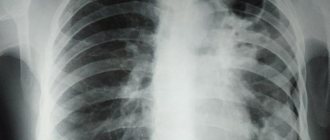
Tuberculosis
The principle of fluorography diagnosis involves the passage of X-rays through the chest tissue. The latter's penetration of dense tissue is recorded on the CCD matrix; darkening indicates pathology.
The radiologist interprets the X-ray photograph. In this case, the nature of darkening in the lung tissue is taken into account; the following indicators can be distinguished:
- Segmental spots are darkening in the shape of a triangle, framed by a light outline. A single manifestation indicates injury to the lung tissue, the presence of a foreign object or endobronchial tumor in the organ area. If there are 2 or more triangles, this may be pneumonia, oncology, fluid in the pleural area, tuberculosis or the spread of metastases.
- A lobar spot outlined with a clear outline is a pulmonary disease, for example, purulent inflammation, bronchiectasis, and others.
- Focal darkening or a spot up to 10 millimeters in size indicates pathologies of the heart and blood vessels, an oncological neoplasm. The focal spot is characteristic of myocardial infarction, but an MRI is required to clarify the diagnosis.
- Focused spots on the surface of the organ indicate different types of pneumonia, the development of bronchial asthma, purulent inflammation in the lungs, helminthic infestations or accumulation of pleural effusion.
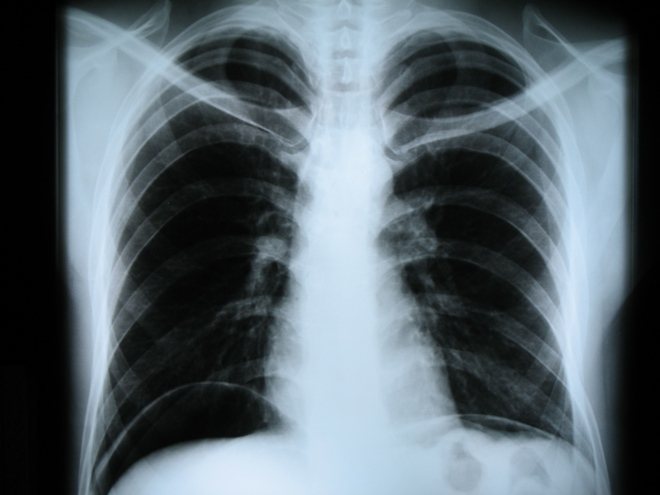
Interpretation occurs in different ways, so interpretation should be done by an experienced radiologist. It is important to know that healthy lungs in photographs have a uniform color, and the heart and bronchi look light.