Biopsy is the gold standard for diagnosis in oncology. This method allows you to obtain a tissue sample, examine it under a microscope and accurately determine the true nature of the tumor, as well as select effective treatment. There are several varieties of this diagnostic method, which are used for neoplasms of various structures and localizations. If breast tumors are detected, a trephine biopsy may be prescribed. This type of procedure has its advantages, disadvantages and implementation features.
- Differences from other methods
- Indications and contraindications for trephine biopsy
- Preparation and conduct of the study
- What information can you get?
- Possible complications
Differences from other methods
Most often, breast biopsy is performed in two ways. The first is a trephine biopsy. To carry it out, you need a special needle, which has a large diameter and is hollow inside. This feature allows you to obtain a column of tissue of a fairly large size.
The second common method is fine needle aspiration biopsy. As the name suggests, in this case a small diameter needle is used, which reduces the risk of bleeding and tissue trauma. Due to safety and high information content, fine-needle biopsy allows you to obtain material from several foci, which is important if there are several nodes in the mammary gland. Among the disadvantages of the method, one can note the possibility of obtaining samples only for cytological examination.
Another type of method is excisional biopsy. It is somewhat different from the types described above. An excisional biopsy is performed after surgical removal of the tumor. In this case, you can select the desired area of the tumor and examine it in detail.
Experts do not compare different types of biopsies and do not identify the best or worst way to obtain the material. Each method has specific indications and areas of application, but each of them has the right to exist and is actively used at the present time.
Book a consultation 24 hours a day
+7+7+78
Indications for trepanobiopsy
Since this study is used only when the presence of a neoplasm has already been proven, the indications for its use are:
- The need to accurately determine the nature (type) of the tumor
- The need to clarify its degree of development and prevalence
- Tracking the dynamics of education growth
- Clarification of the presence and nature of other neoplasms (cysts, polyps, nodes) similar to cancer
- Upcoming surgery to control the condition of the breast
Indications and contraindications for trephine biopsy
Despite its high information content and reliability, a biopsy is not prescribed to all women in a row. As a rule, the procedure is preceded by a comprehensive examination, which includes the following methods:
- Physical examination (chest palpation).
- Mammography.
- Ultrasound of the mammary glands.
- Laboratory tests.
- MRI or CT, if necessary.
If the examination results reveal lumps in the breast and the doctor is unable to reliably determine their nature, then the next step will be to perform a biopsy. When prescribing a study, the symptoms that are observed in a particular patient are also taken into account. The following signs are characteristic of malignant tumors:
- Pathological discharge from the nipples.
- Changes in the relief or color of the skin in the area of the tumor.
- Deformation or retraction of the nipples, etc.
At the same time, there are practically no contraindications to biopsy, due to the fact that this procedure is of utmost importance for the life and health of the patient. If various disorders are identified that can lead to the development of complications after a biopsy, the doctor prescribes the necessary treatment. After stabilization of the condition, the possibility of conducting research is again considered.
Breast biopsy
The mammary gland in women is considered an organ with an increased risk of malignant neoplasms. Breast cancer is a scary diagnosis, but if detected in the early stages, it can be successfully cured without serious consequences for the patient. If suspicious changes occur in the tissue, a breast biopsy is performed. The method involves taking a small sample from a living organ for further examination under a microscope.
In what cases is the procedure prescribed?
A breast biopsy has certain indications. It is necessary to see a mammologist (from the Latin word mamma - female breast) as soon as possible if the following changes are detected:
- A lump is felt inside the gland. It can be large or small, smooth or uneven, painful or not;
- Skin disorders are observed - sores, rashes, peeling, crusts, including on and around the nipples;
- Discharge from the nipples appears outside of the feeding period. This could be mucus, blood, pus, milk-like secretions, etc.;
- Spots appear on the skin of the chest;
- Light or dark spots on an x-ray in the area of the mammary glands;
- Detection of suspicious areas during ultrasound examination;
- Unnatural (uncharacteristic for a particular woman) nipple retraction or bulge.
Breast biopsy has virtually no contraindications. It should not be performed only on pregnant women, patients with an installed biostimulator, or while breastfeeding a child.
Biopsy sampling methods
There are several types of breast biopsies. The choice of method depends on the preliminary diagnosis, the size of the suspicious area, the general condition of the patient, the technical equipment of the clinic and the qualifications of the doctor:
- Fine needle aspiration biopsy. One of the least traumatic methods that does not require long recovery. It involves a puncture (puncture) of the gland with a thin needle, through which a piece of suspicious tissue is sucked out using a special syringe. The manipulation is carried out under ultrasound control;
- Trephine method. It differs from the previous one with a thicker needle, which can be used to obtain more material. The instrument is inserted into the gland several times to take a biopsy from different parts of it. The method is more painful and requires anesthesia;
- Stereotactic biopsy. Necessary for taking changed tissues that cannot be palpated and do not manifest themselves in any way. The material is selected using fluoroscopy or MRI. In this way, it is possible to hit the tool exactly in the desired area;
- Vacuum. Requires hospitalization and serious anesthesia. It is carried out through several incisions, with the introduction of a thick needle with a cutting edge. Using a vacuum device, a rather large, cylindrical piece of biopsy material is cut off and sucked into the needle. Unlike conventional puncture, this method allows you to obtain much more material for research;
- Surgery. Most often it involves complete removal of the tumor, followed by sending it for histological examination to determine malignancy. But there may also be partial taking of material. The intervention is serious and requires long-term rehabilitation.
Preparing for a breast biopsy
Preparation for the procedure can vary greatly depending on the method of its implementation. In any case, it is necessary to first undergo general and biochemical blood tests, urine tests, perform an ultrasound and mammography, and determine hCG for pregnancy.
During a biopsy, the normal functioning of the hematopoietic system is very important. Blood clotting can greatly affect the procedure itself and recovery after it. If the patient is taking medications that affect blood clotting, then under the supervision of a doctor they will need to be discontinued 2-3 days before the test.
There are several general recommendations before taking a biopsy:
- If general anesthesia is necessary, it is important to refrain from eating the evening before the procedure;
- If local anesthesia is used, you need to make sure there is no allergy to the drug;
- It is not advisable to use decorative cosmetics, creams or perfumes on the day of the study;
- You should not drink alcohol at least 24 hours before the biopsy.
The psychological aspect of preparation is also very important. The doctor must explain to the patient that 80% of all neoplasms and other changes in the mammary gland are benign. If possible, it is best for a woman to take a loved one with her to the medical center who can support her morally.
Biopsy procedure
With regular examinations by a mammologist, and preventing the development of a large tumor, a puncture of the mammary gland is most often performed as the most gentle procedure. The patient is placed on a surgical couch, most often facing the doctor. The process of taking a biopsy can be divided into several stages:
- The skin of the breast is treated superficially with an anesthetic. In some cases, infiltration anesthesia is performed (intradermal injection). At the patient’s request, sedation with nitrous oxide or full anesthesia is possible;
- Under the control of ultrasound, MRI or mammography, a puncture is performed with the insertion of a needle into the breast tissue. The doctor visually observes the movement of the instrument and sees which areas he needs to take for examination;
- After the sampling is completed, the needle is removed, and a pressure bandage is applied to the puncture site to stop bleeding;
- When the bleeding stops, apply a regular, non-pressure bandage to keep the wound clean;
- The patient can safely go home within half an hour after the procedure.
After taking the analysis, it is necessary to limit physical activity, try not to wet the puncture site, and avoid visiting saunas for at least a week. If the research method included incisions and suturing, then several days of inpatient observation will be required.
Complications include breast swelling, severe pain, bleeding, and bruising. This can happen either due to incorrect manipulation or due to insufficient care. If your breasts continue to bother you a few days after the biopsy, you should see your doctor.
Decoding the results
Analysis results are usually ready within 5-7 days. The value of a breast biopsy is not only its ability to identify a cancerous tumor. With the help of this study, you can find many other pathological changes, the treatment of which cannot be delayed:
- Diffuse mastopathy (according to modern nomenclature, this disease is called cystic fibroadenomatosis);
- Benign neoplasm (most often fibroadenoma);
- Formation of polyps in the milk ducts;
- Abscesses, cysts and necrosis;
- Mastitis of various etiologies.
If the tumor is malignant, a biopsy will confirm this. With its help, it will also be possible to determine the type of tumor and the structure of its tissues, because they all respond differently to treatment and require specific drugs for chemotherapy:
- Carcinoma;
- Sarcoma;
- Melanoma;
- Paget's disease;
- Colloid, infiltrative, medullary cancer.
How much does a breast biopsy cost?
The cost of a biopsy can vary greatly on a case-by-case basis. The final price will be determined only at the initial appointment, after examination and ultrasound. The cost of the procedure will be influenced by several factors:
- Type of changes in tissues, suspicions, preliminary diagnosis;
- The size and type of neoplasm, if any;
- Biopsy method - puncture, vacuum, surgery;
- Control method – ultrasound, radiography, MRI;
- The need for local anesthesia or general anesthesia;
- Is inpatient observation required?
In our clinic, everything is provided for the comfort and safety of the patient. Experienced doctors, the latest high-tech equipment, complete sterility, disposable consumables, accuracy and reliability of research results - this is what we offer you. Sign up for an initial examination and consultation to take the first step on the path to health.
Preparation and conduct of the study
Trephine biopsy of the breast does not require specific preparation. If the patient is taking any medications, the doctor may stop them a few days before the test. In addition, you should stop smoking and drinking alcohol the night before the biopsy.
Special large-diameter needles are used to conduct the study, so patients often have questions about the pain of the procedure. In order to eliminate pain and discomfort, the breast at the puncture site is first numbed with local anesthetics. If a complex study is planned, then intravenous anesthesia can be used for a short period of time.
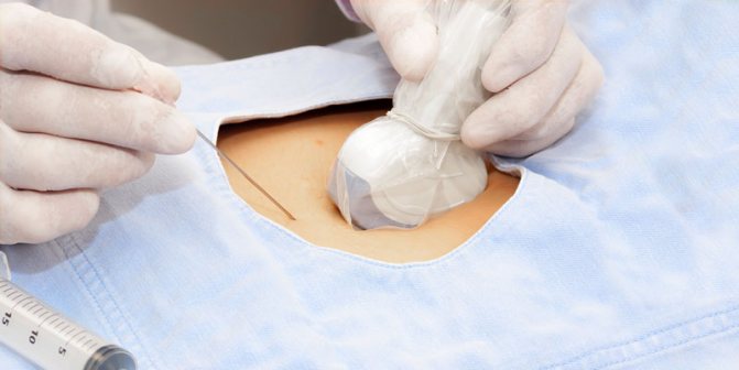
Trephine biopsy is performed on an outpatient basis in an operating room or dressing room in compliance with the rules of asepsis and antisepsis. To obtain a sample of tumor tissue, the doctor may use ultrasound for monitoring purposes. In some cases, a biopsy is performed under digital control, especially if the tumor is located superficially. On average, the procedure lasts 10-15 minutes.
The recovery period lasts about 7-10 days. During this time, it is necessary to limit physical activity, regularly change bandages and treat the chest wound with antiseptic solutions prescribed by the doctor.
How a breast tissue sample is taken
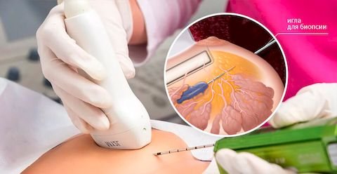
The study is carried out on an outpatient basis, without hospitalization. A thin needle is used, so in most cases no anesthesia is required. This is important for people who are allergic to medications.
A biopsy is performed from days 7 to 14 of the cycle, when the breasts are soft and less sensitive.
To accurately determine the puncture site, an ultrasound sensor is used, which is attached to the chest.
The procedure consists of the following steps:
- The woman sits comfortably on the couch.
- If necessary, local anesthesia is administered.
- Using ultrasound, the location of pathological areas is determined.
- A thin biopsy needle is inserted. A woman may feel slight pressure and discomfort. If the study is carried out correctly, there should be no pain.
- Biomaterial of the neoplasm or fluid is collected, which can be used to fill the cyst.
- The puncture site is treated with an antiseptic.
The biomaterial is sent to the laboratory for research. All manipulations are displayed on the monitor, so one puncture is usually enough to obtain the result. In some cases, a repeat biopsy may be prescribed.
The procedure takes no more than 5-10 minutes. No rehabilitation required. But after the biopsy, you should avoid going to the sauna or bathhouse for 24 hours, and also avoid heavy physical activity.
What information can you get?
The resulting tissue sample is sent to the laboratory for histological examination. The material is first fixed, placed in paraffin blocks, cut and placed on a glass slide. After staining, a pathologist examines the glass under a microscope and determines the type of tumor, the degree of its differentiation and growth characteristics.
A biopsy allows not only to diagnose diseases of a tumor nature, but also to select the correct treatment. To do this, the material is sent for FISH or immunohistochemical study. For example, the drug Trastuzumab from the group of monoclonal antibodies is used in targeted therapy for breast cancer. However, it is effective only if overexpression of the HER-2/neu receptor is detected in the tumor tissue. This indicator can only be determined after a biopsy.
Trepanobiopsy - what is it?
A type of breast biopsy (core-needle biopsy, cutting core biopsy, drill biopsy), which allows you to obtain pieces of the tumor for morphological examination using a special cutting needle or a spring hollow needle equipped with a puncture gun. The material obtained through trephine biopsy allows for a high-quality in-depth histological, immunohistochemical and morphological study of the profile and structure of the tissue samples taken, to determine more subtle structures, in particular sex chromatin, estrogen receptors, etc.
Possible complications
Severe complications that require emergency medical attention rarely develop after a biopsy. Most often, patients are concerned about moderate pain, inflammatory manifestations (redness of the skin, local increase in temperature), and minor bleeding. If you follow all the doctor’s recommendations, these conditions will go away on their own within a few days. With the development of infectious complications, the symptoms are more pronounced - body temperature rises to 38°C or more, pus may be released from the wound, and severe pain is noted. Some patients experience allergic reactions to anesthetics used during trephine biopsy.
The likelihood of developing undesirable reactions will be lower if the diagnostic procedure is carried out by an experienced doctor who has modern equipment and high-quality consumables at his disposal.
Book a consultation 24 hours a day
+7+7+78
Where can you undergo the procedure in Moscow?

Needle biopsy is the safest, most gentle and effective diagnostic method. It allows you to make a diagnosis and begin treatment as accurately and as quickly as possible. Despite the safety and accessibility of this method, high qualifications and experience of the diagnostician are important. Therefore, if you are scheduled for a puncture biopsy of the breast, then you need to take a responsible approach to choosing a medical institution and specialist.
For examination, you can contact the Otradnoe Polyclinic. This is a multidisciplinary large medical center in the North-Eastern Administrative District of Moscow. Our main value is experienced and attentive doctors and friendly staff. Having our own laboratory allows us to obtain research results in the shortest possible time. For more detailed information, make an appointment by phone or through the form on the website.
Why you shouldn't be afraid of a biopsy
Percutaneous puncturing is therefore a minimally invasive method because it does not cause pain. Discomfort during a fine-needle aspiration is similar to an injection, and a trephine biopsy is also an injection and is performed under local anesthesia. There are no nerve endings in the tumor, so there is no pain when the cell tissue is taken.
Another reason for fear before the procedure is the fear that the doctor will miss and insert the needle into the wrong area. Cell collection for diagnosis is performed under ultrasound control, which provides almost 100% accuracy. In addition, at the First Phlebological Center, percutaneous puncture is performed by a surgeon with more than 20 years of specialized experience.
Many people are afraid that a biopsy necessarily precedes surgery. But, in 80% of cases, the neoplasms are benign, fibroadenoma, which does not require surgical intervention.
