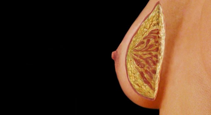
A ductal dilatation occurs when the milk duct under the nipple widens (spreads apart), its walls thicken, and it fills with fluid. The milk duct may then become blocked or blocked by a thick, sticky substance. The condition is often asymptomatic, but some women experience nipple discharge, breast tenderness, or inflammation of a blocked milk duct (periductal mastitis).
With age, the likelihood of dilation of the mammary duct (regardless of the development of inflammation) increases. Dilation of the mammary duct usually goes away on its own without treatment. However, if you have persistent symptoms that persist despite home treatment, you may need antibiotic treatment or surgery to remove the affected milk duct.
Although it is natural to be concerned about breast changes, ductal dilatation and periductal mastitis are not risk factors for breast cancer.
Symptoms
Although mammary duct dilatation is usually asymptomatic, patients may experience:
- Discharge from one or both nipples that is off-white, greenish, or black in color
- Soreness of the nipple or surrounding breast tissue
- Redness of the nipple and sometimes the surrounding area
- A tumor in the breast or a lump near a blocked duct.
- Inverted (inverted) nipple
In addition, a bacterial infection called mastitis can develop in the affected milk duct, causing inflammation in the area around the nipple (the nipple circle) and fever. Signs and symptoms of a dilated breast duct usually go away on their own.
Conditions under which you need to see a doctor
It is imperative that your doctor immediately evaluate changes in the condition of the mammary glands to rule out the possibility of breast cancer. If you have symptoms of dilated mammary duct, especially if there is discharge from the nipple, you should consult a doctor.
Reasons for the development of the disease
Women approaching menopause are at risk. Age-related hormonal changes contribute to the development of this pathology. The ducts are located in the alveoli of the chest. Under normal conditions, milk tubes have a narrow, corrugated structure. All paths connect and go to the nipple. The onset of a dangerous disease can begin suddenly. Among the causes of dilation of the thoracic ducts, a distinction is made between natural and pathological.
Natural causes include:
- Premenstrual period
- Fertility
- Menopause
During menstruation, a woman's body changes. The breasts swell and become especially tender and painful to the touch, as with mastitis. The body is on the eve of fertilization, so the milk ducts undergo slight changes. If pregnancy does not occur, the milk duct narrows and its structures return to normal.
With the conception and further development of the embryo, the woman’s body begins to prepare for lactation after birth. During pregnancy, the mammary glands increase in size, and the excretory ducts dilate for better milk flow. Only after breastfeeding ends do the milk ducts begin to narrow. Limiting breastfeeding should be gradual. It is important to squeeze out the remaining milk to prevent lactastasis.
Due to the hormonal changes that occur during menopause, the structure of the breasts also changes to some extent. Against the background of hormonal disorders, the duct expands. This phenomenon is not pathological, but requires careful monitoring. A woman during menopause should undergo regular examinations, breast ultrasound and mammography. Preventive examinations with a mammologist and gynecologist allow you to detect pathology in time and begin its treatment.
Pathological factors for dilatation of the duct include:
- ectasia
- development of mastopathy
- presence of cysts
- inflammatory process
- tumor formation
A characteristic symptom of mastopathy is an increased amount of estrogen. This causes an increase in the structure of the mammary gland and dilation of its ducts. Large amounts of fluid can also accumulate in the canals. This leads to the formation of cysts and tumors.
Complications
Complications caused by a dilated mammary duct are usually not serious, although they can be uncomfortable. These include:
Discharge from the nipple. Nipple discharge caused by dilation of the mammary duct can be bothersome. A woman may feel embarrassed by wet spots on her clothing caused by fluid leaking from the nipple. Unpleasant sensations in the chest. A dilated breast duct can cause redness, swelling, and tenderness in the area around the nipples. Infection. A bacterial infection (periductal mastitis) may develop in the affected milk duct, sometimes accompanied by pain in or around the nipple or a general feeling of unwellness and fever. If left untreated, abscesses (collections of pus in the breast tissue) may form, which may require surgical drainage to remove. Worry about breast cancer. When a woman discovers breast changes, she may worry that they may be clinical manifestations of breast cancer, especially if a solid tumor forms around a dilated mammary duct. It is imperative that you seek immediate medical attention for these clinical signs and symptoms, but keep in mind that a dilated breast duct does not increase your risk of developing breast cancer.
Intraductal papilloma of the mammary gland - symptoms and treatment
If complaints arise, a woman should consult a mammologist . The first stage of diagnosis is medical history and examination. The doctor records concomitant diseases and factors that could provoke the development of intraductal papilloma, notes the time of onset of symptoms.
The inspection is carried out in two positions:
- standing with arms down: the mammologist assesses the symmetry of the breast, the presence of local changes, palpates the breast;
- lying down with your hands behind your head: a deep layer of tissue is felt.
Be sure to evaluate discharge from the mammary gland. The doctor gently presses on the nipple and takes an imprint for cytological examination. Based on its results, one can draw the first conclusions about the nature of the neoplasm [7].
Radiothermometry and electrical impedance mammography can be used as a screening method at the initial stage of diagnosis . During radiothermometry of the breast, a special sensor measures the electromagnetic radiation of the tissue through the skin and builds a temperature graph. Tumors appear hotter than healthy tissue. Electrical impedance mammography is based on the study of electrical conductivity, which changes in tumor foci. These methods are effective for identifying proliferative processes and make it possible to distinguish intraductal papillomas and breast cancer from fibroadenoma or mastopathy.
The technique is based on the fact that with active cell division, which occurs in hyperplastic diseases, blood supply and tissue nutrition are enhanced. This causes an increase in temperature and electrical conductivity. For intraductal papilloma, radiothermometry is informative in 83% of cases and specific in 90%.
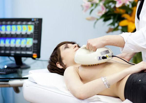
For electrical impedance mammography for cystadenoma, hypoimpedance foci are detected in 70% of cases, and if this figure reaches 100%, then cancer is diagnosed [1].
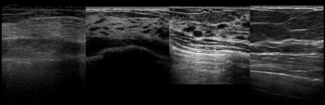
Ultrasound of the breast is performed to search for space-occupying lesions, but for intraductal papilloma this method is not effective enough. Indirect signs of pathology are:
- ectasia, or dilatation, of the ducts;
- soft tissue formation near the nipple;
- hypoechoic nature of the formation (on the monitor it is darker than the surrounding tissues);
- clear contours of the tumor [3].
In women over 45 years of age, ultrasound is not used due to physiological changes in the mammary gland. And mammography will also not show pathology of the ducts, unless the tumor begins to grow into the surrounding tissue.
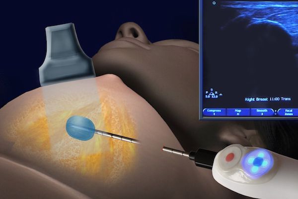
Ductography helps to identify the localization of the process . This is an x-ray diagnostic method in which a contrast agent is injected into the milk ducts. It is prescribed for pathological discharge from the mammary glands, but not in all cases. There are no indications for ductography if:
- The color of the discharge is milky, transparent, yellow-green or brown;
- a nulliparous woman takes medications that affect the function of the pituitary gland;
- the patient recently gave birth or finished breastfeeding [5].
No special preparation is required for ductography. The woman should be warned that it is impossible to squeeze blood out of the nipple before the procedure. The examination takes about 30 minutes. A thin catheter is inserted into the ducts with a special instrument, through which a radiopaque solution is injected. After this, a standard image is taken, as with a mammogram.
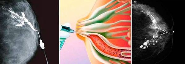
The result of the image is assessed on the same day. Typically, intraductal papilloma does not allow the solution to pass into the distal parts of the duct, so a filling defect appears on the image. Ductography is a safe method for diagnosing pathology of the milk ducts. It is painless, but some patients feel discomfort. Based on the results of the study, the doctor receives an accurate idea of the localization of the pathological formation in order to take a tissue sample for histological diagnosis [5].
Morphological methods help confirm or refute the malignant process [reference: 4:
- trephine biopsy - a piece of tissue is taken from the pathological focus using a device with a special needle. But according to some studies, the technique is not informative enough for intraductal papillomas, because in this type of neoplasm, the cells are heterogeneous, and during sampling only the benign area can be captured. Therefore, there is a risk of false diagnosis;
- vacuum aspiration biopsy - a piece of tissue is taken through a thick needle, which is larger than with trephine biopsy. The method allows you to accurately determine benign and malignant processes [9].
Comparison of histology results obtained after vacuum aspiration biopsy and examination of tissue removed during surgery shows that the diagnoses are the same in most cases.
Treatment of dilated mammary duct
Antibacterial therapy is indicated for this pathology only in the presence of a severe inflammatory process. Therapy should be aimed at correcting immunity, hormonal status, and activating the body's defenses. Complex treatment will help eliminate the symptoms of pathology and normalize the condition of breast tissue.
During treatment, it is important to conduct medical monitoring of the epithelial structures of the breast. With characteristic changes in the ducts, the development of cysts, surgical intervention and surgical removal of cystic lesions is possible.
Homeopathic medicines can effectively restore the structures of the mammary gland. To quickly eliminate the pathological process, doctors recommend the following drugs:
- Mastodinon
- Cyclodinone
- Viburkol.
These remedies relieve breast spasms, reduce pain and suppress active inflammation. The drug Gynekohel helps to cope well with pathology. The product has a safe herbal base and is recommended for age-related changes, hormonal disorders, infectious and inflammatory processes of the reproductive system.
Klimaktoplan can be used with extended tubes. The drug corrects the production of hormones by endocrine organs, regulates the functioning of the thyroid gland and adrenal glands. Dismenorm effectively stabilizes the synthesis of hormones in the female body. This homeopathic medicine corrects the imbalance between prolactin, progesterone and estrogen levels.

Symptoms of a dilated duct
When the milk ducts are dilated, a woman experiences a feeling of heaviness and pain in her breasts. The periosteum swells and a bright fluid may ooze from the nipple. Sometimes the pathology is accompanied by nipple retraction and itching of the skin. Symptomatology is characteristic of many breast diseases. Particular attention should be paid to symptoms such as duct spasms and breast enlargement or bloating.
The first symptom of the disease is increased chest discomfort. The areola is swollen and the skin around it itches. In advanced cases, symptoms of intraductal inflammation are observed. The pain inside the chest intensifies. The main thing is to refer the patient to a doctor in a timely manner.