Free and general T3
T3 is found in the human body in several forms, often in the direction of analysis you can see total and free T3.
Total T3 is a hormone that contains bound and free shares of the hormone in the blood. But some react with protein molecules, and also interact with the tissues and cells of the body in various ways. Such a hormone is called bound, and its remaining molecules form free triiodothyronine. It does not react with any other substances, but simply “travels” throughout the body as part of the blood.
A general blood test for T3 shows the total concentration of bound and free hormone. It must be said that it is not carried out as often as a free analysis, since the results are influenced by many external factors.
However, in some cases such analysis is necessary, for example, when:
- disruption of the pituitary gland and thyroid gland;
- sudden weight gain;
- presence of edema;
- disruption of the menstrual cycle;
- severe PMS;
- painful menstruation.
Reference! The main difference between total and free T3 hormone analyzes is accuracy and information content. Therefore, a free T3 test is most often prescribed.
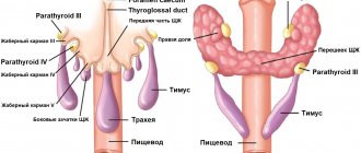
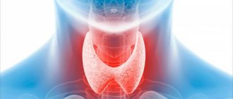
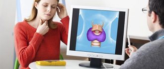
Ultrasound of the thyroid gland: normal
Many patients who plan to have an ultrasound of the thyroid gland would like to understand for themselves what all these words written in the conclusion of the study mean. We will try to explain some of the most important terms used by doctors during ultrasound, as well as their meaning in terms of determining normality and pathology.
The most important ultrasound characteristics of thyroid tissue are:
- contours of the gland;
- gland tissue structure;
- echogenicity of gland tissue;
- presence or absence of focal changes (nodules, cysts);
- blood supply to gland tissue.
The ultrasound condition of the cervical lymph nodes surrounding the thyroid gland is also necessarily described.
Outlines of the thyroid gland
can be clear or unclear. Normally, the contours of the thyroid gland should be clear. The contours become fuzzy (blurred) with the development of inflammation, as well as with the occurrence of malignant tumors of the thyroid gland that grow into the surrounding muscles and adipose tissue.
Fabric structure
can be homogeneous or heterogeneous.
The thyroid gland normally
has a characteristic grainy tissue, which, with some skill, is difficult to confuse with anything else.
Inflammatory diseases of the thyroid gland
, developing as a result of aggression of the immune system (autoimmune thyroiditis, diffuse toxic goiter) are accompanied by the appearance of heterogeneity of thyroid tissue - sometimes like a “honeycomb”, sometimes doctors describe it as “moth-eaten tissue”, but always in the tissue with heterogeneity The structure contains more and less light areas, the tone of which is clearly different. It happens that doctors describe a pronounced heterogeneous structure of the gland, when the difference in the tone of the thyroid gland is large, or a moderately heterogeneous structure of the thyroid gland - this is often found in healthy people who have an increased titer of antibodies to thyroid peroxidase or thyroglobulin.
Echogenicity of thyroid tissue
– this is the same “tone” that is visible on the screen. It should be remembered that the image on the screen of the ultrasound machine is formed by a computer, which analyzes the reflected ultrasound rays coming from the internal organ and, based on this analysis, presents the image in grayscale to the operator. Echogenicity is the tone of gray that the computer represents thyroid tissue. Normally, the echogenicity of the gland tissue is equal to the echogenicity of the parotid salivary gland. With the development of inflammatory diseases, the echogenicity of the thyroid gland is most often reduced, but in the later stages of this process it can even be increased. A pronounced decrease in echogenicity is indicated when the tone of the gland becomes darker than the tone of the surrounding muscles (i.e., almost black) - such changes should always alert the doctor performing an ultrasound of the thyroid gland. The norm of echogenicity may vary slightly, but usually the echogenicity of the thyroid gland is higher than the echogenicity of the muscles, vessels, and esophagus (i.e., the gland looks lighter on the ultrasound machine screen).
Focal changes (nodes)
The thyroid gland does not normally contain. Acceptable are cystic formations up to 3-4 mm in size, which look uniformly black on the screen (i.e., anechoic - without echogenicity) - endocrinologists often call such formations enlarged follicles, accumulations of a hormone-containing gel - colloid. All formations over 4 mm in size that differ in echogenicity from the surrounding thyroid tissue are usually called nodes. Nodes can be:
- isoechoic, i.e. equal in echogenicity to the surrounding thyroid tissue;
- hyperechoic, exceeding the echogenicity of the surrounding thyroid tissue (i.e. lighter);
- hypoechoic, having less echogenicity than the surrounding tissue (i.e. darker);
- anechoic, i.e. completely, completely black (this color is typical for fluid formations, cysts).
A thyroid nodule is always not the norm. The thyroid gland should normally be homogeneous, without nodes. However, modern high-class ultrasound machines used at the North-Western Endocrinology Center make it possible to detect nodes as small as 1 mm. Endocrinologists who perform ultrasound at the endocrinology center understand that it is unreasonable to call every formation in the thyroid tissue measuring 1, 2 or 3 mm a node, since after this, from a formal point of view, it is necessary to establish a diagnosis of “Nodular goiter”. A patient with such a diagnosis then has a lot of problems when visiting other specialists who do not have sufficient knowledge in the field of endocrinology, who, instead of treating, for example, high blood pressure or cardiac arrhythmia, will tell the patient: “Well, what do you want - you have It's a goiter! First, go to an endocrinologist, have him write you a paper saying it’s not the thyroid gland’s fault, and then come to me.” As a result, the patient is forced to make unnecessary trips to the doctor, wasting time, nerves and money. That is why doctors must treat small focal formations very carefully - of course, certain examinations are necessary, but usually no treatment is required as a result.
For each thyroid nodule, the physician performing the ultrasound should describe:
- contours (clear, fuzzy);
- the presence or absence of a dark rim along the periphery of the node (halo rim);
- echogenicity of the node;
- the presence of micro- or macrocalcifications (i.e. calcium deposits that do not have an acoustic shadow = microcalcifications, or have an acoustic shadow = macrocalcifications);
- the presence or absence of cystic transformation of the node (i.e. the appearance of cysts inside the node);
- linear dimensions (it is advisable to describe three linear dimensions of the node, since this makes it possible to calculate the volume of the node and subsequently, with repeated ultrasound, reliably determine the dynamics of its change).
Blood supply to tissue
determined by conducting a Doppler study, which reveals the intensity of blood flow that the thyroid gland has. The norm is the presence of several color signals on the surface of the thyroid tissue. When the thyroid gland is inflamed, blood flow increases, and the entire gland seems to be “bursting with fire” on the ultrasound machine screen. Western researchers even came up with the poetic name thyroid inferno (“thyroid hell”) for this type of blood flow, comparing this picture with the tongues of hellish flames on medieval paintings.
Regional lymph nodes of the neck
Normally, an ultrasound scan of the thyroid gland should not look enlarged. Lymph nodes must have clear, even contours, the length of the lymph node must be at least 2 times the width of the lymph node (the so-called Solbiati index), the gate must be clearly visible in the structure of the lymph node - the place where the lymphatic vessel enters the lymph node. The tissue of the lymph node should not have increased blood flow and, especially, cysts - both of these signs are very alarming and often indicate a malignant lesion of the lymph node.
Ultrasound normal thyroid gland
- this is the standard that every endocrinologist must clearly know, and with which he must compare the image visible on the screen of the ultrasound machine. Of course, it is difficult to give a complete picture of all ultrasound aspects of a normal thyroid gland in a short article. If you doubt whether what was revealed during an ultrasound examination of your thyroid gland in a clinic or general medical center is normal, the most reasonable tactic would be to consult an endocrinologist or an endocrinologist surgeon at the North-Western Endocrinology Center, who will independently perform An ultrasound of the thyroid gland will compare what you see with what is described on your form and explain to you what needs to be done in the future. You will be surprised, but each of our specialists knows how often the “threatening” changes on ultrasound described somewhere, in the end, when carefully examined by experienced specialists using high-quality equipment, turn out to be just another variant of the norm...
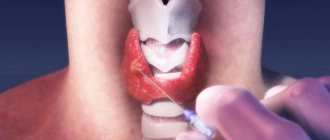
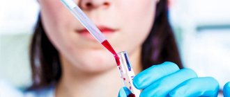
T3 hormone test
Testing for triiodothyronine is carried out for the following indications:
- routine annual physical examination;
- the patient’s initial visit to the endocrinologist due to increasing symptoms of a malfunction of the thyroid gland;
- pathologies of the thyroid gland;
- monitoring the treatment of thyroid disorders;
- pregnancy;
- collecting analysis of the newborn, especially in cases where the mother has problems with the thyroid gland.
A test for the T3 hormone is also prescribed for people:
- with diabetes mellitus;
- with adrenal insufficiency;
- with pernicious anemia;
- elderly;
- women after childbirth;
- having an inherited predisposition to thyroid pathologies.
How to prepare for the analysis?
Some experts recommend taking a thyroid hormone test on an empty stomach, but there is another opinion - this is not required, as it does not change the level of hormones. However, it is not recommended to consume meat and other fatty foods before the study, as this affects the state of the blood serum, and it will be quite difficult for a specialist to isolate the formed elements in it.
In order for the T3 analysis result to be as reliable as possible, you need to:
- Do not eat fatty or meaty foods 24 hours before the test.
- Avoid stress, drinking alcohol and smoking.
- Tell your doctor about the medications you are taking. Some may have to be canceled for a while.
- Tell the doctor about your history of serious illnesses; some pathologies affect the results.
Important! It has been proven that a woman's use of contraceptive drugs affects the concentration of free T3, and according to the test results, the woman could be misdiagnosed with hyperthyroidism.
Norm of T3 hormone in women, men, children
The normal level of T3 hormone in women and men over 18 years of age is:
- general - 1.2–3.1 nmol/l,
- free - 3.1–6.8 pmol/l.
Normal levels of triiodothyronine in children are the same as in adults; they should be in the range of 3.1–6.8 pmol/l. But the level of other thyroid hormones depends on the age of the child.
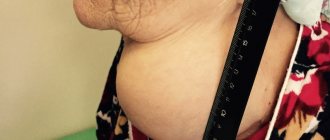
Increased T3 hormone
If T3 is elevated, this means that the thyroid gland has malfunctioned. This may be iodine deficiency, thyroiditis, endemic goiter. Increased concentrations are dangerous due to catabolic disorders, loss of body weight, and deterioration in physical development.
Hormonal imbalance can lead to disruption of many systems at once:
- nervous;
- cardiovascular;
- urinary;
- reproductive;
- digestive;
- hepatobiliary;
- immune.
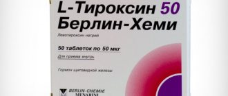
Elevated T3 is the basis for prescribing hormonal therapy, but only an endocrinologist can prescribe drugs.
Normal values
Important! Standards may vary depending on the reagents and equipment used in each particular laboratory. That is why, when interpreting the results, it is necessary to use the standards adopted in the laboratory where the analysis was carried out. You also need to pay attention to the units of measurement.
The article provides normal values accepted in the Invitro and Helix laboratories
T4 free
Norms of free thyroxine according to the Invitro laboratory:
| Age, both genders | Free T4, pmol/l |
| 5 days – 15 days | 13,47-41,32 |
| 15 days – 30 days | 8,71-32,53 |
| 30 days – 6 months. | 11,42-21,89 |
| 6 months - 13 years | 10,80 -16,40 |
| 13 years – 18 years | 10,20 – 15,50 |
| over 18 years old | 9,00 – 19,05 |
Standards in the Helix laboratory:
| Age | T4 free, reference values |
| under 4 months | 11.5 - 28.3 pmol/l |
| 4 months - 1 year | 11.9 - 25.6 pmol/l |
| 17 years | 12.3 - 22.8 pmol/l |
| 7 – 12 years | 12.5 - 21.5 pmol/l |
| 12 – 20 years | 12.6 - 21.0 pmol/l |
| over 20 years old | 10.8 - 22.0 pmol/l |
During pregnancy
| Gestational age | Reference values |
| Until the 13th week | 12.1 - 19.6 pmol/l |
| 13-28 week | 9.6 - 17 pmol/l |
| 28-42 weeks | 8.4 - 15.6 pmol/l |
TSH
A 2013 study shows that TSH levels in adults are optimal in the range of 0.45-4.12 mU/L.
Invitro standards:
| Age | TSH level, mU/l |
| 4 days – 6 months | 0,73 — 4,77 |
| 6 months – 14 years | 0,7 — 4,17 |
| 14 years – 19 years | 0,47 — 3,41 |
| over 19 years old | 0,4 — 4,0 |
Approximate TSH limits during pregnancy:
- 1st trimester: 0.1-2.5 mU/l
- 2nd trimester: 0.2-3.0 mU/l
- 3rd trimester: 0.3-3.0 mU/l
Helix standards:
| Age | Reference values |
| under 4 months | 0.7 - 11 µIU/ml |
| 4 months - 1 year | 0.7 - 8.35 µIU/ml |
| 17 years | 0.7 - 6 µIU/ml |
| 7 – 12 years | 0.6 - 4.8 µIU/ml |
| 12 – 20 years | 0.5 - 4.3 µIU/ml |
| over 20 years old | 0.3 - 4.2 µIU/ml |
In 2021, Russian doctors established a connection between TSH levels and the severity of migraine attacks - the lower the TSH value, the stronger the attack.
T3 free
It is given additionally if the T4 or TSH values are abnormal.
Norms in Invitro:
| Floor | Age | Free T3 level, pmol/l |
| M and F | 4 days - 12 months. | 3,6 – 7,5 |
| 12 months - 12 years | 4,3 – 6,8 | |
| M | 12 years -15 years | 4,4 – 6,7 |
| AND | 12 years -15 years | 3,8 – 6,1 |
| M | 15 years - 19 years | 3,5 – 5,9 |
| AND | 15 years - 19 years | 3,6 – 5,7 |
| M and F | over 19 years old | 2,9 — 4,9 |
The Helix laboratory uses average standards:
- 3.1 - 6.8 pmol/l.