04/26/2018 An ultrasound doctor is a specialist who studies the human body using ultrasonic waves. Ultrasound shows a two-dimensional image of internal organs in real time. Ultrasound diagnostics is one of the most accurate in modern medicine. It helps to correctly diagnose and prescribe appropriate treatment.
The responsibilities of the ultrasound doctor include carrying out the procedure and describing the picture he saw on the monitor of the device, which was obtained as a result of the examination using ultrasound. He also needs to keep reporting documentation and fill out survey forms. To perform his job, the ultrasound specialist must be erudite in topographic anatomy and know:
- about the size of each human organ in normal and pathological conditions;
- about the appearance of all formations - both natural and pathological.
When seeing patients, the ultrasound doctor is required to change gloves every time, record all visitors in a special log and fill out a form for them with the results of the procedure performed.
The ultrasound doctor does not independently make a final diagnosis regarding the presence or absence of a particular pathology. He deciphers the data on the monitor screen connected to the ultrasound machine, comparing them with the norm for the organ being examined. If there is a pathology in the results, the doctor makes a conclusion about what disease it may correspond to. The final diagnosis is made by the patient’s attending physician.
Doctor preparation before ultrasound
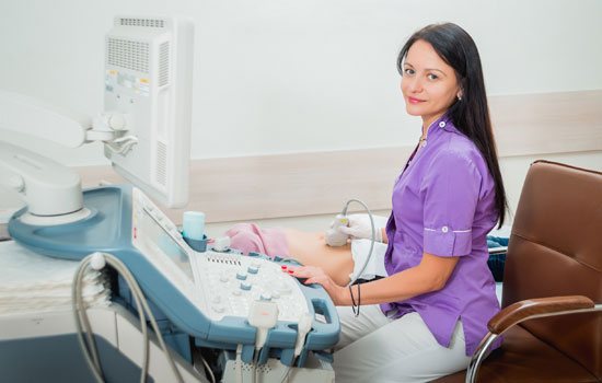
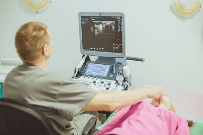
An ultrasound diagnostic specialist can work either alone or with a nurse. If the doctor practices independently, then he has to carry out all the preparation for the upcoming studies himself. First, connect the ultrasound machine and set it up to work. Secondly, control the availability of all consumables, including the gel for the procedure, which is applied to the skin at the test site and along which the sensor moves. A specialized gel (for example, Mediagel ultrasound gel) is necessary to improve the conductivity of sound waves. After monitoring the preliminary procedures, the doctor begins performing an ultrasound.
Ultrasound in gynecology
It is prescribed when necessary to examine the internal organs and pelvic organs - the uterus, ovaries, fallopian tubes, as well as to diagnose pregnancy and the condition of the fetus (at different stages).
An ultrasound examination provides a complete picture of the condition of the female genital organs (uterus, endometrium, ovaries, fallopian tubes). The size of all organs, the presence of tumors in them, blood flow and other characteristics are assessed. Transabdominal ultrasound is recommended as a primary examination and is also performed in the II-III trimesters of pregnancy.
When is an ultrasound performed at an appointment with a gynecologist?
Urgently, on the day of visiting the doctor
- for pain in the lower (and sometimes upper) abdomen, for bleeding (not menstrual), in the presence of pathologies identified during a gynecological examination, cycle disorders.
An ultrasound scan by a gynecologist is also performed as planned.
- during medical examination, in the process of pregnancy planning and preparation for conception, during pregnancy diagnosis, to monitor fetal development.
On what day of the cycle is an ultrasound performed?
- For standard diagnostic ultrasound, cycle days 5-7 are recommended.
- When tracking ovulation, ultrasound is performed several times per cycle, the days of the study are determined by the doctor (calculated depending on the length of the cycle and the timing of the release of a mature egg).
- To diagnose pregnancy, an ultrasound is done 1-2 weeks after the delay (depending on the equipment and technique used).
- During menstrual bleeding, ultrasound is not performed, but if the bleeding is not menstrual, diagnosis is urgent.
- For symptoms of inflammation of the female genital organs, an ultrasound is performed on any day of the cycle.
- To assess the condition of the endometrium, diagnostics are carried out in the first and second phases of the cycle.
- For various diseases of the female genital organs, the ultrasound schedule is determined by the doctor (for ovarian cysts, ovarian dysfunction, after an abortion, etc.).
Why is it important to have an ultrasound performed by a medical specialist?
A gynecologist who conducts both a routine appointment and an ultrasound scan evaluates the patient’s condition comprehensively: taking into account symptoms, existing diseases, and the woman’s condition. After a gynecological appointment, she specifically examines certain organs of the reproductive system and clarifies the diagnosis (if it has already been made).
A specialist doctor with an ultrasound certificate is able to identify pathology without additional tests and studies. In addition, performing an ultrasound directly in the gynecologist’s office, during the initial appointment, significantly saves the patient’s time and allows for a diagnosis and treatment to be prescribed as quickly as possible.
The TN Clinic employs gynecologists certified to perform ultrasound. The presence in the clinic of high-tech expert-class equipment and qualified specialists creates all the conditions for accurate and quick diagnosis of any gynecological diseases right at the reception, in the gynecologist’s office.
Validity period for ultrasound results
Scheduled ultrasound examinations by a gynecologist in the absence of pathologies - 6 months.
Planned ultrasound of the abdominal organs, including the urinary system and heart - also 6 months.
Planned ultrasound of the thyroid gland, breast and other organs - 12 months.
, etc.)
conducting a new examination, as it is necessary to establish why the symptoms arose.
Before surgery on the female genital organs or hospitalization for treatment, it is also necessary
conducting a new ultrasound (valid for no more than 1 month), which is associated with the rapid development of certain diseases and tumors. Doctors need fresh results to assess the scope of a planned operation or treatment.
When visiting a doctor with any health problem and symptoms, the results of past ultrasounds are only relevant as part of the medical history.
Carrying out a new ultrasound is due to several factors:
- assessment of the condition of a particular organ at the moment (which cannot be done based on the results of an ultrasound scan performed 1, 3.6, 12 months ago or more);
- Ultrasound using equipment of a certain class, directly by the doctor caring for a specific patient (it is impossible to determine the accuracy of the results of an ultrasound performed by another doctor);
- some diseases develop very quickly, so ultrasound allows you to assess tumor growth, the amount of inflammation, etc.;
- Without fresh ultrasound results, accurate diagnosis and treatment are impossible.
What organs does the ultrasound specialist examine?
This specialist examines a wide variety of human organs, so he is indispensable and important in all branches of medicine. Today the most requested research is:
- abdominal organs;
- hearts;
- kidney;
- Bladder;
- pelvic organs;
- scrotum;
- uterus and ovaries;
- joints;
- veins and arteries of the lower extremities;
- brachiocephalic arteries;
- skulls;
- thyroid gland;
- salivary glands;
- lymph nodes;
- fetus;
- soft tissues.
Every ultrasound specialist should know what exactly all these structures and organs should be like under normal conditions and in pathology. All results recorded in a special form are handed over to the patient, after which they are transferred to the leading doctor.
Ultrasound services at GMS Hospital
All the most popular and informative studies are carried out in the ultrasound diagnostic rooms of GMS Hospital, including:
- color duplex scanning of blood vessels (USD);
- Ultrasound of internal organs - liver, pancreas, kidneys, gallbladder, spleen, mammary glands, uterus and fallopian tubes, ovaries, heart, joints, lymph nodes, etc.;
- Ultrasound during pregnancy;
- Ultrasound in pediatrics (including neurosonography and examination of the hip joints);
- Cavity ultrasound examinations (TRUS and TVUS)
Where can an ultrasound doctor work?
Today, the specialty of an ultrasound diagnostic doctor is in great demand due to the high role of the research obtained. Many clinics in all cities of Russia need such a specialist. An ultrasound diagnostic doctor can work in both private and public medical centers. Typically, work in establishments of the first type is paid higher. In addition to the outpatient clinic network, an ultrasound diagnostic doctor can also work in hospitals. Doctors with the most experience in ultrasound diagnostics conduct research in scientific and practical centers. The professional path to such institutions is long and usually takes place over many years of practice. Research centers recruit doctors of the first and highest qualification categories.
Requirements for an ultrasound doctor
Basic requirements for an ultrasound physician include:
- Higher medical education, current accreditation certificate for ultrasound diagnostics.
- Ability to perform ultrasound and echocardiography. Decryption experience.
- Possession of skills in diagnosing emergency conditions.
- Ability to work in the clinic’s unified information system.
Ultrasound allows you to accurately determine the location of the tumor, the stage of its development and metastasis.
Institutions for professional development
"Russian Association of Ultrasound Diagnostics in Medicine"
This organization unites practicing ultrasound specialists throughout Russia. The association was formed more than ten years ago with the goal of promoting the professional development of ultrasound physicians. In order to become a member of this organization, you need to submit an application, pay an entrance fee and then make a small annual payment. Joining the Association is very beneficial for any ultrasound doctor, since the company constantly holds thematic conferences, seminars and webinars that enhance professional development. Currently, the Association has several thousand members.
Medical cooperation
Ultrasound diagnostic specialists, like doctors in other fields, organize educational congresses to improve their qualifications and professional level. Most often, such events are held in the form of congresses, sometimes they are even held on an international scale. They are aimed at exchanging experiences between doctors, reading reports, discussing interesting and controversial cases in practice, stimulating and encouraging certain representatives of the profession. Doctors whose report was the most useful and interesting in the field of ultrasound development are awarded. Such events are very important for the training of medical personnel.
Benefits of Ultrasound
The ultrasound method has many advantages, namely:
- relatively low price;
- good information content;
- no contraindications;
- safety;
- the opportunity to examine almost all internal organs and systems of the body;
- speed of the procedure;
- monitoring of structural changes in tissues in real time.
Despite the many advantages of ultrasound, it is performed exclusively as prescribed by a specialized healthcare professional.
The advantages of ultrasound are obvious:
this is the ability to visualize soft tissues and parenchyma when examining organs such as the liver, gall bladder, kidneys, pancreas, thyroid and mammary glands, blood vessels and heart, fetus. Considering the fact that ultrasound is a non-radiation method and relatively inexpensive, studies of these organs can be carried out frequently, which helps study the dynamics of treatment.
Thanks to high quality and computerization, doctors performing ultrasound in Kyiv can evaluate morphological changes in many organs. Due to this, specialists have the opportunity to clarify the size of pathological formations, as well as determine the involvement of “neighboring” organs in the pathological process. Ultrasound machines in Kyiv are equipped with color printers, which can be used to obtain images of organs. This allows you to see any changes and make a comparative description of all data during the study of the organ. Modern ultrasound machines have sensors of various directions, with the help of which a wide range of studies are performed.
When is an ultrasound performed?
Diseases of the liver, gall bladder, pancreas, spleen. Using ultrasound, you can determine structural changes in these organs, determine their size, the presence of an inflammatory process, determine blood flow and the presence of pathological formations. Ultrasound makes it possible to study the gallbladder, determine the presence of stones, polyps, and the presence of various deformations in the form of constrictions, bends. Ultrasound also allows us to determine the presence of free fluid in the abdominal cavity.
Kidney diseases . An ultrasound scan can detect stones in the kidneys, in the lumen of the ureters, and in the bladder. It is possible to determine the presence of congenital anomalies of kidney development (incomplete duplication of the renal-pelvic sinus, kidney dysplasia), the presence of cysts, tumors, and also to diagnose nephroptosis - prolapse of the kidneys.
Ultrasound of the adrenal glands. The spectrum of adrenal pathology includes: adrenal tumors, cysts, adrenal hyperplasia, inflammatory changes, dyscirculatory changes, hematomas.
Thyroid diseases. Using ultrasound, you can determine the size of the thyroid gland, its structure, the presence of nodes, cysts, space-occupying formations, and also evaluate the blood flow in the gland.
Diseases of the mammary glands. Using ultrasound, we can determine the structure of the gland tissue, the presence of pathological space-occupying formations, such as cysts, fibroadenomas, lipomas, and malignant formations. Thanks to ultrasound, we are able to assess the integrity of implants, locate blood flow and examine lymph nodes.
Using ultrasound, we can determine the presence of pathological processes in soft tissues (muscles) and diagnose the nature of subcutaneous formations (lipoma, hematoma).
Gynecological diseases. Ultrasound is used to diagnose pregnancy, inflammatory processes, uterine fibroids, the presence of cysts and neoplasms in the ovaries. Ultrasound monitoring is carried out to monitor the growth of follicles in the ovaries, the ovulation process, determine the thickness of the endometrium (uterine lining), and diagnose polyps and cysts on the cervix.
Ultrasound during pregnancy. It gives us the opportunity to have a fact of pregnancy, a suspicion of an ectopic pregnancy, failure to carry the pregnancy to term, and the further development of the fetus. Using ultrasound, we can perform fetal biometry (size measurement), as well as assess the vital activity of the embryo, and study extraembryonic formations.
Prostate diseases. Ultrasound allows us to determine the size of the gland, study the structure, and the presence of additional focal changes in the prostate gland. Using ultrasound, we are able to identify inflammatory diseases (acute, chronic), as well as benign (adenoma) and malignant formations of the prostate gland.
Diseases of the seminal vesicles . With ultrasound of the seminal vesicles, we can evaluate their shape, size, symmetry, and structure. Ultrasound data allows us to suspect the presence of fibrosis, chronic inflammation, obstruction, and a tumor process.
Urinary tract diseases. By performing an ultrasound examination, we can determine the presence of stones in the bladder or in the lumen of the ureters. It is possible to identify tumor processes in the bladder.
Ultrasound in vascular diseases. When performing an ultrasound examination of the vessels of the lower extremities, we can determine the presence of blood flow in a particular vessel, the presence of blood clots, and varicose veins. Ultrasound also helps us determine the presence of atherosclerotic changes in blood vessels.
With ultrasound of the brachiocephalic region (in other words, Dopplerography of the vessels of the head and neck), we can determine the type and speed of blood flow, the presence of vascular tortuosity, the presence of atherosclerotic plaques, and identify the degree of vascular stenosis.
Ultrasound of joints. Diagnostics in this area makes it possible to visualize cartilaginous, soft tissue structures, and a minimal amount of fluid in the joint cavities and bags. Thanks to ultrasound, we can determine the integrity of the discs and menisci, ligaments, which happens in various traumatic, degenerative and inflammatory processes.
Using ultrasound, you can identify a great variety of diseases. Ultrasound data allows the doctor to obtain additional information to confirm the presence of the disease, as well as to see the extent of organ damage in some diseases.
But you need to remember (!) that if the patient is not properly prepared, then the quality of the ultrasound is significantly reduced, and in some cases it even comes down (!) to a negative result. If a pathological formation is detected and differential diagnosis is difficult, patients are referred to additional examination methods: x-ray diagnostics, computed tomography, magnetic resonance imaging, mammography.
Contraindications for ultrasound are: open wound surface, fistulas and applied bandages at the study sites.