Home — For the public
- Map of medical organizations
- Vaccination
- Clinical examination
- Fluorography
- Addresses and opening hours of clinics
- Emergency rooms
- Oncology
- Where to take an HIV test
- Healthy child's office
- Services
- Prevention of CVD
- Disease Prevention
- World Patient Safety Day
- Newspaper "Medical News"
- specialist
- School of Health
— Disease prevention
- HIV infection
- All about vaccination
- All about proper nutrition
- Hepatitis
- Flu
- Dementia
- Schoolchildren's health
- STD
- Tick-borne encephalitis
- Whooping cough
- Measles
- Legionellosis
- Meningococcal infection
- Oncology
- Acute intestinal infection
- Pediculosis
- First aid
- Pneumococcal infection
- Pneumonia
- Prevention of rabies
- Dependency Prevention
- Rotavirus infection
- Diabetes
- Cardiovascular diseases
- Injuries
- Tuberculosis
- Tularemia
- Physical activity
- Obstructive pulmonary disease
- Exotic infections
- Ecology
- Why is swimming in ponds dangerous?
— Tuberculosis — Frequently asked questions about tuberculosis
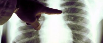
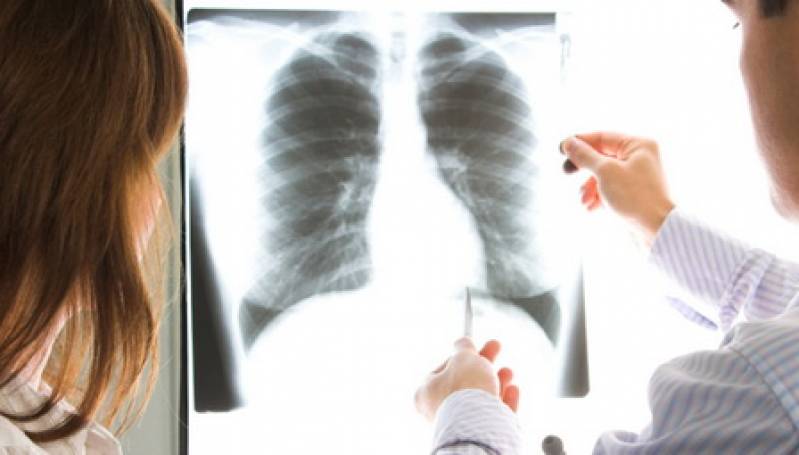
What is fluorography and why is it needed?
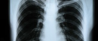
Fluorography is a study of the chest organs performed using x-rays passing through the body and, due to uneven absorption, producing a visible image on a fluorescent screen. Fluorography is one of the main methods for detecting tuberculosis in health care institutions of the general medical network.
Conducting continuous fluorographic examinations of the population ensures early detection of tuberculosis and a sharp reduction in advanced and widespread forms of tuberculosis. Fluorographic examinations are carried out using stationary fluorographic machines installed in clinics or on mobile fluorographic units.
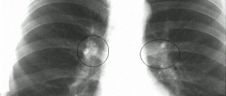
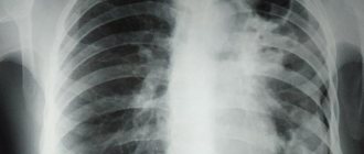
Forms of tuberculosis and their signs
Pulmonary tuberculosis is not clearly visible on fluorography, so the doctor orders an x-ray of the lungs.
X-rays reveal several types of pathology, which are accompanied by various signs. Forms of tuberculosis:
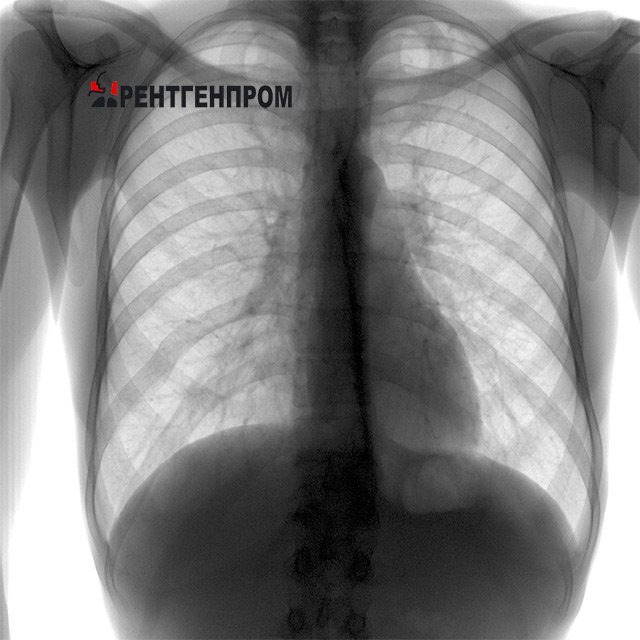
Compaction syndrome. Low to medium density lesion. Tuberculosis.
- tuberculosis of the intrathoracic lymph nodes - manifests itself as an expansion of the root of the lung, the contours are blurred and heterogeneous, connective tissue cords and the presence of calcifications are noticeable;
- disseminated tuberculosis - characterized by multiple areas of darkening. Such areas are no more than 2 mm, so it can be difficult to see. They have clear boundaries and are scattered throughout the lung, and as the disease progresses they merge into large foci;
- focal tuberculosis is the formation of darkening from one to two centimeters, has clear boundaries and is localized in any part of the lung. When the disease worsens, the lesions merge with each other;
- infiltrative - signs of tuberculosis are visualized as whitish formations with jagged edges. It is found in the upper lobes of the lung and has characteristic tracks;
- caseous form of tuberculosis is one of the severe forms. In the picture, the pathological area occupies almost the entire lung. The darkening is uniform, but changes as the disease progresses;
- cavernous - with tuberculosis, a cavity appears with a focus of decay, which is visible as a dark neoplasm with a light spot inside;
- fibrous-cavernous - in addition to the characteristic cavern, areas of fibrosis become noticeable, the pulmonary pattern is deformed, displacement of organs may be noted;
- cirrhotic - darkening in one or both lobes. The lungs are reduced in volume, and the root of the lung is close to the site of pathology;
- tuberculous pleurisy - a disease with typical signs of pathology in the lower lobe of the lung; effusion pleurisy is determined on the image;
- miliary - multiple lesions that overlap each other. Distributed evenly in the two lungs.
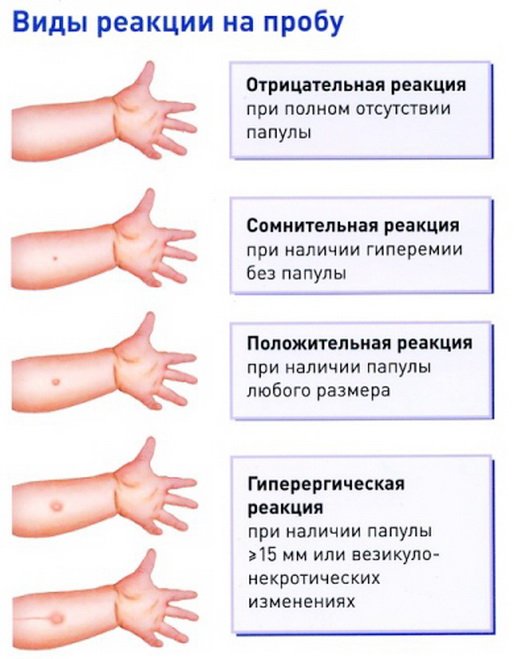
What is Diaskintest?
The Diaskintest test is used as a skin test in the diagnosis of tuberculosis and, unlike the Mantoux test, is highly sensitive in determining a probable tuberculosis infection, including in the initial (latent) stage. The technique for performing and recording the results of this test is identical to the Mantoux test. Diaskintest is used in the age group from 7 to 14 years, in children at risk, which helps reduce the number of cases of late detection of tuberculosis.
For adults, fluorography is prescribed to determine a probable tuberculosis infection.
Contraindications
Digital fluorography is a safe procedure and does not pose any risks to human health.
It's all about the dose of radiation received. During the examination, the patient does not receive a high load, and the radiation is eliminated from the body over time. If the body receives a high load, then pathological processes can begin at the cellular level and the person faces unpleasant consequences, including the development of oncology. For such consequences it is necessary to obtain extremely high radiation, which is simply impossible in a fluorography room.
Fluorography is not performed:
- children under fifteen years of age - the Mantoux test in children is sufficient to confirm the absence of a tuberculosis agent;
- pregnant women and nursing mothers;
- bedridden patients who are unable to support their body vertically;
- patients with respiratory failure.
Other studies are carried out for patients in these categories, and fluorography is done for pregnant women after childbirth.
Magnetic resonance examination
The role of magnetic resonance in the diagnosis of lung diseases is being investigated. This method is increasingly used to examine patients. It provides sufficient contrast between the fatty tissue of the mediastinum, dense formations and vascular structures, allowing the identification of lesions without the introduction of a contrast agent. The disadvantages of magnetic resonance are associated with the inability to determine calcification and the short duration of the image of the lungs, which significantly limits the obtaining of information about their parenchyma.
Diagnosis of lung cancer using FLG
Lung cancer is detected by fluorography images only in some cases.
- Significant size of formations. The images clearly localize the problem area, which usually indicates the last stages of the presence of cancer or the aggravated condition of the patient’s body.
- Location of cancer formation on the surface.
Important! Detecting lung cancer in the early stages is not an easy task, due to the almost complete absence of symptoms. At the initial stage, the disease manifests itself only by the germination of oncological formations of the bronchi, pleura, and blood vessels (or the tumor, due to its growth, begins to compress the tissues surrounding it)
Therefore, the patient does not seek medical help until a clinical picture begins to emerge.
At the initial stage, the disease manifests itself only by the germination of oncological formations of the bronchi, pleura, and blood vessels (or the tumor, due to its growth, begins to compress the tissues surrounding it). Therefore, the patient does not seek medical help until a clinical picture begins to emerge.
Symptoms of tuberculosis
Pulmonary tuberculosis can occur in any person without exception. It appears due to infection with Koch's bacillus. A distinctive feature of this pathogen is that it can remain in the open air for a long time, being resistant to environmental influences.
Before doing chest fluorography, a person needs to know what symptoms may indicate infection with Koch's bacillus. First of all, you should pay attention to such possible signs of the disease as:
- Dyspnea. Most often with tuberculosis it occurs even with minor physical exertion.
- Cough. If it occurs continuously over a long period of time, you should definitely visit a medical facility.
- Wheezing. They are a sign of the presence of an infiltrate in the lungs or any other pathological changes in the structure.
We recommend reading! Follow the link: Pneumothorax of the lung on x-ray diagnostic methods

It is also necessary to pay attention to the occurrence of low-grade fever in the range of 37-37.3 degrees Celsius. If you have several of the above symptoms, you should definitely make an appointment with a pulmonologist or TB specialist.
Radiography
The method makes it possible to photograph pathological changes in the lungs. X-ray examination begins with a survey image in the anterior direct projection. A direct chest x-ray is a photograph in which the central beam passes along the midsagittal plane of the patient's body. All compactions of the lung tissue are visible on the radiograph, but the transparent lung fields are dark.
Radiography in a direct projection is sometimes supplemented with studies in a lateral projection, which significantly expands the understanding of pathological changes in the lungs: the localization of the latter relative to the lobes and segments, their size, and connection with the root of the lung is clarified. By comparing radiographs during treatment, one can judge its effectiveness.
Sight radiography
This method allows you to clarify the nature of changes in the lungs. It is carried out in cases where it is necessary to detail the nature of pathological changes.
Electroradiography (xerography)
The image is obtained on paper using an electroradiographic attachment connected to any X-ray machine. This method achieves high contrast of various formations in the lungs - foci of varying density, cavities, round formations, walls of the trachea and bronchi.
X-ray examination of the lungs is rarely performed using devices with an electron-optical converter (EOC). This method is used for certain indications: control during targeted images, bronchographic, angiographic studies, fistulo- and pleurography. This method also allows you to examine the patient in various positions to identify changes at the apexes of the lungs, behind the shadow of the heart and diaphragm, in the area of the pleural sinuses.
Fluoroscopy allows you to clarify the localization of pathological changes, their mobility and connection with the chest wall, mediastinal organs, determine the mobility of the diaphragm, and the condition of the pleural sinuses. But significant radiation exposure and lack of research documentation reduce the advantages of this method.
Nuances of diagnostics
The effectiveness of the procedure depends on compliance with all the rules for conducting the study, as well as the correct interpretation of the results.
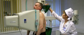
Fluorography consists of the following stages:
- To begin with, a person must expose his torso. All jewelry must be removed, and hair must be secured with an elastic band or hairpin.
- A lead apron is hung on the belt. This is done to minimize the impact of radioactive rays.
- The doctor adjusts the screen according to the patient's height. He, in turn, presses his chest tightly against him with his shoulders brought forward.
- Do not breathe or move while the device is operating. The specialist warns when to freeze, after which the person returns to his usual rhythm.
- Next, the patient gets dressed. The diagnostic result is obtained by the doctor in a few minutes or days, depending on the device. Based on it, a conclusion is drawn describing the state of the respiratory system.
Following the technique of the procedure makes it possible to obtain the most accurate result. Otherwise, you can observe image distortion and a blurry picture, which requires repeating digital fluorography. This fact is undesirable, since large doses of radiation are dangerous to human health.
If the patient has concomitant diseases or disabilities, the procedure becomes more difficult. It all depends on the individual characteristics of the body and the health of the person being examined. In cases where the patient constantly coughs and cannot stabilize the chest, the effectiveness of the procedure is sharply reduced.
General information about the disease and procedure
The bacterium that provokes the development of tuberculosis was discovered by R. Koch in the 80s of the 19th century and named after him. Tuberculosis is transmitted by airborne droplets, household and other common routes. The main specificity of the virus is its extremely high resistance to external conditions and ability to adapt.
A person of any gender, age, place of residence, or social status can become infected. Without proper treatment, bacteria will fill all respiratory tracts, and the infected person will suffocate.
Certain areas or population categories constitute a special risk group:
- prisoners or released from prison;
- military;
- aged people;
- people without a fixed place of residence;
- people with addictions and immunodeficiency.
For these categories, a mobile fluorography unit is of particular value. This is especially true for villages where an infected person lives, and the rest are in contact with him in any case. After all, bacteria can live for a long time in dust, on door handles, and in the air.
The tricky part of the pathology is that at the stages when the initial external manifestations have arisen, it is much easier to diagnose tuberculosis, but it is more difficult to cure, and in a number of particularly advanced cases, it is impossible. The disease develops and remains asymptomatic for a long time. And the incubation period can reach several years. This is why it is so important to carry out regular diagnostics.