Pulmonologist
Prokhina
Maria Egorovna
Experience 38 years
Pulmonologist
Make an appointment
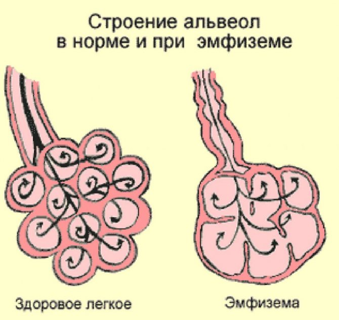
Pulmonary emphysema is associated with impaired gas exchange and pathological expansion of air spaces against the background of significant morphological changes in the tissues of the alveoli. These reasons give the characteristic appearance of the lungs: they swell, acquire a pale tint, the edges are rounded, and the elasticity of the tissues is reduced. It is possible that plugs with mucous or purulent contents may form in the bronchi.
Causes
Factors that can cause this disease are divided into two groups:
- The first group includes phenomena that contribute to the loss of tissue elasticity and strength. These are pathological microcirculation, the consequences of inhaling tobacco smoke, birth defects, severe pollution of the inhaled air and the presence of caustic or aggressive gaseous substances;
- The second group includes factors that provoke an increase in pressure in the respiratory part of the lungs. At the same time, there is a stretching of the alveoli, an increase in the size of the bronchioles and alveolar ducts. This also includes chronic obstructive bronchitis, which creates favorable conditions for the development of the disease. There is a high probability of the inflammatory process spreading to the alveoli with the subsequent development of alveolitis and destruction of the partitions separating them.
Prevention
Prevention of pulmonary emphysema involves minimizing factors influencing the development of the disease:
- to give up smoking;
- prevention and timely treatment of lung diseases;
- eliminating exposure to harmful substances on the lungs;
- general strengthening of the immune system.
It is extremely important to start treatment as early as possible, that is, even at the stage of development of the primary disease. Therefore, if you are sick, you should not postpone going to the doctor.
Medical experts warn that lung diseases should not be self-medicated. If you suspect that you or your loved ones have pulmonary emphysema, consult a doctor as soon as possible so that a specialist can make the correct diagnosis and prescribe adequate treatment.
Symptoms
Most symptoms of emphysema are associated with shortness of breath and coughing up sputum, which are caused by pathological changes in the tissues of the lungs.
- At the initial stage of emphysema, shortness of breath appears only with intense physical exertion and active movement, and the cough contains a small amount of mucous secretions.
- At the second stage, or stage of pulmonary failure, the patient experiences severe shortness of breath, cough with a small amount of sputum, changes in the shape of the fingers, hemoptysis, and noticeable cyanosis of the face and extremities.
- At the third stage, called the stage of pulmonary heart failure, severe shortness of breath and cough with a large amount of purulent mucous discharge are noted. Due to constant tension in the patient's throat, the neck veins and liver size increase, swelling is noticeable, and venous pressure increases. The face becomes swollen, the chest takes on a rounded, convex shape.
Often, the signs of pulmonary emphysema can be used to judge the nature of the disease:
- if bronchial obstruction is impaired, patients complain of shortness of breath, dry cough, feeling of lack of air, wheezing and whistling when breathing, forced participation of the abdominal and shoulder muscles in inhalation and exhalation;
- with an increase in pulmonary volume, patients experience expansion of the chest, weakening of breathing, protrusion of the intercostal spaces and a characteristic “box” sound when breathing;
- in persons with respiratory failure, shortness of breath, skin cyanosis, deformation of the fingers, enlargement of the right ventricle of the heart, tachycardia, swelling of the neck veins, shallow breathing with a short sigh and a long exhalation are noted.
Complications of emphysema
Rapid progression of the disease without treatment can cause serious complications. This:
- pneumosclerosis;
- pulmonary dysfunction, making full breathing impossible;
- preumothorax;
- circulatory disorders;
- gas exchange problems causing changes in the kidneys, liver and other organs.
Are you experiencing symptoms of emphysema?
Only a doctor can accurately diagnose the disease. Don't delay your consultation - call
Emphysema
Pulmonary emphysema (Emphysema pulmonum) is an animal disease characterized by pathological expansion and increase in the volume of alveoli or accumulation of air in the interalveolar connective tissue.
There are alveolar (acute and chronic) emphysema. When lung tissue ruptures and air enters the interlobular connective tissue, interstitial pulmonary emphysema develops. Most often, alveolar emphysema is observed in horses, hunting and sled dogs; interstitial emphysema is usually recorded in cattle.
Alveolar pulmonary emphysema (Emphysema pulmonum alveolarae) can be local (local), developing as a compensatory phenomenon for abscesses, bronchopneumonia and other lung lesions, when healthy areas of the lungs are overstretched with increased breathing, and general, involving the entire lung.
Etiology . Acute alveolar emphysema in animals occurs when the alveolar tissue is overstrained during frequent and intense breathing, when the pulmonary alveoli, filled to the limit with air, are strongly compressed during exhalation.
This kind of overexertion occurs during prolonged running of horses at sporting competitions, during intensive and long-term work of working horses without rest, during excessive use of sled dogs and hunting dogs, when the elastic properties of the lung tissue are weakened, the alveoli are stretched and enlarged. At the same time, the lung tissue retains its structure and only increases in volume.
Chronic alveolar emphysema develops as a continuation of acute emphysema, if the animal has not fully recovered, and etiological factors continue to affect the lung tissue. Most often, chronic alveolar emphysema in animals develops as a secondary disease in chronic diffuse bronchitis, peribronchitis and bronchospasms (asthmatic bronchitis, bronchial asthma). The disease is also recorded with stenosis of the larynx, trachea and bronchi, chronic pneumonia and pleurisy. Allergic factors play a certain role in the occurrence and development of alveolar pulmonary emphysema (inhalation of mold spores by horses, plant pollen by dogs, etc.).
The cause of interstitial emphysema is the penetration of air into the interlobular connective tissue when the walls of the bronchi and caverns rupture during excessive physical exertion. In cattle, it occurs when the lungs are injured by foreign piercing objects from the proventriculus (mesh).
Predisposing causes of pulmonary emphysema are: general weakening of the body, fatigue, cardiovascular failure, kidney disease and central nervous system.
The development of the disease is facilitated by a lack of vitamins, microelements (cobalt, iodine, manganese), carbohydrates in the body, a long-term lack of exercise, and an excess of nickel and silicon oxide in feed.
Pathogenesis . Primary in the process is irritation of bronchial interreceptors by infectious and mycotic factors, cold, and chemicals. These irritations coming from the interoreceptors of the bronchi, especially small ones, lead to a breakdown of nervous regulation in them, leading to a number of functional disorders of the neuromuscular apparatus of the bronchi and trophic processes in the lungs. The emerging functional disorders in the lungs of animals are especially typical for the initial forms of emphysema and are manifested by symptoms of spasm of the small bronchi, leading to a decrease in the functional capacity of the lungs, an increase in expiratory activity and, possibly, a change in the production of histamine by the lung tissue. Under the influence of the above factors, the alveoli can expand 5-15 times more than their normal volume. The existing functional disorders gradually turn into anatomical ones. At this time, the exhalation activity of the sick animal increases even more. Stretched alveolar walls, squeezing the capillaries embedded in them, create difficulties for blood circulation and gas exchange in the lungs, leading to congestion in the pulmonary circulation, causing congestive catarrh in the bronchi in the animal. In chronic pulmonary emphysema, prolonged stretching of the walls of the alveoli leads to the development of atrophic processes in them with profound morphological changes, up to rupture of the alveoli. Catarrhal phenomena cause swelling of the bronchial mucosa, causing its swelling and leading to a narrowing of the bronchial lumen. All this makes breathing even more difficult, increasing the animal's shortness of breath. If the walls of the alveoli rupture, air can penetrate into the interstitial tissue and under the skin. The resulting difficulties in blood circulation in the pulmonary circle lead to hypertrophy of the right ventricle of the heart. If there is insufficient compensation from the right ventricle of the heart, the animal may experience symptoms of heart failure. Thus, with pulmonary emphysema, pulmonary respiration is impaired, oxidative and metabolic processes in the body of the sick animal are disrupted. Clinically, these processes are manifested by a decrease in the alkaline reserve of the blood. With chronic pulmonary emphysema, these processes in the body are usually irreversible.
Pathological changes. With total emphysema, the lungs are enlarged in volume, swollen, fluffy, their elasticity is reduced, pale pink in color, occupies the entire pleural cavity and imprints of the ribs can be seen on its surface, a slight cracking sound (crepitus) is heard when palpated, the pit does not level out from pressure for a long time, the cut surface is dry.
Chronic alveolar emphysema, when autopsied in a dead animal, is characterized by bubbles in the lungs filled with air, sometimes quite large - up to the size of a pigeon's egg (bullous form). The chest is enlarged (barrel-shaped). In the heart there is hypertrophy of the right ventricle, and with decompensation - its expansion.
Interstitial emphysema is expressed by the accumulation of air in the interlobular connective tissue and under the pleura. When opening a corpse, we find multiple thin-walled air bubbles or chains of them, easily moving when stroking or palpating. Interstitial emphysema is more common in horses, cattle and dogs.
The altered bronchi contain mucous-purulent plugs.
Clinical signs . In patients with acute alveolar emphysema, we register rapid fatigue even after slight physical exertion. Severe shortness of breath is characteristic: during breathing, sharp movements of the costal walls and abdominals are visually visible, the animal's nostrils are dilated, the anus protrudes when exhaling, sometimes breathing is accompanied by groans, dogs breathe with their mouths open. With careful observation, you can notice a prolongation of the expiratory phase. A little driving of the animal sharply enhances these signs. During auscultation in the anterior parts of the lungs, hard vesicular breathing is usually heard, with severe bronchitis - increased, hard vesicular breathing and dry melodious, less often moist rales, as well as mixed shortness of breath.
We often notice a dull, short and weak cough typical of emphysema.
Percussion of the pulmonary field produces a loud box sound. We note a symptom characteristic of alveolar emphysema - a shift of the caudal border of the lungs back by 1-2 ribs; in some sick animals this border extends beyond the last rib. The area of cardiac dullness decreases. Body temperature is normal and in rare cases subfebrile.
In most sick animals, we register a compensatory increase in cardiac activity: increased heart rate, increased heart sounds, especially the second (diastolic) in the pulmonary artery. If the course of the disease is favorable, after eliminating the animal’s physical stress and giving it rest, the symptoms of acute alveolar emphysema may disappear within a few days.
With chronic alveolar emphysema, there is a gradual increase in characteristic expiratory shortness of breath. The exhalation of a sick animal becomes tense and prolonged. It occurs in two phases: first, there is a rapid collapse of the chest, and then after a short period of time we notice a powerful contraction of the abdominal wall. During exhalation, retraction of the intercostal spaces is noticeable, at the border of the chest and abdominal walls there is a clearly defined depression (ignition groove) and protrusion of the anus. Despite the intense breathing, the stream of air exhaled by the animal is weak. In horses suffering from chronic alveolar emphysema for a long time, the chest becomes barrel-shaped. During percussion, a loud box sound is clearly detected throughout the entire pulmonary field; the percussion border of the lungs is pushed back 1-4 intercostal spaces. When auscultating the lungs, weakened vesicular breathing, weakening of the cardiac impulse, increased diastolic heart sounds, and increased heart rate are heard. After physical exertion, the symptoms of expiratory dyspnea in a sick animal sharply intensify.
Interstitial emphysema is usually characterized by an acute and rapid course of the disease. When air penetrates into the interlobular connective tissue of a sick animal, a sharp deterioration in the general condition occurs with an increase in signs of asphyxia: progressive shortness of breath, cyanosis of the mucous membranes, and symptoms of cardiovascular failure appear. When auscultating the lungs, fine bubbling rales and the sound of crepitus are heard. Under the skin in the neck, chest, sometimes back and croup, upon palpation we detect crepitus of air bubbles (subcutaneous emphysema). The body takes on a puffy outline.
By performing an X-ray examination of the lungs, we establish clearing of the pulmonary fields and moderate mobility of the diaphragm displaced back. In emphysema with clear symptoms of peribronchitis and pneumosclerosis, the shadow of the diaphragm has a convexity in the upper part, facing backward, and in the lower part, a convexity facing forward. The excursions of the diaphragm are unevenly wavy, with the greatest sweeps in the lower convex part. The chyle pattern is enhanced.
The diagnosis is made based on the history and clinical symptoms of the disease. X-ray examination reveals clearing of the pulmonary field in emphysematous areas of the lungs, increased bronchial pattern, and backward displacement of the dome of the diaphragm. In horses, a compensatory increase in the number of red blood cells and the amount of hemoglobin in the blood is often recorded, and in some animals - eosinophilia and monocytosis.
Differential diagnosis . When carrying out differential diagnosis, veterinary specialists need to exclude pneumonia, pleurisy, hydrothorax, hemothorax and pneumothorax.
Treatment . Animals with acute alveolar emphysema must be given rest and kept in a clean, well-ventilated area, in the shade in clean air in the summer. The diet consists of low-volume, easily digestible feed. To expand the lumen of the bronchi and reduce shortness of breath, a 0.1% solution of atropine (horses 10-15 ml per injection), a 5% solution of ephedrine (horses 10-15 ml per injection) is administered subcutaneously for 5-7 days in a row. or aminophylline (0.1-0.2 g per dog per dacha). In the presence of allergic factors, it is necessary to administer calcium chloride, sodium and potassium bromide, novocaine, chlorpromazine, propazine, suprastin, pipolfen. When carrying out complex drug treatment for sick animals, cardiac and general tonics (caffeine, cordiamine, strophanthin, glucose, etc.) are used.
If there is a threat of pulmonary edema in the animal, the animal is given bloodletting.
Patients with chronic alveolar emphysema, if they do not represent significant breeding and decorative value, are discarded or used with minimal physical activity. Drug treatment should be aimed at weakening or eliminating the consequences of chronic bronchitis, for which expectorants, inhalations, and iodine preparations are prescribed, as in the treatment of chronic bronchitis.
Treatment of animals with interstitial emphysema is carried out as for acute alveolar emphysema. In this case, the main treatment should be aimed at mitigating and eliminating cough in order to prevent further penetration of air into the interlobular connective tissue of the lungs and normalize the function of the cardiovascular system.
Prevention of pulmonary emphysema consists of proper use of draft animals (horses, oxen) during work. Long and fast gaits should not be allowed in horses; For transport animals, the draft force when carrying heavy loads must correspond to the state of health of their body and their endurance. Follow the established rules for training sports horses and dogs. During medical examination of animals, it is necessary to conduct a thorough examination of the lungs and bronchi; All animals with beginning symptoms of emphysema are freed from stress, treated, and then transferred to light work. It is important for animal owners to promptly prevent the occurrence of bronchitis, as they are often complicated by emphysema.
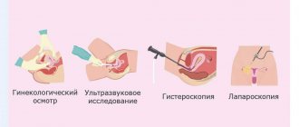
Diagnostic methods

An accurate diagnosis can be made to the patient by:
- X-ray, in which transparent areas filled with air are visible in the lungs;
- spirometry, which allows you to clarify the volume of inhaled and exhaled air. With emphysema, the second indicator is much greater than the first;
- peak flowmetry helps to clarify the expiratory flow rate when taking bronchodilators and without their use.
An additional method for diagnosing pulmonary emphysema is a blood test, which can be used to monitor the presence of an inflammatory process in the body and its intensity.
The set of necessary diagnostic measures depends on the condition of a particular patient and the need to detail the picture of his health.
What happens in the lungs?
The development of emphysema is associated with irreversible changes in the wall of the bronchi and lungs under the influence of prolonged inflammation and prolonged narrowing of the airways. The elastic properties of the lungs are disrupted: after exhalation, a larger amount of air begins to remain in them than should be normal, which causes overextension (inflation) of the lungs. Such excess air does not participate in breathing and the overstretched lung tissue does not function fully. Which, in turn, is accompanied by a loss of the ability to sufficiently contract and difficulty exhaling, as a result of which the supply of oxygen to the blood and the removal of carbon dioxide from it is disrupted. Compensatory, in order to improve the removal of carbon dioxide, shortness of breath occurs.
Read also: Top best hookahs for home 2018
Also in the bronchi and lungs, the amount of connective tissue begins to progressively increase, which, as it were, “replaces” the air areas of the lung tissue, and also contributes to a long-term narrowing of the bronchi, regardless of the existing inflammation.
As a result of these changes, numerous air sacs of different sizes are formed in the lungs, which can be scattered throughout the lung (diffuse form of emphysema). Sometimes swollen areas of the lungs are combined with normal lung tissue (local form of emphysema). Bullous emphysema is also distinguished separately (a bulla is an emphysematous (swollen) area more than 1 cm in size).
Treatment
The list of main objectives of the treatment course for pulmonary emphysema includes the elimination of complications, elimination of the infectious process and its symptoms, restoration of normal function of the lungs and respiratory tract. Therefore, when developing a course of treatment, preference is given to the following groups of drugs:
- compositions for restoring bronchial patency;
- drugs for removing phlegm;
- means of eliminating respiratory failure;
- medications to restore heart function;
- drugs to combat the associated infection.
Questions and answers
How is pulmonary emphysema treated?
Patients with emphysema are prescribed a conservative course of treatment, which allows them to cope with symptoms, ensure normal functioning of the respiratory tract and eliminate possible complications. The duration of treatment depends on the severity of the patient's condition and the symptomatic picture. It is possible to adjust the previously chosen tactics in the direction of adding or eliminating some of the medications taken.
Is there a cure for emphysema?
We can talk about the chances of curing the disease only on the basis of research results and timely seeking medical help. In the early stages, it is possible to almost completely eliminate the signs of pathology and return the patient to the ability to breathe fully. In advanced cases with numerous complications, it will not be possible to cope with the disease without consequences.
What is the main symptom of emphysema?
The disease is indicated by shortness of breath after physical exertion or without it (in the later stages), as well as a cough with a large volume of sputum with purulent or mucous contents. Other symptoms - wheezing and whistling, protrusion of the chest, puffiness and swelling of the face - may vary depending on the nature of the pathology.
In adults
Emphysema in adults develops mainly due to the harmful effects of environmental factors. And also as a result of the harmful effects of tobacco smoke. Smoking greatly complicates the disease process.
Emphysema is more common in men. The age category is most often from sixty years. It is in old age that all diseases worsen and this is due to the same influence of unfavorable factors.
In adults, as a result of chronic lung diseases, a severe obstructive process occurs in the pulmonary alveoli. What are the main signs of emphysema in adults? The main symptoms of the disease in adults include:
- cough:
- sputum production;
- body temperature may increase;
- swelling of the lower extremities;
- weight loss;
- weakness.
In adults, in the absence of proper treatment, the acute process of the disease passes into the chronic stage. The chronic stage of the disease leads to a long course and the development of complications. Respiratory and cardiac failure are often noted.
Diagnosis in adults contributes to early detection of the disease. Treatment with drug therapy can achieve good results. Surgical intervention helps to improve the disease process and even leads to recovery.
Read also: Pregnant woman smokes and drinks


