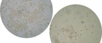
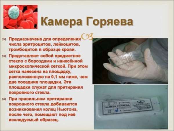
The Goryaev chamber is a device for measuring the number of cells and particles in a certain volume of liquid (blood, urine), consisting of a special thick slide and cover glass.
The slide has a recessed area in the middle, onto which a grid of 225 large squares is applied. Of these squares, 25 are evenly divided into 16 smaller squares. Thanks to this grid volume, the counting is performed more accurately than with other manual counting devices.
Camera maintenance
After using the instrument, the slide is disinfected by immersing it in a 4% formaldehyde solution for an hour or in a 70% ethyl alcohol solution for half an hour. After this, the chamber is thoroughly washed with distilled water. Excess liquid is removed with a soft cloth. It is worth paying attention to the fact that the camera cannot be wiped with a cotton swab because the fibers cling to it. Glass is stored in a dry place.
Egorov's rule
When using the camera, those FEs that are located inside the grid square and on the top and left lines of its perimeter are counted. In this case, particles touching the right and bottom lines of the perimeter are not included during the calculation of this cell.
Goryaev's camera and its characteristics
The device is made of thick glass, inside of which there is a recess.
It has a grid on it, visible only under a microscope, according to which the laboratory doctor counts the cells. Slides are placed on top of the chamber so that the material being examined is compressed on both sides. The Goryaev camera is produced in 2 versions:
- double-mesh (consists of 2 chambers);
- four-grid (consists of 4 chambers).
The price of the device depends on its quality. The manufacturer must polish the glass and apply the mesh correctly using a laser or vacuum deposition.
Goryaev's grid consists of 225 squares. They come in big and small. The doctor moves the microscope over them from left to right, down, then right to left until all fields are completed.
Maintenance of Goryaev's camera
In order for the device to show the correct results, it must be looked after. The camera must be thoroughly cleaned before use. To do this, use a bandage that is moistened with alcohol. The sterilizing liquid must be of high quality so that there is no sediment left on the surface of the glass. The glass is ready for use if it has rainbow rings on both surfaces.
Important! You should not use cotton wool to wipe the camera, as it contains fibers that may remain on the surface of the glass, making examination difficult.
Goryaev's camera comes standard with cover glasses. If they are missing or broken, it is possible to use regular glass of appropriate size.
When working with the camera and slides, the laboratory technician must wear gloves. This prevents fingerprints from getting in the way of the microscope.
After completion of work, the chamber must be disinfected. To do this, it is immersed entirely in a solution of ethyl alcohol. Disinfection time is 30 minutes. If another sterilizer is used, the time is extended to 1 hour. After completing this stage, the device is treated with water and wiped dry.
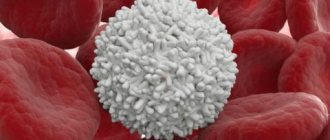
Determination of leukocytes in the blood
Method of counting leukocytes
Counting the number of leukocytes begins with diluting the blood in a 3-5% solution of ethanoic acid with methyl blue 20 times. The working material is wiped dry with gauze before use.
The cover slip must be wiped until colored Newton's rings appear. Next, the chamber is filled with diluted blood, with the first two drops released onto filter paper. Liquid enters the chamber due to the capillary property of water. It is important to ensure that nothing flows into the grooves when filling. If this happens, the liquid is carefully removed with filter paper. Then leave the grid alone for a minute, waiting for the FEs to stop their movement.
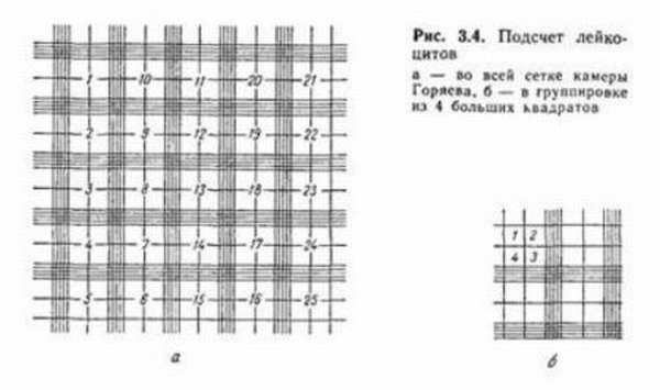
Leukocyte counting is carried out within one hundred large squares at low magnification, that is, with a 10x eyepiece and an 8x objective, according to Egorov’s rule. The resulting figure is used in the formula.
X = (n × 250 × 20) / 100 or X = n × 50
X – number of leukocytes.
n is the number obtained as a result of counting in the chamber.
The number of leukocytes is determined taking into account the number of large grid squares - 100 and blood dilution - 20.
Counting leukocytes in the Goryaev chamber - camera design and principle of counting indicators
The Goryaev chamber is a device for measuring the number of cells and particles in a certain volume of liquid (blood, urine), consisting of a special thick slide and cover glass.
The slide has a recessed area in the middle, onto which a grid of 225 large squares is applied. Of these squares, 25 are evenly divided into 16 smaller squares. Thanks to this grid volume, the counting is performed more accurately than with other manual counting devices.
Camera maintenance
After using the instrument, the slide is disinfected by immersing it in a 4% formaldehyde solution for an hour or in a 70% ethyl alcohol solution for half an hour.
After this, the chamber is thoroughly washed with distilled water. Excess liquid is removed with a soft cloth.
It is worth paying attention to the fact that the camera cannot be wiped with a cotton swab because the fibers cling to it. Glass is stored in a dry place.
Egorov's rule
When using the camera, those FEs that are located inside the grid square and on the top and left lines of its perimeter are counted. In this case, particles touching the right and bottom lines of the perimeter are not included during the calculation of this cell.
Method of counting leukocytes
Counting the number of leukocytes begins with diluting the blood in a 3-5% solution of ethanoic acid with methyl blue 20 times. The working material is wiped dry with gauze before use.
The cover slip must be wiped until colored Newton's rings appear. Next, the chamber is filled with diluted blood, with the first two drops released onto filter paper.
Liquid enters the chamber due to the capillary property of water. It is important to ensure that nothing flows into the grooves when filling. If this happens, the liquid is carefully removed with filter paper.
Then leave the grid alone for a minute, waiting for the FEs to stop their movement.
Leukocyte counting is carried out within one hundred large squares at low magnification, that is, with a 10x eyepiece and an 8x objective, according to Egorov’s rule. The resulting figure is used in the formula.
X = (n × 250 × 20) / 100 or X = n × 50
X – number of leukocytes.
n is the number obtained as a result of counting in the chamber.
The number of leukocytes is determined taking into account the number of large grid squares - 100 and blood dilution - 20.
Leukocyte norm and deviations
The norm of leukocytes in humans is 4.0 – 9.0 × 10⁹/l. Or, in other words, there are approximately 6,000 white blood cells in 1 cubic mm of blood.
Deviations from the norm can be influenced by external physiological factors:
- eating before donating blood,
- stress,
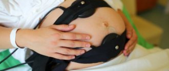
- pregnancy or menstruation in women,
- hypothermia or overheating,
- a lot of physical activity before taking tests.
If the white blood cell count exceeds 9.0 × 10⁹/L (increased white blood cells), the condition is called leukocytosis. A similar picture can be seen in malignant blood diseases, infections, radiation damage and poisoning.
Medicines can also increase the number of white blood cells. Tissue necrosis, excessive bleeding, including as a result of operations, renal coma, heart disease - all this can lead to leukocytosis.
A drop in the white blood cell count below 3.9 × 10⁹/L is called leukopenia. However, some people have a constant white blood cell count of 3.5 × 10⁹/L.
It is generally accepted that such people retain a reserve of these cells in their tissues that is 50 times greater than in their blood.
Functional leukopenia also occurs after the administration of analgesics, sulfonamides and other drugs, after long muscular work, as a result of infection and viruses (typhoid, influenza, measles).
Leukocyte formula
All types of leukocytes make up the leukocyte formula. It can help identify certain pathologies. Formula standards vary depending on the age and period of a person’s life. The shift is clearly visible in women during pregnancy or after childbirth.
Different methods are used to calculate the ratio of the types of these cells, but they are all related to the fact that heavier cells (basophils, for example) are located closer to the edge of the smear, and lymphocytes, like light cells, remain in the middle.
- Filipchenko's method. In this method, counting is carried out along the transverse straight border of one end to the other. The smear is mentally divided into three parts.
- Schilling method. Leukocytes are counted in four areas of the smear.
The received data is recorded in a table. Individually, the number of each type of white blood cell is calculated using formulas. Those elements that come into view are noted. Counting is carried out until the sum of all counted cells reaches 100.
Method for counting leukocytes in urine
The number of leukocytes in the urine is calculated in the Goryaev chamber according to Nechiporenko. This test is needed to monitor kidney disease. Periodic urine testing helps monitor the correctness of treatment and the effectiveness of the prescribed therapy.
Urine collection
Only the first morning urine in the middle of urination is studied. It is important for the patient to follow the basic rules for collecting this biomaterial. No special additional preparation is required for the study.
Calculation method
10 ml of freshly collected urine is centrifuged for three minutes at 2.5 thousand rpm. Before this, it is necessary to ensure that the urine has a slightly acidic reaction, in order to avoid partial breakdown of cells.
Using a pipette with a narrow, drawn-out end, remove the top layer. From 0.5 ml of urine to 1 ml is left in the test tube, depending on the volume of sediment.
The precipitate is carefully mixed with the supernatant liquid. The resulting solution is filled into Goryaev’s counting chamber according to a similar principle to that used when calculating blood PE. The camera is left alone for 3 to 5 minutes.
White blood cells should be counted in large squares across the entire reticle with a 7x eyepiece and 40x objective.
The resulting figure is used in the formula:
X = (a/0.9) × (1000/V)
X is the number of leukocytes in 1 ml of urine.
a – the calculated amount divided by the volume of the chamber in 1 μl of urine sediment.
v – Amount of urine taken for research.
1000 – amount of sediment.
It is important! Leukocyte norm: in 1 ml of urine no more than 2 × 10³ white blood cells.
Loading…
Source: https://dlja-pohudenija.ru/serdcze/metodika-podscheta-lejkoczitov-v-kamere-goryaeva-normy-pokazatelej-i-otkloneniya
Leukocyte norm and deviations
The norm of leukocytes in humans is 4.0 – 9.0 × 10⁹/l. Or, in other words, there are approximately 6,000 white blood cells in 1 cubic mm of blood.
Deviations from the norm can be influenced by external physiological factors:
- eating before donating blood,
- stress,
- pregnancy or menstruation in women,
- hypothermia or overheating,
- a lot of physical activity before taking tests.
If the white blood cell count exceeds 9.0 × 10⁹/L (increased white blood cells), the condition is called leukocytosis. A similar picture can be seen in malignant blood diseases, infections, radiation damage and poisoning.
Medicines can also increase the number of white blood cells. Tissue necrosis, excessive bleeding, including as a result of operations, renal coma, heart disease - all this can lead to leukocytosis.
A drop in the white blood cell count below 3.9 × 10⁹/L is called leukopenia. However, some people have a constant white blood cell count of 3.5 × 10⁹/L. It is generally accepted that such people retain a reserve of these cells in their tissues that is 50 times greater than in their blood. Functional leukopenia also occurs after the administration of analgesics, sulfonamides and other drugs, after long muscular work, as a result of infection and viruses (typhoid, influenza, measles).
Work No. 1. Counting red blood cells in Goryaev’s chamber.
⇐ PreviousPage 2 of 4Next ⇒
Equipment: microscope, Goryaev counting chamber, 4 ml of 3% sodium chloride solution, capillary from a Sali hemometer, scarifier, cotton wool, alcohol.
Purpose of work: learn to determine the number of red blood cells in human blood and evaluate the results.
Progress:
1. The cover glass is ground against the glass of the Goryaev chamber until Newtonian rings appear.
2. Pour 4 ml of 3% sodium chloride solution into a test tube.
3. Draw blood (0.02 ml) into a capillary tube from the Sali hemometer to the blood mark.
4. Carefully blow blood into a test tube with 3% sodium chloride solution and rinse the capillary with the same solution. Mix the contents of the test tube thoroughly.
5. Using the end of a round glass rod, take a drop of blood from the test tube, tilt it, and fill Goryaev’s chamber.
6. Place the camera on the microscope stage at low magnification, find the grid of the counting chamber and turn the microscope tube to high magnification.
7. Count the number of red blood cells in 5 large squares, divided into 16 small ones, which are located diagonally on the grid. When counting, Egorov’s rule is used: “red blood cells lying both inside the square and on its left and upper borders are considered to belong to a given square.”
8. The number of red blood cells is calculated using the formula:
where A is the sum of red blood cells in 80 small squares,
1/4000 mm3 – the volume of Goryaev’s chamber over one small square,
200 – blood dilution in 3% NaCl,
80 is the number of small squares in which red blood cells are counted.
Results:
Conclusions:
DETERMINATION OF HEMOGLOBIN CONCENTRATION
IN BLOOD BY SALI METHOD
The amount of hemoglobin in the blood is determined using the Sali colorimetric method. There is a special device called a colorimeter for this purpose. It is possible to determine the amount of the test substance in a colorimeter in cases where there is a parallel relationship between the concentration of a given substance in solution and the intensity of coloring. The simplest type of colorimeter is the Sali hemometer, which is usually used to determine Hb in the blood. It consists of three test tubes of the same diameter. One of them is empty, and the other, sealed at both ends, contains a standard solution of hematin hydrochloride, corresponding to the highest normal limit of Hb 166.7 g/l. All three test tubes are inserted into a small stand, the back wall of which is made of milky glass. A pipette with a capacity of 0.02 ml (or 20 mm3) is attached to the hemometer.
Work No. 2. Determination of the amount of hemoglobin in the blood using the Sali method (hematine method).
Equipment: Sali hemometer, pipette, scarifier, 0.1 N hydrochloric acid solution, cotton wool, alcohol.
Purpose of work: learn to determine the amount of hemoglobin using Sali’s method and evaluate the results.
Procedure: pour 0.1 N hydrochloric acid solution into the middle tube of the hemometer to the mark. Using a Sali hemometer pipette, draw 0.02 ml of blood (to the mark) and blow it to the bottom of the tube. The contents of the tube are mixed and left for 5-10 minutes to convert hemoglobin into hydrochloric acid hematin. Then distilled water is added dropwise to the contents of the test tube until the color of the resulting solution is the same as the color of the standard (adding water, mix the solution with a glass rod). The number at the level of the meniscus of the resulting solution indicates the hemoglobin content in the test blood in g%. To convert to g/l, the resulting figure is multiplied by 10. Normal hemoglobin concentration (absolute content) in men is 130-160 g/l, in women – 120-140 g/l. Compare the result with the norm and evaluate the result.
Results:
Conclusions:
CALCULATION OF RED CYTE INDICES.
The color index characterizes the relationship between the amount of hemoglobin in the blood and the number of red blood cells. The color index allows you to assess the degree of saturation of red blood cells with hemoglobin. The color index of a healthy person is 0.85-1.05. If the color index is higher than normal - hyperchromia. If below normal - hypochromia.
Work No. 3. Calculation of erythrocyte indices.
Purpose of the work: to determine the degree of saturation of erythrocytes with hemoglobin, to evaluate the result.
Progress:
1. Color index (CP) or (Fi) farb index ( Farb
– color,
index
– indicator) relative content of hemoglobin in red blood cells, calculated using the formulas:
CP= __________3*Hb (g/l)__________________
The first 3 digits of the number of red blood cells in millions.
Normally, CP = 0.8-1.0 (in newborns - 0.9-1.3; up to 1 year - 0.75-0.8; after 1 year - as in adults).
2. Average hemoglobin content in an erythrocyte - SGE (MCH, HbE):
The calculation of the SGE is carried out using the formula:
HGE = hemoglobin content (g/l)_
number of red blood cells per l
Normally 24-36 pg (10-12g), (in adults - 30 pg; in newborns - 33.3 pg; 5-6 months - 26.1 pg)
Results:
Conclusions:
⇐ Previous2Next ⇒
Recommended pages:
Leukocyte formula
All types of leukocytes make up the leukocyte formula. It can help identify certain pathologies. Formula standards vary depending on the age and period of a person’s life. The shift is clearly visible in women during pregnancy or after childbirth.
Different methods are used to calculate the ratio of the types of these cells, but they are all related to the fact that heavier cells (basophils, for example) are located closer to the edge of the smear, and lymphocytes, like light cells, remain in the middle.
- Filipchenko's method. In this method, counting is carried out along the transverse straight border of one end to the other. The smear is mentally divided into three parts.
- Schilling method. Leukocytes are counted in four areas of the smear.
The received data is recorded in a table. Individually, the number of each type of white blood cell is calculated using formulas. Those elements that come into view are noted. Counting is carried out until the sum of all counted cells reaches 100.
Chamber structure
Goryaev's chamber is nothing more than a glass slide (but much thicker than usual), divided into three parts by transverse grooves. The middle part of the glass contains a special grid for counting. The outermost parts of the chamber are used to grind the cover glass - thus, a closed chamber is created in the center with capillary spaces on the sides, through which it is filled with liquid.
As for the grid, Goryaev’s chamber is divided into 225 large squares of equal size - they are arranged in fifteen rows. The 25 large squares are further divided into smaller ones, 16 each. The length of each side of this small square is 0.05 mm.
Method for counting leukocytes in urine
The number of leukocytes in the urine is calculated in the Goryaev chamber according to Nechiporenko. This test is needed to monitor kidney disease. Periodic urine testing helps monitor the correctness of treatment and the effectiveness of the prescribed therapy.
Urine collection
Only the first morning urine in the middle of urination is studied. It is important for the patient to follow the basic rules for collecting this biomaterial. No special additional preparation is required for the study.
Calculation of color index (color coefficient)
The color indicator expresses the average hemoglobin content in one red blood cell. Normally, the color coefficient is 1.0 with 100% hemoglobin and 5,000,000 red blood cells in 1 mm3 of blood. It is calculated based on the following proportion: the found amount of hemoglobin (in units) is related to its normal amount as the found number of red blood cells is to their normal number.
For example, if the found hemoglobin is 88 units, and the number of red blood cells is 4,8000,000 per 1 mm3, then the color index is 0.91.
If the number of red blood cells is less than 1,000,000, it is necessary to divide by the first two digits of the calculated number of red blood cells. The normal color index is 0.9-1.1.