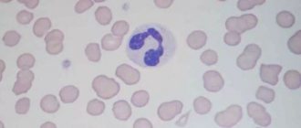
Lymphocytes are the main cells of the immune system, which are responsible for all of our internal defenses. If we decipher the word “lymphocytes” itself, it consists of two ancient Greek words – lymph and cytos. Lymph is a container, and cytos is a cell. Lymphocytes are representatives of white blood cells, which are the main cells of the immune system. From the moment they appear in the bone marrow until they must perform their particularly important function - to stand on our internal genetic purity - a certain period of time passes.
Read more about blood tests in our article
“Hematological tests: norms and interpretation of results.”
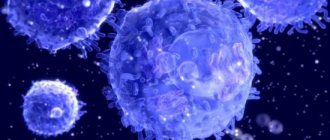
Types of lymphocytes
When the bone marrow releases a young lymphocyte into the bloodstream, it is not yet ready to perform its most delicate functions. Some of the young cells, lymphocytes, go to the thymus, or thymus gland, and there they must undergo maturation or “training.” What does "training" mean? A young lymphocyte must become familiar with the antigens that our body has encountered throughout life, starting from prenatal development. After maturation, thymus-dependent lymphocytes (T-lymphocytes) will begin to recognize antigens, understand that these are genetic strangers and will be ready to carry out their tasks.
T lymphocytes.
There are several types of T-lymphocytes.
The first type is killer T-cells. Killers are killers. They are the ones who are sharpened to recognize a genetic stranger and try to kill him right away.
The second type is T-helper cells, or helper cells. It is the T helper cells that will include the following cells that will produce antibodies or protective protein structures for us.
The third type is T-suppressors - those cells that will stop the immune response.
And so, if a genetic alien enters the human body, the killers will be the first to recognize it and will try to kill it immediately. What can act as a genetic stranger? These are viruses, bacteria, parasites, fungi, and cancer cells that can appear every second. Cells of the body in which a failure occurred at the genetic level. If a person has food intolerance, then, unfortunately, they can also turn into genetic strangers and also deplete the lymphocyte component of the immune system.
B lymphocytes
These are also lymphocytes, which, having emerged young from the bone marrow into the bloodstream, rush to the lymph nodes, to the spleen, or to the liver, where they will also mature. The main function of B cells is to produce representatives of humoral immunity, or blood immunity, for us. It is the B cells that will produce antibodies that will fight viruses, bacteria, and other genetic invaders.
NK cells or natural killer cells.
This is a group of cells that will primarily be focused on protecting us from cancer. First of all, natural killers will work in antitumor defense and antiparasitic defense. NK cells are the main types of lymphocytes.
Activated lymphocytes (CD3+HLA-DR+, CD3-HLA DR+)
The study includes the determination of absolute and relative values of lymphocyte subpopulations (T, B, NK cells) expressing the surface activation marker HLA-DR. It is recommended for use to monitor the dynamics of the cellular component of the immune system after a comprehensive immunological examination.
Synonyms Russian
Immunophenotyping, cellular immunity, multicolor cellular analysis by flow cytometry, lymphocyte activation marker.
English synonyms
Human Immune System, Immunophenotyping, Multicolor Flow Cytometry Cell Analysis, Human Leukocyte Differentiation Antigens, HLA-DR activation marker.
Research method
Flow cytometry.
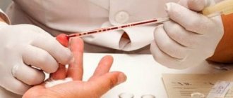
What biomaterial can be used for research?
Venous blood.
How to properly prepare for research?
- Eliminate alcohol from your diet for 24 hours before the test.
- Children under 1 year of age should not eat for 30-40 minutes before the test.
- Children aged 1 to 5 years should not eat for 2-3 hours before the test.
- Do not eat for 12 hours before the test; you can drink clean still water.
- Completely avoid (in consultation with your doctor) taking medications for 24 hours before the test.
- Avoid physical and emotional stress for 24 hours before the test.
- Do not smoke for 30 minutes before the test.
General information about the study
Assessment of the cellular composition (immunophenotyping) of human blood lymphocytes—the main component in assessing immune status—is performed by flow cytometry.
Immunophenotyping is the characterization of cells using monoclonal antibodies or any other probes that make it possible to judge their type and functional state by the presence of a particular set of cellular markers.
Immunophenotyping of leukocytes involves detecting differentiation markers, or CD antigens, on their surface. Leukocytes express a range of surface and cytoplasmic antigens unique to their subpopulation and developmental stage. CD antigens (cluster of differentiation antigens) are antigens on the surface of cells, markers that distinguish one cell type from another. The differentiations of these antigens have been studied and standardized, and they are assigned specific numbers. CDs can be recognized using appropriate monoclonal antibodies. Using fluorescently labeled monoclonal antibodies that bind to specific CDs, flow cytometry can be used to count the content of lymphocytes belonging to subpopulations of different functions or developmental stages.
Flow cytometry is based on photometric and fluorescent measurements of individual cells as they traverse a beam of monochromatic light, usually laser light, one after another along with a fluid stream.
CD3
This marker allows you to identify mature resting (intact) T cells and count the total number of T lymphocytes. Quantitative assessment of the CD3+ lymphocyte subpopulation has diagnostic value in the following cases:
— primary and secondary immunodeficiencies;
— acute viral infections, including HIV;
- intracellular bacterial and parasitic infectious diseases (for example, tuberculosis, leprosy, leishmaniasis);
— malignant neoplasms;
— transplant rejection reactions and graft-versus-host disease;
— lymphoproliferative disorders (acute T-lymphoblastic leukemia).
In diabetes mellitus, a decrease in the percentage and absolute number of CD3+ lymphocytes is quite often observed in patients.
Anti-HLA-DR
The HLA-DR molecule is also an activation marker and belongs to MHC class II. It is a transmembrane glycoprotein consisting of a- and b-subunits with a molecular weight of 36 and 27 kDa. Anti-HLA-DR reacts only with the HLA-DR epitope and does not cross-react with the HLA-DQ and HLA-DP molecules. It is expressed on B lymphocytes, monocytes, macrophages, and activated T lymphocytes.
There is evidence that the HLA-DR molecule is expressed on approximately 10% of PC T-lymphocytes, but when cells are activated by mitogen, the number and density of its expression increases sharply. It has been suggested that the HLA-DR molecule on T cells may act as a receptor involved in signal transduction by activated T lymphocytes. This suggests its possible role as a “professional” APC involved in the maintenance of immune memory.
HLA-DR may also be present on thymic epithelial cells, on cells of B-lymphocyte-dependent fields of the spleen and lymph nodes, and B-cell lymphoma. This antigen is co-expressed with CD1a antigen on Langerhans cells.
The method allows you to evaluate small cell populations, as well as study their functional activity:
- activated T lymphocytes (CD3+HLA-DR+CD45+);
- B lymphocytes and activated NK cells (CD3-HLA-DR+CD45+).
When is the study scheduled?
It is recommended for use to monitor the dynamics of the cellular component of the immune system after a comprehensive immunological examination.
What do the results mean?
Changes in various cell populations of lymphocytes, up or down, develop during various pathological processes in the body, such as infections, autoimmune and oncological diseases, immunodeficiencies, in the postoperative period, and during organ transplantation.
Below is a table with clinical situations that can lead to changes in the subpopulation composition of lymphocytes.
| Lymphocyte subpopulation | Increasing the indicator | Decrease in indicator |
| Activated T lymphocytes (CD3+HLA-DR+CD45+) |
| Has no diagnostic value. |
In combination with clinical data, symptoms, and other laboratory research methods, the above changes are a diagnostic sign of the occurrence of these pathological processes in the human body.
Important Notes
- The results of this study must be compared with clinical data and indicators of other laboratory tests.
- It should be noted that the assessment of indicators over time significantly increases the clinical significance of the study.
Literature
- Khaitov, R.M. Allergology and immunology: national guide / ed. R.M. Khaitova, N.I. Ilyina. – M.: GEOTAR-Media, 2009. – 656 p.
- Khaitov, R.M. Manual of Clinical Immunology. Diagnosis of diseases of the immune system: a guide for doctors / R.M. Khaitov, B.V. Pinegin, A.A. Yarilin. – M.: GEOTAR-Media, 2009. – 352 p.
- Zueva E.E. Immune system, immunogram: recommendations for prescription and use in the diagnostic and treatment process / E.E. Zueva, E.B. Rusanova, A.V. Kurtova, A.P. Ryzhak, M.V. Gorchakova, O.V. Galkina - St. Petersburg. – Tver: Publishing House “Triada” LLC, 2008. – 60 p.
- Ketlinsky, S.A. Immunology for doctors / S.A. Ketlinsky, N.M. Kalinina. St. Petersburg : Hippocrates, 1998. – 156 p. Yarilin, A.A. Immunology: textbook / A.A. Yarilin. – M.: GEOTAR-Media, 2010. – 752 p.
- Khaitov, R.M. Immunology: atlas / R.M. Khaitov, A.A. Yarilin, B.V. Pinegin.M. : GEOTAR-Media, 2011. – 624 p.
- Khaitov, R.M. Immunology: textbook / R.M. Khaitov. – M.: GEOTAR-Media, 2009. – 320 p.
- Khaitov, R.M. Assessment of human immune status in normal and pathological conditions / R.M. Khaitov, B.V. Pinegin // Immunology. – 2001. – N4. – P. 4–6.
- Whiteside, TL Role of Human Natural Killer Cells in Health and disease / TL Whiteside, RB Herberman // Clinical and Diagnostic Laboratory Immunology. – 1994. – Vol. 1, No. 2. – P. 125–133.
- Ginadi, L. Differential expression of T-cell antigens in normal peripheral blood lymphocytes: a quantitative analysis by flow cytometry / L. Ginadi, N. Farahat, E. Matutes // J. Clin. Pathol. – 1996. – Vol. 49, No. 1. – P. 539–544.
- Mercer, JC Natural killer T-cells: rapid responders controlling immunity and disease / JC Mercer, MJ Ragin, A. August // International J. Biochemistry & Cell Biology. – 2005. – No. 37. – P. 1337–1343.
- Nikitin, V.Yu. Activation markers on T-helpers and cytotoxic lymphocytes at various stages of chronic viral hepatitis C / V.Yu. Nikitin, I.A. Sukhina, V.N. Gypsy [and others] // Vestn. Ross. Military medical acad. – 2007. – T. 17, No. 1. – P. 65–71.
- Boettler, T. T cells with CD4+CD25+ regulatory phenotype suppress in vitro proliferation of virus-specific CD8+ T cells during chronic hepatitis C virus infection / T. Boettler, HC Spangenberg, C. Neumann-Haefelin // J. Virology. − 2005. − Vol. 79, N 12. - P. 7860–7867.
- Ormandy, LA Increased Populations of Regulatory T Cells in Peripheral Blood of Patients with Hepatocellular Carcinoma / LA Ormandy, T. Hillemann, H. Wedemeyer // J. Cancer Res. − 2005. − Vol. 65, N 6. - P. 2457–2464.
- Sakaguchi, S. Naturally arising FoxP3-expressing CD4+CD25+ regulatory T cells in immunological tolerance to self- and non-self / S. Sakaguchi // Nature Immunol. − 2005. −Vol. 6, N 4. - P. 345–352.
- Romagnani, S. Regulation of the T cell response / S. Romagnani // Clin. Exp. Allergy. –2006. − Vol. 36. - P. 1357–1366.
- Khaidukov S.V., Major and minor populations of human peripheral blood lymphocytes and their normative values (method of multicolor cytometric analysis) / Khaidukov S.V., Zurochka A.V., Totolyan A.A., Chereshnev V.A. // Med. immunology. — 2009. -T. 11 (2-3). - pp. 227-238.
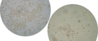
Norm of lymphocytes in the blood
Unfortunately, there are often situations when lymphocytes in the blood can deviate from the norm - increase or decrease. This can happen in various situations. Before we talk about when the number of lymphocytes changes in the blood, we need to talk about how much is their normal component.
If we talk about cell clones, then:
T cells make up 75% of the total number of lymphocytes;
B cells about 15%;
Natural killers are about 10%.
And speaking about the norms of lymphocytes, it must be said that at different periods of a person’s life the norms differ.
Lymphocytes begin to be produced by the bone marrow already in utero, and this process continues throughout life. The lifespan of these cells also varies - from several months to sixty years. And these will all be lymphocytes.
The number of lymphocytes in the blood during pregnancy
Accordingly, there are periods of life, especially in the female body, when the concentration of lymphocytes will also decrease. First of all, I mean pregnancy. In order to prevent immunological rejection from occurring, since the fetus contains 50% of foreign cells, the body arranges everything in such a way that the production of its own lymphocytes decreases. That is why we are talking about a physiological, immunosuppressive state in women during pregnancy. There is a decrease in immunity so that the woman can safely carry the fetus.
Online consultation with infectious disease specialist, allergist-immunologist Natalia Nikolaevna Gordienko
Cost of online consultation: 3500 rubles
Online consultation
During the consultation, you will be able to voice your problem, the doctor will clarify the situation, interpret the tests, answer your questions and give the necessary recommendations.
Increase and decrease in lymphocytes in blood tests
And so, you and I have already understood that lymphocytes are the main cells of our immune system. And depending on how their quantity in the blood changes, changes in the blood formula will be called.
Reduced number of lymphocytes in the blood
— lymphopenia.
Increased number of lymphocytes in the blood
– lymphocytosis.
What causes our immune system and bone marrow to release increased numbers of lymphocytes into the blood? Since these are the main cells that are responsible for our genetic protection, it means that we need to understand that “not everything is in order in the state.”
I would also like to draw your attention to the fact that lymphocytosis and lymphopenia are not diseases. These are conditions of the human body in which a violent reaction occurs or does not occur in the immune system. And any change in the number of lymphocytes will always indicate that it is necessary to check what reasons led to this or that change.
Prevention
The reason for the malfunction in the level of lymphocytes lies in the child’s immune system. To protect your baby from the return of the disease, it is important to exclude some moments from the baby’s life:
- frequent stress;
- poor nutrition;
- strong physical activity;
- lack of alleys in the fresh air;
- infection of a young body;
- long exposure to the hot sun.
Don’t forget about preventive appointments with doctors. Even if there are no symptoms, it is better to prevent the development of the disease at its inception.