Endocrinologist, nutritionist, nutritionist
Evdokimova
Elena Viktorovna
13 years of experience
Endocrinologist, nutritionist, nutritionist, member of the Russian Association of Endocrinologists. Member of the European Association for the Study of Diabetes (EASD)
Make an appointment
Pathology in the form of breast enlargement in men is called “gynecomastia”. Its main cause is hormonal disorders in the body, which cause intensive growth of fatty and glandular tissue in the chest area. In addition to the undesirable cosmetic effect, in the absence of medical control and treatment, the disease can have serious consequences in the form of the development of neoplasms of a benign or malignant nature.
Etiology
Literally translated from Latin, “gynecomastia” means female breasts. The pathology is observed in men and has the appearance of a benign formation, the structure of which is formed due to the cells and ducts of glandular tissue or the growth of the fat layer. In the first case, they talk about the so-called true gynecomastia, and in the second, about a false type of disease. The increase can reach 12 cm, which causes the patient severe psychological discomfort and significant inconvenience in everyday life. Unilateral enlargement of the glands is rare; more often the disease affects both glands. Externally, it manifests itself in the form of sagging tissues, reminiscent in shape of a woman’s breasts.
What is more informative: ultrasound or mammography?
Both examination methods are equally effective and allow you to detect dangerous diseases in the early stages, reduce the risk of surgical intervention, and also save the patient’s life. In some cases, these methods can be used together and complement each other.
Mammography, unlike ultrasound diagnostics, refers to x-ray examinations and involves radiation exposure to the body. Ultrasound is a completely safe method that can be performed repeatedly, since it does not pose a risk of radiation.
| Ultrasound of the mammary glands | Mammography |
| During the procedure the patient lies | During the procedure the patient stands |
| The examination is painless | The chest is slightly compressed with special plates |
| Ultrasound is more informative for women with dense breasts, as well as for patients with implants. Gives a more complete picture when examining cysts, lipomas and metastases. Allows you to examine lymph nodes. | Mammography is performed to detect tumors inside the milk ducts, microcalcifications and other pathological formations. Prescribed if the clinical symptoms of the disease are not confirmed by ultrasound. |
The choice of method for examining the patient should be made by the doctor.
Decoding the results
After completing the ultrasound protocol, you will receive a conclusion. The final diagnosis is made by your attending physician, assessing the overall clinical picture, all data obtained, including laboratory tests and the collected medical history.
Advantages of the Miracle Doctor clinic
| All types of ultrasound diagnostics | Modern equipment |
| We offer breast screening with Doppler ultrasound, color Doppler mapping and elastography. | The clinic is equipped with advanced expert-class equipment, which is highly sensitive. |
| Experienced doctors | Affordable prices |
| Ultrasound is performed by specialists who have at least 10 years of practical experience and regularly improve their skills. | The cost of breast ultrasound is average in the capital. At the same time, our patients receive objectively more accurate and high-quality images. |
If you need an inexpensive breast ultrasound in Moscow, call the clinic. We will make an appointment for you on a convenient date and time, and you will be able to get into the diagnostic room without queues or other formalities.
Reasons for development
The intensive growth of adipose and glandular tissue has serious internal reasons. Among the influencing factors it is worth noting:
- uncontrolled long-term medication use;
- exposure to toxic substances on the body;
- hormonal imbalances;
- diseases with systemic symptoms.
Each of these phenomena triggers the process of intensive production and entry into the blood of female sex hormones - estrogens. Against the background of a stable or reduced amount of the male hormone androgen, hormonal imbalance is observed, which is expressed in the impact on the so-called target organs that are most sensitive to changes in the body. Among them is the mammary gland, the growth of which is sharply activated. The mechanism of development of gynecomastia, taking into account the influence of the hormonal factor, is completely similar to the process in the body of young girls during adolescence, when a sharp increase in female hormones stimulates the process of breast growth.
In the body of a healthy man, the hormone content is balanced, and the amount of estrogen cannot affect the growth of breast tissue. In addition, their volume is controlled by another hormone - testosterone, which determines the characteristics of the male figure and the functioning of the body of representatives of the stronger sex. But in the event of a hormonal imbalance and a weakening of the influence of testosterone, estrogens trigger a process of external and physiological changes, including the process of development of secondary sexual characteristics characteristic of the opposite sex.
How to treat gynecomastia?
Physiological types of gynecomastia
most often do not require correction with medications, and over time they often disappear on their own.
Gynecomastia in adolescents
usually disappears within 2-3 months after detection due to increased testosterone production during puberty.
Pathological gynecomastia
eliminated by treating the underlying disease that caused hypertrophy of the mammary glands (for example, treatment of thyrotoxicosis - excessive function of the thyroid gland, after which gynecomastia caused by thyrotoxicosis is also eliminated). To reduce the amount of estrogen in the body of men, anti-estrogenic therapy is used with the drug tamoxifen, a blocker of the effects of estrogen on the mammary glands, as well as testosterone preparations (Sustanon, Omnadren), or human chorionic gonadotropin (hCG).
If gynecomastia is caused by taking medications
, after their cancellation it undergoes regression.
Pathological gynecomastia after prolonged fasting
and subsequent resumption of feeding usually does not require treatment and goes away on its own after normalization of the patient’s diet.
Long-term gynecomastia can be effectively treated with hormonal drugs that suppress estrogen concentrations, such as clomiphene, tamoxifen, and danazol. If the process lasts in adults for more than two years, and also in the absence of conservative therapy, plastic surgery is often resorted to, removing excess fatty and glandular tissue of the mammary gland surgically.
Surgery to remove the mammary gland can also be used in adolescents if breast enlargement remains greater than 5 cm for more than 3 years. Such an operation is not strictly necessary from a clinical point of view and is more of a cosmetic nature, however, in order to prevent the teenager from developing psychological complexes associated with his own body, most parents insist on having it performed.
Treatment of false gynecomastia
consists primarily of correcting body weight, after which the issue of surgical intervention is decided.
Treatment of gynecomastia without surgery is also ineffective in cases where the pathological process is caused by a tumor lesion of the mammary gland. In this case, surgical intervention is inevitable, and it consists of excision of excess breast tissue and restoration of its contour. Surgery to remove gynecomastia - mastectomy - is well tolerated by male patients and does not require a long hospital stay.
After removal of gynecomastia, it is recommended to wear shapewear for several weeks, which helps to shrink the skin and form a new muscle contour. Restrictions on physical activity may continue for a month.
The first ultrasound of the operation area is performed after 2 months, then (if the results of the first ultrasound are normal) the study is repeated every 6 months.
Forms and stages of development
The gradual growth of breast tissue allows us to talk about the gradation of the disease. To establish the degree of its manifestations at a specific point in time, a histological examination of the cells of the pathological formation is carried out. Experts distinguish:
- proliferating stage. The duration is approximately 4-6 months, and the main symptoms include intense tissue growth.
- an intermediate stage lasting about a year. The formation of the “female” gland is completed, its size stabilizes.
- fibrous stage. Its main symptom is the proliferation of glandular and connective tissue, which gives the breast a characteristic appearance.
Based on the diagnostic report on the stage of the detected disease, the doctor selects the optimal form of therapy, taking into account the age and general condition of the patient’s body.
In order to further clarify the nature of the disease, the following classification of gynecomastia has been adopted:
- physiological - considered normal in infancy, adolescence or old age, therefore at these stages of the patient’s life it does not require strict monitoring and intensive treatment. There are subtypes of pathology based on age: transient gynecomastia of newborns, adolescent form and gynecomastia of older patients. Transient gynecomastia occurs in 70-80% of babies and goes away within the first three weeks of life. Its cause is the entry of estrogen from the mother into the fetus through the placenta. Pubertal gynecomastia is observed in teenage boys aged about 13-15 years, which is associated with hormonal changes, a sharp increase in the number of estrogens and the maturation of the body. Signs of the disease disappear on their own approximately a year after their appearance. The cause of gynecomastia in elderly patients is suppression of testosterone, against the background of which the amount of estrogen begins to predominate and influence physiological processes in the body.
- false gynecomastia. The disease is characterized by intense growth of adipose tissue. more often observed in patients suffering from various forms of obesity. The degree of breast enlargement varies depending on the constitution of the body and other diseases.
- True gynecomastia is caused by a hormonal imbalance that causes breast growth.
- idiopathic pathology. The diagnosis is made when it is impossible to accurately indicate the cause of the disease.
Some experts refuse the proposed classification, combining physiological, idiopathic and true forms of the disease based on the similarity of their histological picture. However, most doctors use the accepted official form of describing gynecomastia due to their differences in their nature and characteristics of the course. In addition, women are diagnosed with gynecomastia. This usually happens when it is necessary to note in the anamnesis the fact of breast growth during a period of the patient’s life, which is not characterized by such changes.
Which doctor should a man see for gynecomastia?
If you suspect the development of gynecomastia in a man, you can contact the following specialists:
- therapist;
- urologist;
- endocrinologist
Most often, this pathology is treated by an endocrinologist, since the root cause of breast growth is the improper functioning of the hormonal system.
I am an endocrinologist, candidate of medical sciences, associate professor Georgiy Nikitich Romanov, an experienced specialist with more than 20 years of work experience. I was able to help many patients overcome their illness and live a normal life. As a specialist, I have extensive up-to-date knowledge that helps me successfully treat patients with true gynecomastia.
I not only conduct appointments at the clinic, but also offer paid consultation services. You can contact me through any of the presented instant messengers or social networks: VKontakte, Direct, Skype, Whatsapp, Telegram, Viber.
Signs
External signs of gynecomastia are determined by the stage of its development:
- At the proliferation stage, there is a slight increase in the size of the mammary glands. If the pathology is detected at this stage and the patient is offered treatment, the disease goes away within a short time;
- at the intermediate stage, breast tissue grows rapidly, and milk secretion is released from the nipple. This condition causes significant discomfort to the patient. Most cases of gynecomastia are diagnosed at the second stage, because symptoms of the disease become obvious;
- at the fibrotic stage, intensive proliferation of adipose and connective tissue causes characteristic lumps to appear in the breasts. Its structural changes are considered irreversible even with prolonged intensive treatment.
Symptoms of gynecomastia are often observed in athletes taking steroid drugs. The main signs of the disease include itching, burning, discomfort in the nipple area, and the release of a small amount of milk secretion.
Prevention
Prevention of physiological gynecomastia is difficult due to the fact that it is part of normal biological processes in the body. This condition depends on natural hormonal fluctuations and quite often goes away on its own, requiring neither prevention nor treatment.
To prevent pathological gynecomastia, it is necessary to carefully monitor the hormonal system, avoid taking medications that affect the production of hormones, give up alcohol and drugs Source: Risk factors, the role of steroid hormones and their receptors in the development of gynecomastia. Vovchuk I.L. Scientific notes of the Crimean Federal University named after V.I. Vernadsky. Biology. Chemistry, 2014. Prevention of the disease is complicated by the fact that most often it is not the cause, but a consequence of the development of other diseases in the body.
Sources:
- Gynecomastia. Beltsevich D.G., Vanushko V.E., Kuznetsov N.S., Kats L.E. Endocrine surgery, 2012. p. 18-23
- Etiology and age factors of gynecomastia. Akimov D.V. Tumors of the female reproductive system, 2012. p. 34-38
- Pathomorphological and molecular biological characteristics of gynecomastia. Mnikhovich M.V., Ben Ammar M.A., Kulikov E.P., Zhakota D.A. Bulletin of Volgograd State Medical University, 2021. p. 32-37
- Modern ideas about the epidemiology, etiology and pathogenesis of gynecomastia. Yashina Yu.N., Rozhivanov R.V., Kurbatov D.G. Andrology and genital surgery, 2014. p. 8-15
- Risk factors, the role of steroid hormones and their receptors in the development of gynecomastia. Vovchuk I.L. Scientific notes of the Crimean Federal University named after V.I. Vernadsky. Biology. Chemistry, 2014
The information in this article is provided for reference purposes and does not replace advice from a qualified professional. Don't self-medicate! At the first signs of illness, you should consult a doctor.
Diagnostics
Characteristic external symptoms allow the doctor to accurately make a diagnosis and prescribe additional measures to clarify the nature of the disease. You can get a comprehensive picture:
- interviewing the patient with a request to describe his own feelings and complaints of malaise;
- external examination of the mammary glands with palpation of the contents, as well as studying the condition of other organs, signs of pathology of which can be detected visually;
- biochemical blood test for gynecomastia, on the basis of which the doctor can easily determine the fact of hormonal imbalance;
- mammography, the image of which clearly shows the structure of the tissue and the features of its development;
- computed tomography and magnetic resonance imaging, thanks to which it is possible to determine the cause of development and the type of overgrown tissue;
- biopsy of overgrown tissues with their mandatory histological examination, necessary for the timely detection of a malignant process.
The sooner the correct diagnosis is made to the patient, the greater the chance of achieving significant treatment results and completely eliminating the external manifestations of the pathology.
What does a breast ultrasound show?
The examination allows you to determine:
- early stage breast cancer;
- neoplasms: cysts, tumors, lymphangiomas, etc.;
- breast diseases: mastitis, mastopathy (nodular, diffuse), fibroadenoma, etc.;
- minimal disturbances in tissue structure;
- status of lymph nodes in real time;
- infectious processes that appear after injuries, illnesses or breast augmentation surgery;
- abscesses accompanied by edema;
- disturbance of blood flow in vessels, etc.
In addition, an ultrasound test can be prescribed for men with injuries to the chest, as well as hormonal imbalances, diseases of the thyroid gland, problems with the pituitary gland, the main manifestations of which are swelling and pain in the mammary gland. Ultrasound allows you to diagnose the slightest deviations from the norm, thanks to which a specialist can prescribe treatment in a timely manner.
The study shows tissues that differ in density. Using the method, the mammary gland, ducts, skin, nipples, subcutaneous fat, pectoral muscles, and lymph nodes are assessed in real time. Modern equipment makes it possible to identify the exact location of tumors reaching a size of less than 4 mm. The result of the examination is a description of the structures of the gland, stage of development, functional activity, nature and size of tumors, if any.
Preparing for a breast ultrasound
To obtain objective results, the examination is carried out from the 4th to the 13th day of the menstrual cycle, since during this period the milk ducts narrow. It is not recommended to apply cosmetics - creams and lotions - to the skin of the décolleté, even if they are quickly absorbed. No other additional preparation is required. During the examination, you will be asked to remove jewelry and clothing from the waist up.
How is the procedure done?
1. The patient needs to undress to the waist and lie down on the couch. You will need to place your hands behind your head.
2. The doctor will apply a hypoallergenic gel to the patient’s chest, which improves the glide of the sensor and the passage of sound waves.
3. An examination is performed. The doctor presses on individual areas of the chest with a special sensor and records the information received. If necessary, the patient will be asked to lie on her side, stand up, or sit down to better visualize the tissue.
During the examination, the specialist also pays attention to the lymph nodes: axillary, supra- and subclavian, parasternal, located between the sternum and the mammary gland. If the doctor finds any abnormalities, the patient will be asked to undergo additional diagnostics, including the following methods:
- Dopplerography - assessment of the condition of the vessels supplying the mammary glands, as well as the speed of blood flow in them;
- color Doppler mapping - detailed visualization of veins, arteries and breast tissue;
- elastography - assessment of the density of breast tissue, neoplasms and areas with pathologies.
Depending on the characteristics of the examination, the procedure can last from 20 minutes to an hour. After this, the patient will receive a written report and photographs.
How often can you undergo ultrasound diagnostics?
Women are recommended to have an ultrasound scan of the mammary glands every year, and if breast disease is suspected, they should be examined at least once every six months. Ultrasound diagnostics has no contraindications and can be performed several times in a row in a limited period of time to clarify the diagnosis or check the condition of the tumor.
Treatment methods
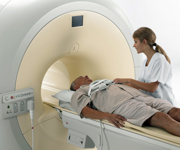
Depending on the severity of the disease according to the diagnostic results, the age and condition of the patient, the doctor decides on the choice of drug or surgical treatment.
A course of taking medications allows you to adjust your hormonal levels and eliminate the imbalance in favor of female hormones. It is possible to achieve good results by taking medications no later than 4 months from the moment the first symptoms appear. The basis of the course is anti-estrogenic drugs and vitamin complexes.
Surgical treatment is possible only in the absence of contraindications: acute infections, third degree obesity or cardiovascular diseases. The main method of surgery for gynecomastia is mastectomy, which allows you to completely or partially remove overgrown tissue. The priority is considered to be endoscopic or subcutaneous methods, which are characterized by minimal trauma and a good cosmetic effect.
A separate group includes cases where gynecomastia can be treated without surgery and a long course of medications. In this case, treatment is designed to reduce risk factors: reduce the content of fatty tissue in the body, normalize hormonal levels. Fats, fried and smoked foods, table salt, all types of alcohol, as well as coffee and chocolate are excluded from the diet. Dynamic observations allow us to evaluate external changes in the body and make a decision on the advisability of choosing a different treatment option for gynecomastia.
The condition of the organ is normal
Normally, the skin of the mammary glands is visualized as a smooth hyperechoic line up to 7 mm thick. The presence of a fatty layer changes this picture: the dermis takes on the appearance of two hyperechoic lines separated by a thin hypoechoic stripe. Unlike the breast, the boundary between the skin and the underlying tissues is never visualized.
The nipples are visualized as round, delimited formations of low echogenicity. Acoustic shadows may be observed behind them. The echogenicity of subareolar structures is always high.
The retromammary region includes fatty tissue, ribs, intercostal muscles and pleura. When scanned they appear like this:
- adipose tissue - in the form of hypoechoic lobules between hyperechoic lines;
- the pectoralis major and minor muscles are like multidirectional hypoechoic layers parallel to the skin with a transverse hyperechoic septum, bordered by hyperechoic fascial lines;
- ribs - in the form of oval hyperechoic formations with a persistent acoustic shadow;
- intercostal muscles take on the appearance of hypoechoic masses with a typical muscle pattern;
- the pleura is the deepest hyperechoic line.
Breast normal values vary depending on age.
| Man's age | Norm |
| Puberty | Heterogeneous structure of the gland due to active hormonal status. The skin is poorly differentiated from subcutaneous fat. Elements of adipose tissue are visualized as areas with low echogenicity, fibrous tissue - as granular fragments with medium echogenicity. |
| Average age | The skin is visualized as a hyperechoic line 0.5–1.5 mm thick. Adipose tissue - in the form of a single layer 2–3 cm thick with low echogenicity. Against its background, fibrous tissue is determined in the form of fine-grained structures with high echogenicity. |
| Elderly age | The skin can have different thicknesses. It is defined as a thin hypoechoic layer of fat. Almost the entire body of the gland is filled with rounded hypoechoic structures with a pronounced hyperechoic rim - fat lobules. |
| The main sign of healthy mammary glands is the absence of pathological inclusions and various types of formations. | |
Male breast anatomy
The male breast is identical in its anatomical structure to the female breast, the only difference is that the gland does not grow during puberty and remains so throughout life. A man also has a mammary gland, but it is usually called the mammary gland.
The male mammary gland, unlike the female mammary gland, does not have a lobular structure, but just like the female mammary gland, it has hormone-sensitive receptors. Therefore, an imbalance of estrogen and testosterone in the body of a sexually mature man can cause phenomena such as gynecomastia, which translated from Greek means “feminine breasts.”


