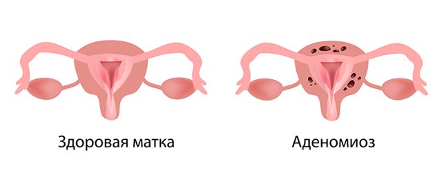
Adenomyosis of the uterus is a gynecological disease of a benign nature, in which endometrial cells grow into the walls of the organ, leading to disruption of its functions and infertility. The exact causes of the pathology have not yet been established, but doctors have identified a number of predisposing factors that, under a combination of unfavorable circumstances, can become a trigger for the development of adenomyosis. At the initial stages of development, the disease does not manifest itself in any way. But as it progresses, pronounced clinical symptoms are disturbing, which should be a reason to visit a gynecologist as soon as possible and undergo a comprehensive examination. One of the methods for diagnosing adenomyosis is ultrasound examination (US). Echosigns of uterine adenomyosis are manifested by changes in the density of organ tissue, the presence of areas with hypoechogenicity, hyperechogenicity and heteroechogenicity. The treating gynecologist interprets the ultrasound results and, based on the data obtained, will select the most effective treatment plan. The medical clinic is equipped with high-precision ultrasound diagnostic equipment of the new generation, with which you can determine the diagnosis at the earliest stages.
What is adenomyosis
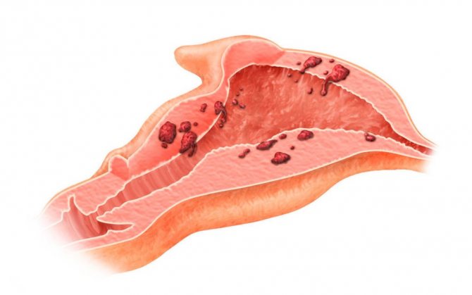
Adenomyosis is a benign hormone-dependent tumor formed by endometrial cells that grow uncontrollably, affecting the myometrium to varying depths. Depending on the nature of the course and the type of changes inside the walls of the uterus, the following forms of the disease are distinguished:
- Focal. In the focal form, endometriotic tissue grows into the myometrium in the form of locally located focal areas.
- Nodal. This type is characterized by damage to the walls of the reproductive organ by glandular epithelial cells in the form of multiple nodules.
- Diffuse. With diffuse adenomyosis, the growth of pathological cells occurs to different depths, sometimes forming fistulas in the pelvic area.
- Diffuse nodular. Combines diffuse and nodular types of adenomyosis.
Adenomyosis occurs in 4 stages:
- First. Pathological processes are limited to the myometrial mucosa; any suspicious symptoms indicating the disease are completely absent.
- Second. Endometriotic cells spread to the muscle tissue of the reproductive organ, the clinical picture is absent or mild.
- Third. The pathological process affects the entire thickness of the muscle wall, right down to the serous membrane. Certain symptoms are concerning.
- Fourth. The muscular layer of the uterus is completely affected by endometrial tissue, which can also grow into the tissue of organs located nearby. The clinical picture is clearly expressed.
Classification of pathology
In medicine, there are 3 types of adenomyosis: local adenomyosis, focal adenomyoma and diffuse adenomyosis. What kind of disease the patient has can be determined by the extent and depth of the process:
- Local (focal, focal) adenomyosis
- the endometrium grows in one specific place of the uterus. - Adenomyoma
is a form of localized adenomyosis, but it is more extensive and results in a significant increase in uterine mass or a benign tumor similar to uterine fibroids. With adenomyoma, the endometrium grows deeply into the uterine tissue. - Diffuse adenomyosis
- the process spreads throughout the uterus.
Classification of the type of adenomatosis is important when choosing treatment tactics for the disease. The first two cases do not require hysterectomy (removal of the uterus). In case of diffuse adenomyosis, the organ will have to be removed.
How does adenomyosis develop?
Adenomyosis is one of the types of endometriosis. To understand the mechanism of pathology, it is important to know the structure and basic functions of the uterus. The body of the uterus consists of smooth muscle. But muscle tissue itself is not enough to bear and give birth to a child. During each menstrual cycle, the body produces a special layer to which the fertilized egg attaches and develops further. This layer is called the endometrium. The endometrium, in turn, consists of 2 membranes:
- Functional. Designed for bearing a fetus.
- Basal. The shell from which the functional layer grows.
If fertilization does not occur during the cycle, the functional membrane is rejected and leaves the uterus along with menstrual bleeding. In the next cycle, the functional layer is formed again. But under the influence of negative factors, a malfunction occurs; the endometrium damages the basal layer, as well as the membrane, and in some places begins to grow into the body of the uterus. The reproductive organ reacts to the failure by forming thickenings of muscle fibers around the pathological foci, trying to limit further spread. As a result, the body of the uterus increases in size, which leads to disruption of its functioning.
Unusual manifestations of adenomyosis
a) “Swiss cheese” appearance ( Fig. 9 )
b) adenomyoma and polypoid adenomyoma ( Fig. 10 and Fig. 11 )
- Local fusion of glands, forming a mass of adenomyosis.
- It may appear as a mass deep in the myometrium, most often in the uterine body.
- Sometimes it may protrude from the endometrium (submucosal adenomyoma) or protrude through the endometrium and grow as a polypoid mass (polypoid adenomyoma).
c) cystic adenomyoma ( Fig. 12 )
- A large hemorrhagic cyst caused by menstrual bleeding in the ectopic endometrium.
- May be interstitial, submucosal or subserosal.
- High signal intensity on T1 from the contents, hypointense signal on T2 in the periphery (adenomyosis).
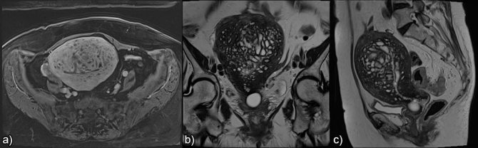
Fig. 9 Swiss cheese appearance a) axial T1 3D FS, b) coronal T2 and c) sagittal T2 showing a poorly demarcated, thickened transition zone with a large number of glandular cysts, nodules and linear grooves in the myometrium.
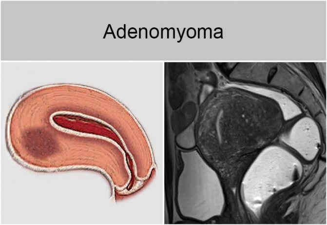
Fig. 10 Adenomyoma. Sagittal T2-weighted image, circumscribed hypointense mass in the thickness of the myometrium with unclear contours, small hyperintense foci in the structure and minimal mass effect.
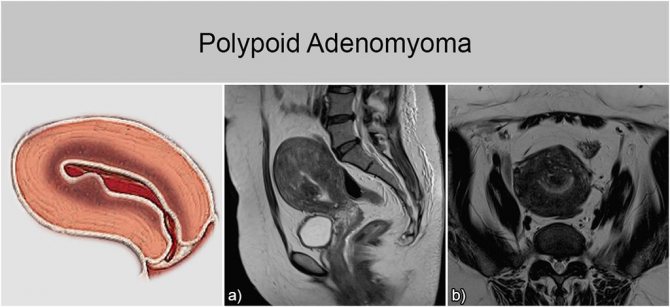
Fig. 11 Polypoid adenomyoma: a) sagittal T2 and b) axial T2-weighted images, a spherical section of the transition zone with unclear contours, protruding in the uterine cavity.
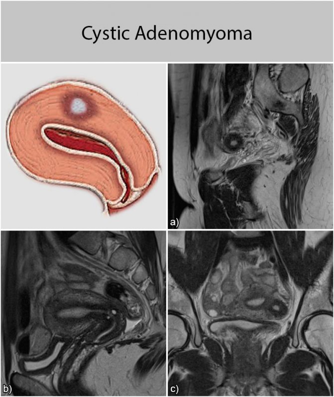
Fig. 12 Isolated or juvenile cystic adenomyoma: a) and b) sagittal c) coronal T2-weighted images, spherical formation with a central cavity having a hyperintense signal, not associated with the uterine cavity; otherwise the uterus is normal.
What are the features of using ultrasound?
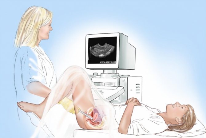
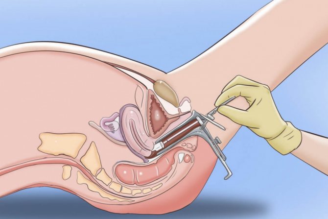
If the development of adenomyosis is suspected, the gynecologist, after an initial examination, gives a referral for an ultrasound examination, which is performed transvaginally. This ultrasound examination method is not used in girls and young women who are not yet sexually active.
For an ultrasound to be informative and effective, you need to prepare for it. Basic rules of preparation:
- 2–3 days before the procedure, exclude from the diet foods that contribute to increased gas formation.
- The evening before the test, drink a laxative and have a bowel movement.
- Before manipulation, empty your bladder.
If there are contraindications for transvaginal ultrasound, transabdomial ultrasound is prescribed. To confirm the diagnosis, the following diagnostic procedures may be additionally prescribed:
- hysteroscopy;
- Ultrasound of the uterus with Dopplerography, elasography;
- hysterosalpinography;
- Ultrasound of the ovaries.
Manifestations of adenomyosis on an ultrasound echogram
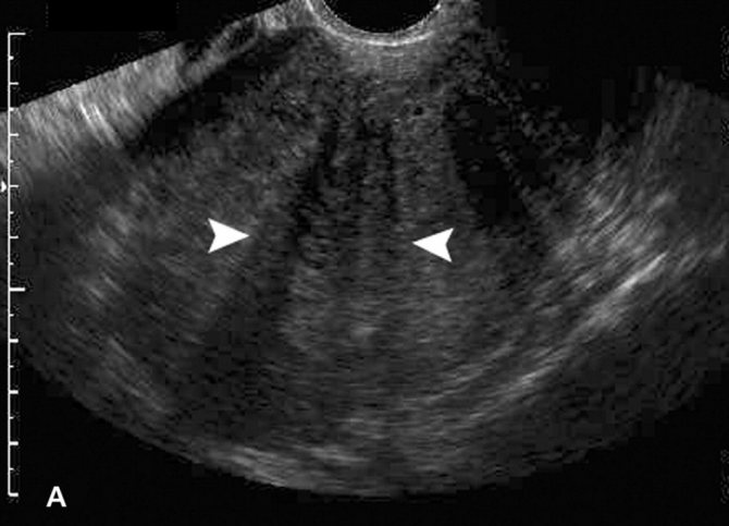
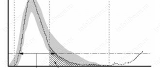
Signs of uterine adenomyosis on ultrasound are characterized by noticeable deviations from generally accepted physiological norms. Pathology is best visualized at the beginning of the second cycle of the luteal phase, when endometrial functions are most active.
Characteristic echo signs of adenomyosis on ultrasound:
- heterogeneity of myometrial tissue;
- blurred boundaries between the germ and muscle layers;
- rounded shape of the organ;
- asymmetry in wall thickness;
- the presence of endometrial spiral vessels in the myometrium;
- deviation from the norms in length, width, weight by more than 10%;
- pockets of compaction, voids more than 2 mm inside the muscle layer.
Adenomyosis on ultrasound is manifested by changes in the density of uterine tissue, as well as the presence of areas with hypoechogenicity, hyperechogenicity, and heteroechogenicity. Also, during an ultrasound examination, pathological spots with impaired density ranging in size from 1 to 50 mm are detected.
Symptoms of adenomyosis

Signs of adenomyosis begin to actively appear from stage 2. At first they are almost invisible. The patient does not feel any pain, and the presence of pathology can only be indicated by prolongation of menstruation and increased bleeding (this happens only in some cases).
Starting from stage 2, the following signs of uterine adenomyosis are observed:
- Pain in the lower abdomen that occurs a few days before menstruation and disappears 2-3 days after menstruation.
- Painful sexual intercourse.
- Discharge a few days before and after menstruation: spotting, brownish in color.
- Manifestations of anemia: pallor and flaking of the skin, headaches, weakness, drowsiness, fainting.
- Bleeding in the middle of the menstrual cycle.
- Disruptions of the menstrual cycle: as a rule, the volume of discharge increases, and the periods themselves last longer.
Reference! Pain with adenomyosis is cutting in nature. They are capable of spreading to the left or right side of the groin area, perineum, and rectum.
Decoding the ultrasound protocol
The results of the ultrasound are recorded in a protocol, which will be deciphered by the gynecologist. When studying the data obtained, the specialist first of all pays attention to the parameters of the organ and their compliance with the standards.
Sonographic signs of the absence of adenomyosis:
- Size – corresponds to physiological standards.
- The shape is pear-shaped.
- The wall thickness is uniform and symmetrical.
- The contours of the boundaries between the muscle and growth layers are clear, even, without bulges.
- The structure of the myometrium is unchanged, homogeneous.
- The thickness of the functional layer of the myometrium corresponds to the phase of the cycle, there is no deformation of reflection according to M-echo.
If echo signs of uterine adenomyosis are visualized, the doctor evaluates the form, localization and stage of the disease, after which he draws up an individual treatment plan or prescribes surgery.
Signs of adenomyosis development on ultrasound
Ultrasound signs of adenomyosis in the initial stages of development are clearly visualized on the monitor:
- The presence of tubes from the endometrium to the myometrium.
- Formation of circles or ovals, as well as notches in the basal layer.
- Various defects in the endometrial layer.
As adenomyosis progresses, signs of adenomyosis on ultrasound become more pronounced:
- asymmetry of the walls;
- increase in size;
- uneven increase in the posterior and anterior walls.
Adenomyosis as a pathology of the endometrium
Uterine adenomyosis is a common disease of benign etiology, diagnosed in women of childbearing age. Pathology is considered as one of the stages of endometriosis. According to the location of endometriosis, it can be:
- genital;
- extragenital.
Genital, in turn, is internal and external. When internal, the body of the uterus and the supravaginal cervical part are affected. Externally, pathological processes occur in the following areas:
- body of the uterus and part of the cervix;
- retrocervical zone;
- the fallopian tubes;
- vagina;
- ovaries;
- Douglas space.
Reasons for development
The root causes of the development of the pathology have not been fully established. But doctors put forward various theories with the help of which they try to explain the etiology and pathogenesis of adenomyosis. A popular theory is the implantation of separated endometrial cells during retrograde reflux of menstrual blood through the fallopian tubes.
There are theories that speak of the spread of pathological cells by hematogenous and lymphatic routes. The genetic factor may also play a significant role, given the concordance of the manifestation of adenomyosis in twins. Many medical studies have been conducted in search of the exact root cause of the pathology, but doctors have not received a clear answer to this question.
What are the causes of adenomyosis
Adenomyosis, or endometriosis, has been known for a long time. To this day, scientists cannot accurately name either the reasons for its development or decide on uniform approaches to the treatment of uterine adenomyosis.
One theory suggests that adenomyotic heterotopias develop from endometrial cells that enter the abdominal cavity during gynecological operations. Retrograde reflux of menstrual blood can occur during menstruation. Other scientists believe that adenomyosis develops from the remnants of the Wolffian and Müllerian ducts, which are formed during embryogenesis. There is an opinion that foci of adenomyosis arise in the place where the uterine endothelium has metastasized through the blood or lymphatic vessels.
The theory of hormonal dysfunction has the largest number of supporters. They believe that due to a violation of the hormone ratio, proliferation of the coelomic epithelium begins, which is located among the endothelium of the peritoneum. This causes adenomyosis of the uterus. Its treatment has also not been fully studied.
Risk factors
- Risk factors for the occurrence of pathology include:
- higher than normal estrogen levels;
- early onset of menstruation, their abundance, duration more than 8 days;
- surgical intervention on the uterus, appendages;
- abuse of bad habits;
- obesity;
- immune dysfunction;
- chronic stress, nervous overload;
- unfavorable environmental conditions;
- genetic predisposition;
- history of abortions.
Risk factors for developing adenomyosis
Gynecologists have identified factors that increase the risk of developing this condition:
- Age from 40 to 50 years (before menopause);
- A history of several births, although this disease is also often found in nulliparous women, detected during an examination for infertility.
- Surgical operations of the uterus - caesarean section, surgery to remove fibroids, abortions.
Diagnostics

If you have characteristic symptoms, you should contact a gynecologist who will diagnose and treat the pathology. To confirm the diagnosis, a referral is given for a comprehensive examination, the results of which will help determine further tactics for managing the patient.
Bimanual examination
During a bimanual examination, one hand of the doctor is placed on the stomach, the other is inserted into the vagina. The procedure allows you to determine the increase in the size of the uterus, poor mobility, which is the result of the process of formation of adhesions.
Inspection in the mirrors
A gynecological examination in the speculum will provide the specialist with information about the presence of endometriotic lesions in the vagina and on the surface of the vaginal cervix.
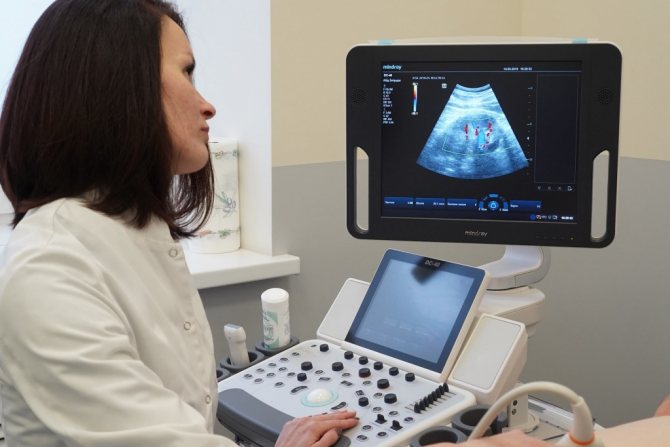
Ultrasound diagnostics
Ultrasound examination of the pelvic organs is an accessible, effective, safe, painless diagnostic method, which, if adenomyosis is suspected, is recommended to be performed over time on days 8–10 and 20–24 of the cycle. The procedure will help determine the degree of pathological changes in the structure of the reproductive organ. At stage 1 of adenomyosis, the uterus on ultrasound is slightly enlarged in size, and in the projection of the walls, small cystic formations with a diameter of up to 2 - 3 mm are determined. The presence of cystic inclusions makes the structure of the myometrium heterogeneous, its echogenicity is slightly increased.
At stages 2–3, endometrial cells spread to the muscle tissue towards the serous layer, which indicates a diffuse form of pathology, which is characterized by the following signs:
- heterogeneous structure of the organ;
- uneven wall thickness;
- linear striation of the median M-echo, blurring of its contours;
- increase in organ size.
Ultrasound picture in Doppler mode
To assess the condition and functionality of the uterine vessels during ultrasound, color Doppler mode is used. This type of scan allows the doctor to see blood flow and detect signs of adenomyosis.
If the walls of the uterus are damaged by pathological endometrial cells, Doppler ultrasound can detect a slight increase in vascular resistance and increased blood flow in areas affected by the disease.
Possible diagnostic errors
Errors in ultrasound diagnosis of uterine adenomyosis occur quite rarely, but still they are not excluded, because the signs of the disease often coincide with the clinical picture of other diseases. For example, myomatous nodes can be confused with overgrown endometrial tissues, which are located in unusual places. Specific bending of the feeding vessels around the formation in color or power Doppler mapping mode will help to distinguish myomatous nodes.
The nodular form of adenomyosis can be confused with uterine sarcoma, but the malignant neoplasm is characterized by characteristic hypervascularization in the CD mode with a low-resistant type of blood flow. An ultrasound doctor conducting an examination of a patient with suspected adenomyosis must be able to recognize the clinical signs of all existing gynecological pathologies and have comprehensive information about the echographic picture of each. This will help to avoid mistakes and inaccuracies in making a diagnosis, and also to prevent the development of serious complications associated with the lack of timely treatment.