Expecting a baby is a wonderful time for any woman. The child is still in the mother’s womb, but the mother wants to see her baby as quickly as possible and make sure for herself that his health is not in danger.
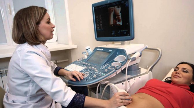
Carrying out the so-called “screening” - a method of examining the fetus in utero using 3D and 4D ultrasound allows medical specialists to assess the general health of the embryo, determine the sex of the unborn child and at the same time show the woman a picture of the development of her unborn child.
3D ultrasound
Three-dimensional ultrasound is a method of ultrasound examination that allows you to see an object of interest from three dimensions . The ability to examine the embryo from all sides, determine its length and width makes this type of diagnosis a unique find in the field of gynecology.
This type of examination of the baby is a very important link in the long stage of its formation in the mother’s womb. 3D ultrasound can be an excellent tool for obstetricians to determine all the important points related to the health of a developing child. This examination should be carried out only in highly specialized medical institutions where first-class specialists work. The examination is carried out in all 3 trimesters of pregnancy. The 1st and 3rd screenings are considered significant.
3D ultrasound - how it works
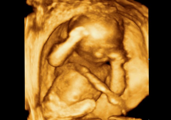
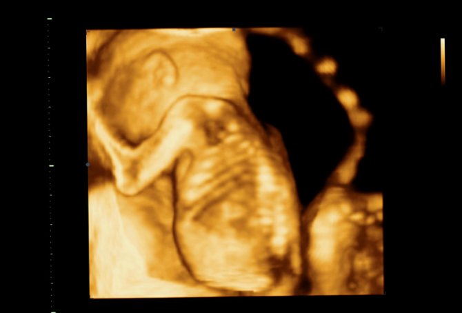
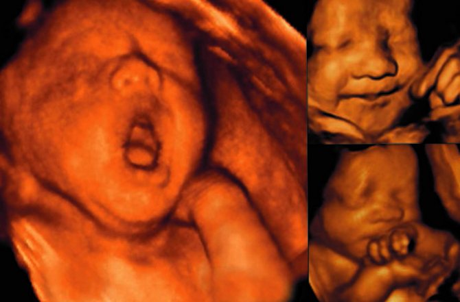
The technique of three-dimensional ultrasound examination has no fundamental differences from standard ultrasound, which is based on the penetrating ability of ultrasound, its distribution in the tissues of the body depending on the density and composition of the medium. Only the image provided by a classic ultrasound does not allow future parents to detect the baby’s spine and large bones without medical assistance. While a 3D ultrasound image can be compared with an ordinary photograph in which the child’s face is visible, it is possible to count his fingers.
You need to prepare for the fact that the duration of the procedure will increase compared to a standard ultrasound. It will take at least 40 minutes.
When is a 3D ultrasound performed?
The first ultrasound that is significant for a pregnant woman is performed at the 20th week , the other at the 8th month . The latest screening allows you to determine:
- Fetal position.
- Presence of pathologies (if any).
- Features of embryo diligence.
- Possibility of entwining the fetus with the umbilical cord.
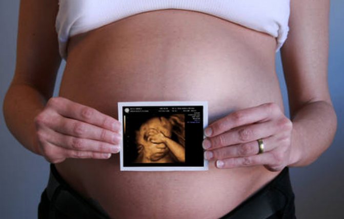
According to medical workers, a study of this nature is more important for parents, since they can see the baby’s face and discern his facial features. The ultrasound results are recorded on disk so that parents can later remember these happy moments of the desired “meeting” with the baby.
The woman's bladder should always be empty during the procedure.
4D ultrasound
Today, such a study as 4D ultrasound is a highly informative ultrasound diagnostic . The essence of it is that not a static picture is displayed on the monitor screen, but an image in three-dimensional format, recorded in real time in video format.
Throughout the entire period of pregnancy, there are certain time intervals when expectant mothers must undergo examination, part of which is monitoring of fetal development in the intrauterine state. To undergo screenings, doctors developed an appropriate ultrasound schedule.
4D should begin around the 20th week of pregnancy . This period was not determined by chance; the picture obtained at the 20th obstetric week will turn out to be more realistic.
At an earlier stage, instead of the child, with such an ultrasound it will be possible to examine only bones and internal organs.
A timely 4D ultrasound allows you to examine all the smallest details of the unborn child’s body. It will allow you to determine the sex of the child and examine the fingers and toes.
This ultrasound is carried out at the 32nd week, in this case it will already be possible to see all the limbs, the baby’s body itself; The device can even record the facial expressions of the fetus.
4D ultrasound does not require any special preparation; the fullness of the pregnant woman’s bladder does not play a role during its passage.
Contraindications for the procedure
At what time it is better to do a 3D ultrasound during pregnancy, the doctor should determine when he analyzes the results of the diagnostic studies already performed. As a rule, if the picture of fetal development is clear and assessed as positive, additional studies (in particular 3D ultrasound) are not prescribed. It must be said that this procedure has no specific contraindications.
Yes, during a 3D ultrasound, stronger ultrasound waves are indeed emitted. However, this does not happen throughout the entire procedure (for example, a whole hour). A lot of time is spent on processing the information received rather than on the scanning itself.
Importance of the study
Screening helps to identify various pathologies at an early level of their appearance. This diagnostic method makes it possible to promptly identify anomalies and developmental defects in the unborn child:
- Down syndrome.
- Cleft lip.
- Wolf's mouth.
- Heart disease and so on.
The pros and cons of 4D ultrasound are exactly the same as the 3D format.
Ultrasound in three dimensions
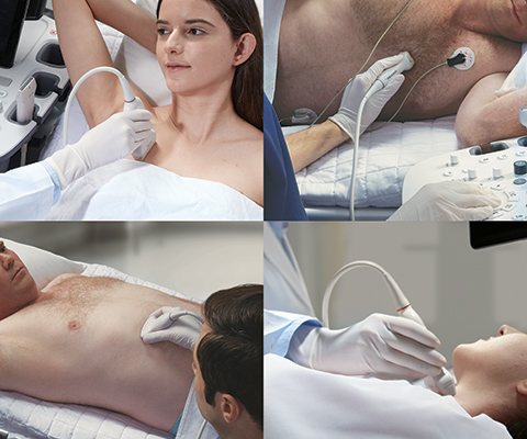
Ultrasound scanner HS60
Professional diagnostic tools.
Assessment of tissue elasticity, advanced 3D/4D/5D scanning capabilities, BI-RADS classifier, options for expert cardiological studies.
Over the past decade abroad and in the last two years in our country, a new method of ultrasound diagnostics - three-dimensional ultrasound - has become increasingly popular among both patients and doctors. How does this study differ from the traditional, two-dimensional one?
The first ultrasound devices began to be developed in 1955, and at the moment it is difficult to imagine medicine without this diagnostic method. The safety of the method has been tested many times by scientists around the world. Ultrasound is recognized as a safe and reliable method of fetal imaging.
The first “three-dimensional” device appeared in 1989 in Austria. Unfortunately, the quality of the images was very poor, and it took up to 30 minutes to obtain one static 3D image. Naturally, the method has not found wide application in medicine. Only in 1996, thanks to a breakthrough in computer technology, a scanner with the ability to perform three-dimensional reconstruction in real time appeared. And from now on, the 3D ultrasound technique is increasingly used in medicine, especially in the field of obstetrics!
Devices for two-dimensional and three-dimensional ultrasound look identical in appearance and differ only in the presence of a special built-in module (a set of high-tech electronic boards) and special sensors. Understanding this is very important, since only new functions are added, while the scanning frequency (usually 3.5 MHz), intensity and power of the ultrasonic wave remain the same
- the same as with conventional ultrasound examination. That is, in a physical sense, three-dimensional ultrasound is no different from two-dimensional ultrasound, but in diagnostic terms it expands its capabilities.
A few words about the sensor the doctor uses to conduct a three-dimensional examination. Externally, it differs from the sensor you are used to only in that it is several times larger. This is due to the fact that inside its body there is a conventional two-dimensional sensor, which is constantly moving using a special mechanism. Multiple scans - two-dimensional images - are transferred from the sensor to a powerful computer located inside the scanner, where they are summarized using the aforementioned built-in module. The resulting three-dimensional (volumetric) image is displayed on the device screen.
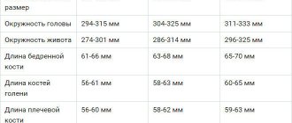
To be fair, it must be said that modern two-dimensional ultrasound machines enable specialists to obtain the maximum amount of information necessary to determine the condition of the mother and child. Unfortunately, not every medical institution has a fleet of devices that meets modern diagnostic requirements. Considering that the two-dimensional ultrasound technique has been used and improved for several decades (since the 50s of the last century), it can be stated that specialists have clearly developed methods for standardizing the data obtained from ultrasound examination. So, each period corresponds to certain sizes of the head, limbs, internal organs of the fetus, including certain structures of the brain, heart and others. Three-dimensional examination data provides additional information, especially for the diagnosis of certain developmental defects: limbs, face, spinal column. Thus, suspicion of the presence of such defects can also be considered a medical indication for conducting a three-dimensional study.
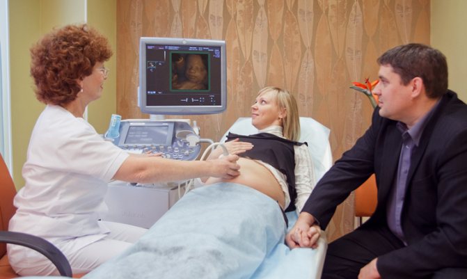
3D ultrasound is a natural technical development of 2D ultrasound, not only adding precision to the examination, but also allowing the expectant mother to get to know the baby before birth. The most optimal option for ultrasound examination is a combination of both methods. In this case, the doctor first of all receives all the necessary information using a traditional study, supplements it with the help of three-dimensional vision and confirms his opinion about the well-being or unfavorability of the course of this pregnancy. Parents have the opportunity to see the baby not in the form of incomprehensible black and white “floating” lines and dots, but in the form of a three-dimensional image in real time, reminiscent of “old” video recording. If you use this “miracle of technology” for all 3-4 ultrasound examinations required during pregnancy, then by the time the baby is born, a whole video will have been shot about it. It is probably unnecessary to talk about how parents react to footage of such a film, both at the time of its creation, and when watching the videotape and remembering what the child was like when he was in his mother’s tummy. In this regard, it is also interesting that it will be easier for you to explain to your baby where he was when he was not there yet. And existing older children, having been in the ultrasound room, treat the upcoming appearance of a brother or sister with greater responsibility and understanding. 3D ultrasound combined with video recording is a beautiful and modern part of family documentation.
When undergoing a three-dimensional ultrasound, it is necessary to take into account that the examination time may be slightly longer than with a standard two-dimensional one. The quality of the resulting image when using three-dimensional ultrasound depends on the position of the fetal body, the location of its limbs, the umbilical cord and the placenta. Difficulties in obtaining volumetric images may be due to a small amount of amniotic fluid, even in cases where their relatively small amount is not yet pathological (oligohydramnios). Significant problems in the quality of the picture usually arise if the pregnant woman is overweight or if she has scars on the anterior abdominal wall after abdominal surgery. The success of a three-dimensional study (obtaining high-quality images of the fetus) often depends on motor activity - the more active the fetus, the greater the chance of seeing more interesting pictures of intrauterine life. If the fetus is motionless and is located inconveniently for the researcher, then the doctor may suggest interrupting the examination for a while to wait for a suitable position of the child. During this time, it is advisable to drink some sweet drink (for example, sweet tea), which usually increases the motor activity of the fetus after 10-15 minutes.
The information obtained and the type of three-dimensional picture depends on the stage of pregnancy at which the examination is carried out. During pregnancy from 3 to 8 weeks, 3D images are not effective. From the 10th to the 16th week you can see the entire fetus, its posture, arms, legs, umbilical cord (without distinct small details). In some cases, during these periods it is possible to visualize the face of the unborn child, however, it should be understood that the face of the fetus during these periods bears little resemblance to the face of a person. The optimal pregnancy period for 3D ultrasound is from 15 to 30 weeks. At such a time frame, it is possible to obtain images of live facial expressions in the fetus. After 23–25 weeks, the fetus becomes so large that obtaining an image of it as a whole is no longer possible, so on the screen you can see the head and shoulders, arms, legs, and torso in turn. With upcoming repeat births, the gestational age at which 3D ultrasound can be successful increases to 34-38 weeks.
To summarize all of the above, three-dimensional ultrasound in obstetric practice has already taken its strong place next to two-dimensional research. Being a modern high-tech method, it improves the diagnosis of various anomalies and gives a new look at fetal malformations already identified by the usual method. And a visit to the doctor’s office with the whole family makes an invaluable psycho-emotional contribution to preparing for the arrival of a new person. If for some reason you do not have the opportunity to undergo all the ultrasound examinations necessary during pregnancy in combination with a three-dimensional technique, then do not miss this at least in the third trimester, when the baby will be almost the same as when you personally “met” in the delivery room.
Ultrasound scanner HS60
Professional diagnostic tools.
Assessment of tissue elasticity, advanced 3D/4D/5D scanning capabilities, BI-RADS classifier, options for expert cardiological studies.