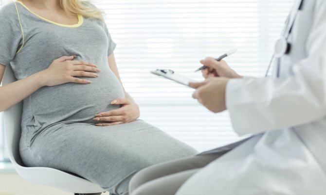
Our mothers had to live through 9 months of pregnancy, having a very vague idea of how their body was changing and how the baby was developing. Modern medical technologies have made it possible to make the period of waiting for a child more comfortable for a woman, relieving her of many worries.
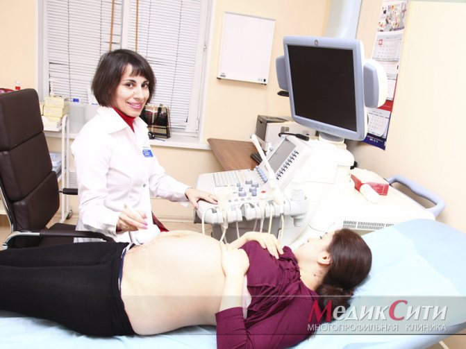
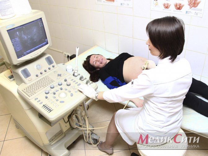
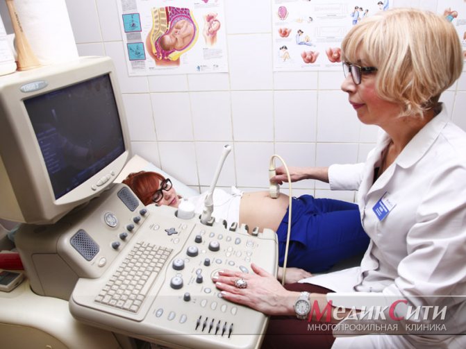
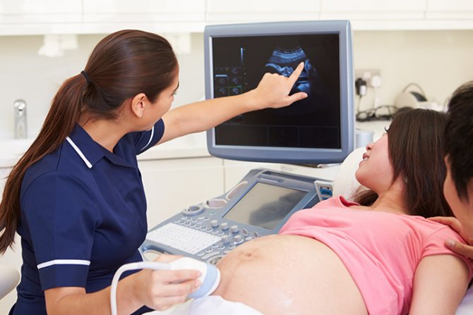
Ultrasound examination is the main method for studying the anatomical and functional state of the fetus throughout pregnancy. Ultrasound allows you to determine the presence and duration of pregnancy, find out the sex of the child, and most importantly, with the help of ultrasound scanning, doctors can monitor the development of the embryo and take timely measures if any trouble is detected.
At MedicCity, patients have the opportunity to undergo ultrasound examinations during pregnancy using premium Voluson 10 equipment. The scanner is indispensable for diagnosing fetal development during pregnancy, identifying congenital pathologies and other potential dangers. Our highly professional obstetricians and gynecologists with extensive experience in this field, doctors and candidates of medical sciences, armed with first-class equipment, will do everything possible to ensure that the period of waiting for a child proceeds as smoothly as possible!
1 fetal ultrasound
2 fetal ultrasound
3 Fetal ultrasound
General preparations
|
| Preparation for an ultrasound is required only in the early stages of pregnancy |
The preparatory stage depends on the ultrasound method and the stage of pregnancy.
Two methods are used in the first trimester
:
- with the transvaginal ultrasound method, no preparation is required, ordinary hygiene measures are sufficient,
- When using the transabdominal method, when the sensor is located on the anterior surface of the abdomen, gases and food debris present in the intestines may interfere with the examination of the uterus, because Ultrasound waves will be reflected from these structures, so the following measures
: 2-3 days before the ultrasound, it is not recommended to eat foods that lead to fermentation and the formation of gases in the intestines (vegetables, some fruits, baked goods, dairy products, brown bread, carbonated drinks), - the study is performed on an empty stomach, the last meal is possible 8-10 hours before the procedure,
In the second and third trimesters, ultrasound is performed transabdominally, but no preparation is required, because the intestine is pushed to the periphery of the abdominal cavity by the enlarged uterus and cannot interfere with the examination.
Our doctors
General Director Anshina Margarita Beniaminovna Make an appointment
Director of the Rizhinashvili clinic Semyon Iosifovich Make an appointment
Obstetrician-gynecologist-reproductologist Zhordanidze Diana Omarovna Make an appointment
Obstetrician-gynecologist-reproductologist Torchinov Aslanbek Ruslanovich Make an appointment

Obstetrician-gynecologist, reproductive specialist. Efimova Maria Sergeevna Make an appointment
Obstetrician-gynecologist-reproductologist Anna Sergeevna Fedorova Make an appointment
Obstetrician-gynecologist Samoilova Tatyana Evgenievna Make an appointment
Urologist-andrologist of the highest qualification category Gablia Mikhail Yurievich Make an appointment
Obstetrician-gynecologist-endocrinologist Sharyafetdinova Failya Abdulkhaevna Make an appointment
Obstetrician-gynecologist Pushkina Valeria Vadimovna Make an appointment
Obstetrician-gynecologist Margarita Yuryevna Skvortsova Make an appointment
Obstetrician-gynecologist Slobodchikova Valeria Yuryevna Make an appointment
Obstetrician-gynecologist Peskova Irina Evgenievna Make an appointment
Perinatologist Nelly Idrisovna Kokhno Make an appointment
Obstetrician-gynecologist Olga Borisovna Kuharkina Make an appointment
Obstetrician-gynecologist Pitskhelauri Elena Germanovna Make an appointment
Hemostasiologist Lopukhin Vadim Olegovich Make an appointment
Oncologist-mammologist Victoria Borisovna Akimova Make an appointment
Ultrasound diagnostics doctor Olga Ivanovna Babaeva Make an appointment
Ultrasound diagnostics doctor Brilliantova Nadezhda Nikolaevna Make an appointment
Ultrasound diagnostics doctor Natalya Sergeevna Gorovaya Make an appointment
Cardiologist Maria Viktorovna Cherkasova Make an appointment
Endocrinologist Anzhelika Vladimirovna Manovitskaya Make an appointment
Gastroenterologist Golysheva Svetlana Valerievna Make an appointment
Obstetrician-gynecologist-reproductologist Olga Aleksandrovna Iskorostinskaya Make an appointment
Urologist-andrologist Iskorostinsky Evgeniy Vladimirovich Make an appointment
Psychologist Barskaya Ekaterina Sergeevna Make an appointment
Therapist Alexandra Vladimirovna Kabakova Make an appointment
Anesthesiologist-reanimatologist Isaev Oleg Vladimirovich Make an appointment
Psychiatrist, sexologist Teona Otarievna Kacharava Make an appointment
Head of the Embryology Laboratory. Ph.D. Sergeev Sergey Alexandrovich Make an appointment
Embryologist Kalinina Irina Ivanovna Make an appointment
Embryologist Kalyuzhny Sergey Andreevich Make an appointment
Embryologist Matveeva Elona Olegovna Make an appointment
Embryologist Vladimir Viktorovich Ovsyankin Make an appointment
Indications for ultrasound of pregnant women
|
| Ultrasound for pregnant women is performed routinely in each trimester. |
In obstetrics and neonatology, the ultrasound research method is considered harmless and safe for the mother and fetus, therefore it is used for dynamic monitoring of the course of pregnancy and the development of the child. Ultrasound can be prescribed routinely and for special indications.
- Pregnancy study as planned.
During the normal course of pregnancy, if the expectant mother is not worried about anything, and the doctor is satisfied with the results of her tests, the woman must undergo three scheduled ultrasounds. Timing of these screening studiesregulated in the order of the Ministry of Health of the Russian Federation No. 572n dated November 1, 2012:
- 1st at 11-14 weeks of the first trimester,
2nd at 18-21 weeks of the second trimester,
- 3rd at 30-34 weeks of the third trimester.
- Additional Research for special indications.
Reasons for sending a woman for an ultrasound scan during periods of pregnancy not established by law may be indications from the mother or fetus:- Indications for an unscheduled ultrasound on the part of the mother
: pain or discomfort in the lower abdomen,
the appearance of bloody vaginal discharge, discharge with blood clots,
- Indications for an unscheduled ultrasound on the part of the mother
- leakage of amniotic fluid,
- threat of premature termination of pregnancy,
- suspicion of ectopic pregnancy,
Ectopic (tubal) pregnancy - acute infectious diseases or exacerbation of chronic diseases of a woman (decompensation of diabetes mellitus, exacerbation of chronic pyelonephritis, cholecystitis, etc.),
- suspicion of multiple pregnancy,
- abnormalities of the placenta,
- history of complicated pregnancy (miscarriage, frozen pregnancy),
- discrepancy between the size of the uterus and the gestational age,
- pathology of the cervix,
- multiple pregnancy,
Multiple pregnancy - suspicion of a disease of the female reproductive system (tumors, acute inflammatory diseases, cysts, polyps),
- a burdened hereditary history of the development of genetic diseases in a woman (Down's disease),
- abdominal trauma in a pregnant woman,
- monitoring the condition of the uterine scar after cesarean section,
- negative Rh blood factor,
- dynamic monitoring of pregnancy during IVF.
- Indications for unscheduled ultrasound of the fetus
:- suspicion of atypical attachment of the fertilized egg,
- the appearance of symptoms of insufficient fetal activity in the second and third trimester of pregnancy,
- threat of intrauterine infection of the fetus,
- signs of fetal hypoxia,
- suspicion of the development of genetic abnormalities or fetal malformations.
Fetal brain malformation (cephalocele)
The purpose of screening ultrasound is to assess the general condition of the unborn child and mother, measure the size of the fetus, assess the location and condition of the umbilical cord and placenta, study uterine, placental and vascular blood flow, timely diagnosis of possible hereditary and congenital pathologies of the fetus, pathological abnormalities in the course of pregnancy at various stages, dynamic monitoring the health of women and babies.
Ultrasound in early pregnancy
|
| Ultrasound of a pregnant woman in the first trimester of pregnancy |
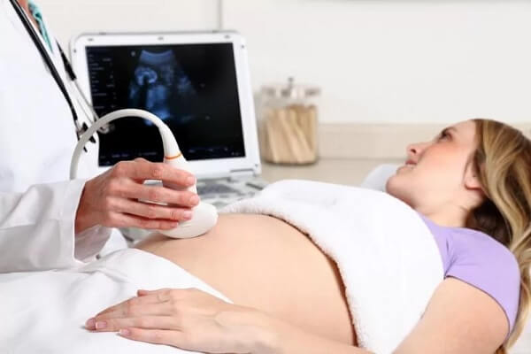
With the help of transvaginal ultrasound, the onset of intrauterine pregnancy can be reliably determined no earlier than the 4-5th week, with the transabdominal method - later, at the 8th week. With a vaginal examination at the 6th obstetric week, the fetal heartbeat can already be determined.
But whether it is necessary and whether it is dangerous to perform ultrasound in very early stages of pregnancy is a controversial issue among obstetricians around the world. Carrying out ultrasound during this period
justified not so much in order to ensure the presence of a fetus, but in order to:
- exclude ectopic pregnancy,
- determine the number of formed embryos,
- eliminate the risk of miscarriage.
Unfavorable prognostic signs
with ultrasound in very early stages of pregnancy are:
- deformation of the ovum,
- impossibility of identifying an embryo at 6 weeks of gestation,
- absence of fetal heartbeats at 6-7 weeks,
- lag in the increase in the size of the ovum from the gestational age,
- absence of fertilized egg in the uterine cavity,
Identification of threatening signs allows you to make a timely decision about the possibility of continuing the pregnancy or the need to terminate it without serious complications.
At the same time, ultrasound examination in the early embryonic period cannot be considered completely safe. The embryo is still too small, and the rate of cell division and the likelihood of disruption of this process are very high. The detrimental effect of ultrasound on the process of formation of fetal organs has no evidence base, but obstetricians prefer not to take risks and, unless absolutely necessary, do not prescribe ultrasound for pregnant women until the end of the main process of organogenesis. The older the fruit, the lower the risk of damage.
The safe period for ultrasound examination of pregnant women around the world is considered to be 10-14 weeks of the first trimester - the first planned ultrasound.
This is a very important screening test, sometimes called a “genetic ultrasound,” which provides essential information about the condition of the mother and fetus. The study can be carried out both vaginally and transabdominally, both methods make it possible to find out
:
- the presence of intrauterine pregnancy (determination of a gestational egg in the uterine cavity),
- gestational age, this is necessary to determine the expected date of birth,
- stage of fetal development, compliance with the timing of pregnancy,

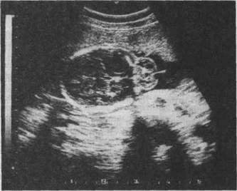
Normal photograph of a fetus at 12 weeks - presence of signs of a “frozen” pregnancy,
- number of embryos,
- presence of fetal heartbeat,
- position of the fetus, determine its motor activity,
- intrauterine anomalies of fetal development,
- presence or absence of chromosomal abnormalities
: according to the measurement results: the width of the collar zone is an accumulation of lymph in the neck area between the skin and soft tissues that cover the spine, thickening of this zone is a symptom of chromosomal disorders (Down syndrome), - determining the presence of nasal bones, because their absence within 11-14 weeks indicates the likelihood of Down syndrome;
The first screening ultrasound makes it possible to exclude such pathological conditions
, How:
- uterine fibroids,
- various neoplasms,
- abnormal placenta previa,
- abnormal aging of the fetus,
- chorionic insufficiency,
- ectopic pregnancy.
Exclusion of ectopic pregnancy is an important goal of ultrasound. If a woman has abdominal pain or spotting before 11 weeks of gestation, an unscheduled ultrasound is performed very early, which sometimes allows surgical intervention to be avoided. If there are no threatening symptoms, an ultrasound scan is performed as planned. The fact of an ectopic pregnancy can only be reliably determined using ultrasound.
How is the examination carried out?
Depending on the duration, the study is carried out using the transabdominal or transvaginal method. In the first case, the doctor treats the abdomen with a safe hydrogel, which is necessary for better transmission of ultrasound and sliding of the sensor. The patient lies on the couch, exposing her stomach.
In the second case, a device is used, the sensor of which is inserted into the vagina. At an early stage, this method allows you to study the cervical canal and cervix.
To get more useful information about the timing, benefits and price of the study, sign up for a paid pregnancy ultrasound at the FertiMed Center for Reproduction and Genetics. To do this, just call the numbers listed on the website.
Don't waste time! Make an appointment now!
By clicking the “Submit Application” button, you consent to the processing of your personal data in accordance with the terms
Privacy Policy and User Agreement
* - required fields
How long can you determine the sex of a child?
Mom and dad of the baby always look forward to the moment when they find out who will be born - a boy or a girl. It is also important for doctors to know the sex of the baby, especially if there are risk factors for the transmission of genetic diseases, in order to timely plan for the possibility of prolonging or terminating the pregnancy.
Sex cells are formed in the 5th week of pregnancy, but the gonads begin to form only in the 7th week. Despite the fact that the sex has already been genetically determined, it is not yet possible to see this.
At the 8th obstetric week, ovaries form in girls, and testicles form in boys, which almost immediately begin to produce testosterone (male sex hormone).
The external genitalia begin to appear at 10-11 weeks, but at this time it is still impossible to distinguish a boy from a girl. And only by the 12th week does the genital tubercle turn into the penis or clitoris.
From this moment (12 weeks) it is already possible to determine the sex of the baby, but only on top-class devices with 3D or 4D reconstructions.
| Determining the sex of a child (boy) at 12 weeks of pregnancy using 3D ultrasound |
With a regular two-dimensional ultrasound, the sex of the baby is determined after the 20th week, i.e. during the 2nd mandatory ultrasound. At a later date, closer to childbirth, it can be difficult to determine gender, because Due to its large size, the fetus becomes less mobile in the uterine cavity.
| Determining the sex of the baby by ultrasound (20th week): boy on the left, girl on the right |