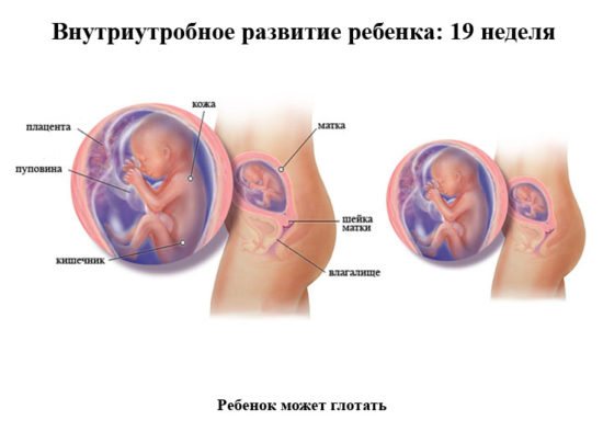
For a pregnant woman at 19 weeks, a kind of milestone comes - almost half of the difficult path to the birth of a new life has already been completed. An enlarged belly at the 19th week of pregnancy is perceived as usual; the need for care over an unborn baby increases every day. The stage of daily mutual communication between future parents and the child begins, since he is already actively making himself known through movements, reacting to intonation, hunger, stroking and other external stimuli. An ultrasound at 19 weeks of pregnancy will help monitor the process of intrauterine development.
Information content of ultrasound at 19 weeks
With the help of ultrasound diagnostics, at this period it is possible to ascertain the degree of formation and development of the functional systems of the fetal body, and to assess the condition of the woman’s reproductive organs. The baby at the 19th week of pregnancy is already developed so much that an ultrasound will tell about:
- The activity of movements and their purposefulness
- Formation of tooth buds
- Development, first of all, of the nervous, urinary and muscular systems
- Child's field (with body position available for viewing)
- Body proportions, anatomical features
- The size of the fetus - its weight and height
- The number of heartbeats and respiratory acts
- The presence of pathologies such as: hydrocele of the brain, Down syndrome, developmental retardation, malnutrition, abnormal body constitution, fetoplacental insufficiency)
- Condition of the umbilical cord
- Degrees of placenta maturity
- Condition of the uterus
- Quantity and quality of amniotic fluid
An addition to the standard ultrasound procedure is vascular Doppler. This study is carried out in the same way as ultrasound. Doppler ultrasound allows you to evaluate blood circulation in the functional system “uterus-placenta-fetus”: the direction of blood movement through the vessels, its speed.
Discharge in the nineteenth week of pregnancy
By mid-pregnancy, there are no significant changes in the nature of vaginal discharge. They become slightly more liquid than before under the influence of the hormone progesterone.
Normally, leucorrhoea has a uniform consistency and does not smell. A barely noticeable sour smell is allowed. The color of the discharge is transparent or white.
The discharge of a healthy woman is not accompanied by unpleasant sensations in the lower abdomen and perineum, and does not cause itching and redness of the mucous membrane of the external genital organs. If leucorrhoea causes itching, burning in the vaginal area, and is accompanied by an unpleasant, noticeable odor, you should consult a gynecologist. Perhaps it's an infection.
Changes in vaginal discharge may be caused by thrush or candidiasis, ureaplasmosis, trichomoniasis, gonorrhea, chlamydia and other equally unpleasant diseases.
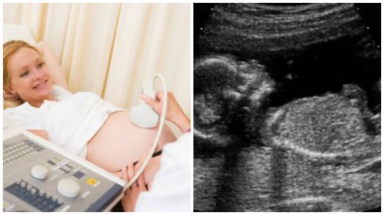
At 19 weeks, many women undergo a second trimester ultrasound. This is a mandatory procedure that should be performed no earlier than 16 and no later than 22 weeks of pregnancy.
During an ultrasound, you can see the child and observe how he behaves. At this stage of development, the fetus makes body movements, raises its arms, turns its head from side to side. This is very interesting to watch, despite the fact that the baby makes all these movements involuntarily.
An ultrasound at the nineteenth week allows you to determine the sex of the unborn child. During this period of time, various pathologies of its development can be diagnosed, including hydrocephalus. During the procedure, the specialist determines the exact size of the child, the length of its limbs, the diameter of the head circumference, determines its location, and assesses the condition of the amniotic fluid.
What will you need for an ultrasound?
Carrying out ultrasound diagnostics does not require special preparation. The procedure itself is also not a complex manipulation. At this stage, there is no need to fill the bladder, since there is already enough amniotic fluid to examine the structure of the areas of interest. Eating food before the procedure does not in any way affect the results of the ultrasound.
Many clinics do not indicate the need to bring anything with you; otherwise, you will be warned about this in advance. Most often, for the procedure, the patient may need a diaper and napkins. The diaper will need to be placed on the couch, since the ultrasound is performed in the supine position. Wipes are used to wipe away excess gel.
Why is ultrasound performed?
Ultrasound of a 19-week pregnancy is a very important study, which is not recommended to be neglected even by an absolutely healthy woman. Based on the size of the fetus and its parts, a specialist can diagnose delayed or advanced development, which affects the management of pregnancy and the expected date of birth. By assessing the structure of internal organs, including the brain, the doctor will report the presence of possible developmental anomalies.
During an ultrasound at 19 weeks of pregnancy, the fetal face is clearly visualized, on which the so-called markers of intrauterine pathology are easily distinguishable. The amount of amniotic fluid, the structure of the umbilical cord and the blood vessels of the placenta must be assessed - this is an important sign characterizing the condition of the baby in the womb. Additionally, the condition of the cervix and its appendages is examined.
If a pregnant woman is at risk, in addition to an ultrasound, she is recommended to undergo a so-called triple test . In this case, the level of three hormones is determined in the woman’s venous blood: hCG, estriol and alpha-fetoprotein. In rare cases, the concentration of inhibin is also measured. Such tests are prescribed only if pathology is suspected based on the results of the first screening at 10-14 weeks or if there is an increased risk of chromosomal abnormalities of the fetus.
Description of the diagnostic procedure
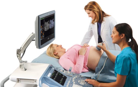
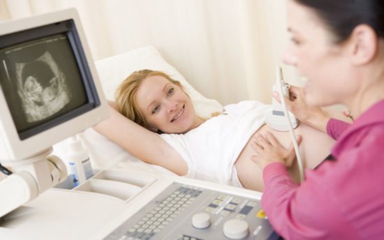
The manipulation is done transabdominally - through the anterior abdominal wall. A pregnant woman lies with her back on the couch and removes her belly from clothes. The doctor applies the gel to the abdomen and slides a sensor over it, which is attached to the monitor of the device. The fetus is visualized on the screen, the doctor measures all the necessary indicators.
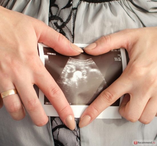
Most often, there are 2 monitors in the office so that the expectant mother can also watch her baby. The presence of the future dad is also allowed, who can take a photo of the monitor with the image of the baby.
Indicators of normal ultrasound results at 19 weeks of pregnancy

The fetus at the 19th week of pregnancy significantly changes its appearance and other indicators. The results of any medical study are compared with norms.
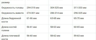
A set of medical norm parameters:
- Weight – 230-300 g
- Height – 20-25 cm
- Heart rate – 120-160 beats/min
- Systole-diastolic ratio in the umbilical cord artery – 4.55-4.67
- Biparental size – 41-46 mm
- Head circumference – 14-18 cm
- Abdominal girth – 12-16 cm
- Fronto-occipital size (lzt) – 5.4-6 cm
- Coccygeal-parietal size (ktr) – 14-15 cm
- Thickness of the collar space (tvp) – 5 mm
- Nasal bone thickness – 5.2-5.5 mm
- Shoulder length – 21-26 mm
- Forearm length – 21-26 mm
- Thigh length – 25-35 mm
- Shin length – 23-31 mm
- The baby’s presentation is cephalic, but another position is not considered a deviation, since normally it can still change more than once before the 30th week
- Amniotic fluid is clean, without suspension, volume up to 500 ml.
- The location of the placenta is the anterior and lateral wall of the uterus, at a distance of at least 7 cm from the internal os (exit of the uterus)
- The degree of maturity of the placenta is zero
- Condition of the placenta – no infarctions or calcinosis
- Internal and external pharynx – closed
- Cervical length – at least 3 cm
- Uterine tone is absent.
If any indicators are outside the normal range, the doctor will prescribe therapy that is appropriate in each specific case. Most abnormalities and conditions are treatable with timely diagnosis. For this reason, it is recommended not to neglect routine examinations at different stages of pregnancy.
What to do if your stomach hurts in the nineteenth week of pregnancy
If you experience any unusual discomfort in the abdominal area at any stage of pregnancy, you should contact a specialist. It is worth knowing that pain is not always a warning sign.
Mild aching pain in the lumbar region and lower abdomen indicates muscle strain and gradual separation of the pelvic bones. Lower back pain can also be caused by excessive physical activity on the body during the day. If it occurs, you need to lie down for 15-20 minutes.
Severe cutting pain inside can be caused by errors in nutrition. It is worth remembering that you should not take any medications even for diarrhea in case of pregnancy without prior consultation with a specialist.
The appearance of paroxysmal, cramp-like pain in the lower abdomen at week 19 should be very alarming. This symptom is a sign of increased uterine tone, causing abortion. If such pain occurs, you must take a lying position and be sure to call an ambulance. If the diagnosis of uterine hypertonicity is confirmed, the patient will need to be hospitalized. This measure will help avoid a life-threatening miscarriage.
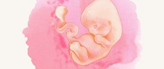
Fetus at 19 weeks gestation
All parameters that can be assessed using ultrasound diagnostics are determined by the rapid development of the child. At week 19, the following processes occur in his body:
- The baby’s movements become more conscious and orderly, thanks to the strengthening of neuromuscular connections. The child can move his jaws, push away from the walls of the uterus, and suck his fingers.
- The body takes on more proportional shapes: the limbs have the correct length ratio, the head also begins to look natural in relation to the size of the body. The phalanges of the fingers have formed.
- The skin begins to lighten a little due to the accumulation of subcutaneous fat. But the skin still looks wrinkled. The sebaceous glands produce a special lubricant that envelops the baby’s entire body and protects it from damage and infections, from the constant influence of amniotic fluid, and ultimately helps it to be born. In addition, the entire body is covered with vellus hair, which is also intended to perform a protective function.
- Centers responsible for analyzers and sense organs are formed. Receptors are actively developing.
- The digestive system will improve and it will be able to digest nutrients. Therefore, original feces are formed in the intestines.
- The child’s daily routine changes - the duration of the activity phase increases to 6-7 hours. The rest of the time he remains in a state of sleep.
- In the respiratory system, the formation of bronchioles occurs.
- The reproductive system is practically formed, and it is already possible to determine gender with great certainty. Most of the genital organs, both internal and external, are already laid. In girls, the rudiments of eggs have already been formed in the ovaries.
- The heart is fully formed.
- The immune system begins to form, the child’s body is already able to independently produce substances that can protect against infections and viruses.
At week 19, all systems are close to the final stages of their formation, the baby becomes an original organism that will soon be ready for birth.
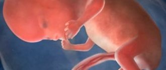
Fetal growth and development
The baby continues to grow and develop rapidly. At the 19th week of pregnancy, its body length should be from 13 to 15 cm, and it weighs about 200 grams.
Baby size now
During this period, new changes occur in the body of the growing fetus:
- The arms and legs continue to grow in length, so they now look more proportional to the body.
- The process of intensive accumulation of subcutaneous adipose tissue continues, especially in the neck, around the kidneys and some other internal organs.
- The neck becomes stronger, so the child increasingly actively turns his head in different directions.
- Blood supply to the lungs improves.
- The baby moves intensely in the tummy, so some women already feel its first movements. If the mother is pregnant for the second time, then she could feel the movements of the fetus in the previous week, and if it is the first, then it is quite possible that the movements will become noticeable a little later, in the 20th week. If the little toddler is already noticeably moving, then at a doctor’s appointment the woman can find out how often the baby should move at a given stage in order to monitor the regularity of her sensations throughout the remaining period of pregnancy.
All movements of the fetus become more coordinated and, so to speak, expedient, because the baby is actively training his muscles.
Changes in a woman's body

The nineteenth obstetric week of pregnancy is characterized by the following features in the body of the expectant mother:
- In the second trimester, a normal weight gain of about 250-300 grams per week occurs; by week 19, the total weight gain should be in the range of 4-7 kg.
- This week the belly is already noticeably enlarged and causes some inconvenience: restrictions appear in sleeping positions, and the gait changes. The woman feels the development of the baby thanks to his activity, which at this stage already manifests itself in the form of light tremors. Some women may experience protrusion of the navel - this is an individual phenomenon and will go away after the birth of the child. Abdominal pain at this stage is an alarming symptom, and if it occurs, you should urgently consult an obstetrician-gynecologist.
- The 19th week is characterized by an increase in the size of the uterus to 320-350 g, and this is already 5-7 times its original weight. The upper border of the uterus is located approximately 2 cm below the navel. The growth of the uterus can provoke pain in the pelvic bones - the birth canal gradually begins to prepare for childbirth.
- The breasts continue to increase in volume, the halos darken, and when pressed, discharge from the nipples may appear.
- From time to time, a woman may experience cramps, especially in the calf muscles. Cramps most often occur during sleep. This occurs due to increased load on the lower limbs. Another likely reason for this could be a lack of calcium.
- There is a tendency for hemoglobin levels in the blood to decrease due to an increase in blood volume. This can affect a woman’s well-being in the form of dizziness, weakness, and fatigue. Breathing may be difficult and your heart rate may increase.
- Vaginal discharge may become more intense, but normally should not change color, character and smell, that is, it should be: transparent whitish, uniform in consistency, odorless.
- Dry skin may appear, which will lead to stretch marks. To avoid this, it is important to maintain water balance, eat fortified foods, use moisturizing hygiene products, and wear special bandages.
Every week a woman’s body prepares more and more thoroughly for the process of childbirth. Most changes are manifested by discomfort and inconvenience, but after the birth of a child, all systems will gradually return to their previous state.
Examination price
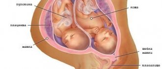
The cost of ultrasound in the second trimester on average ranges from 600-850 rubles. For the study of uteroplacental-fetal blood flow using Doppler, you need to pay about the same amount. Such a complete examination is advisable, since Doppler allows you to find out the functionality of the cardiovascular system. In case of multiple pregnancy (if twins or more are expected), the price for ultrasound and Doppler examinations will increase by 150-200 rubles. If it is not possible to take a photograph yourself, you can purchase an ultrasound image directly from a sonologist. This service will cost on average 150-300 rubles.
Ultrasound is ideal for prenatal screening. It does not cause discomfort and does not hide any health risks for the expectant mother and her child. In addition to the above, an important advantage is its high effectiveness - with its help, at 19 and other weeks of pregnancy, you can learn in detail the process of formation and stages of development of the child’s body systems, and assess the functionality of the woman’s reproductive organs.