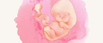
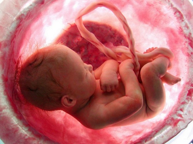
Childbirth at 35 weeks of pregnancy
However, children born at 35 weeks of pregnancy are considered to be born prematurely and require supportive measures in a hospital setting in the first days of their life. Although they can already breathe on their own. Also, such babies are already able to maintain a constant body temperature on their own; for this they already have enough subcutaneous fat, although it is undoubtedly less than in children born after 36 weeks.
The usual practice is that if a baby is born at 35 weeks of pregnancy, he will have to spend a couple of weeks in the pediatric department until his weight increases slightly, after which he and his mother will be safely discharged home. The prognosis for early birth at this stage is very favorable; by the age of 2–3 months, such children are no different from their peers who were born full term
There is a high probability of giving birth at 35 weeks of pregnancy in women with multiple pregnancies. Thus, twins often do not wait until full term and are born several weeks earlier than the expected date of birth. And although in terms of height and weight, such children are initially inferior to babies born at full term (and children of twins, even if the pregnancy lasted a full 40 weeks, are still slightly smaller at birth than their peers who do not have twin brothers and sisters, although the total weight Having twins at 35 weeks of pregnancy is already quite a burden for the mother), this does not affect the health of each child of the twins.
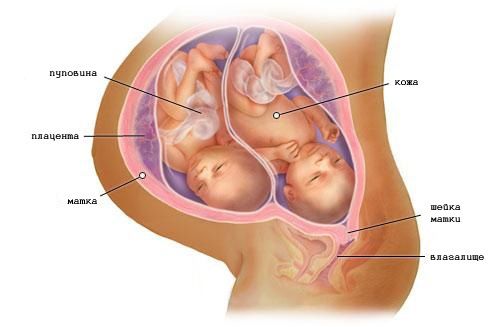
35 weeks pregnant: twins
In any case, now you should be constantly prepared so that the sudden onset of labor does not take you by surprise. Your bag for the maternity hospital should already be completely packed, and all your loved ones should know where it is. Wherever you are (even if you leave home for a minute to the nearest store), you should always have documents with you: a passport and exchange card, as well as a health insurance policy, if you have taken out one.
Understand that, theoretically, labor could begin at any minute. To understand that you should prepare for them in the near future, read the article about the harbingers of childbirth. Although these signs are conditional, real labor begins only in one of two possible scenarios: contractions or rupture of amniotic fluid (this process is described in detail in our article on the signs of the onset of labor).
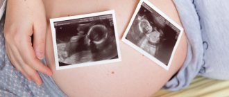
Research results
The 35th week of pregnancy in diagnostic terms differs little from the standard third screening study. The thirty-fifth week protocol includes:
- information about a pregnant woman
- fetometric measurements of the fetus
- assessment of anatomical structures
- data on the state of provisional entities.
The section about a pregnant woman contains information about her age, gestational age, last menstruation, and also describes the area of the appendages, the condition of the walls of the uterus and its pathology (if any).
Fetal fetometry
Fetometric measurements are important at any stage of pregnancy. The scope of measurements depends on the period, but the principles of measurements are general. All measurements are carried out in a strictly sagittal or transverse projection, measurements are taken several times and the smallest result obtained is accepted as true.
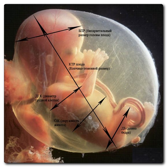
The diagnostic value of fetometry is to determine the duration of pregnancy, the correspondence of fetal development to the term, and determine intrauterine growth retardation and malformations.
There are special tables containing standard values for all fetometric indicators for each period of pregnancy. The tables contain average values, minimum (fifth percentile) and maximum acceptable values (ninety-fifth percentile).
Table of fetometric values
Values are presented at 35 weeks.
| Index | Values, cm | ||
| 5th percentile | 50th percentile | 95th percentile | |
| Biparietal head size | 7,8 | 8,5 | 9,4 |
| Fronto-occipital head size | 10,1 | 11,0 | 12,0 |
| Average breast diameter | 7,42 | 8,44 | 9,52 |
| Average abdominal diameter | 8,3 | 9,5 | 10,6 |
| Head circumference | 29,9 | 31,8 | 33,7 |
| Abdominal circumference | 28,22 | 30,71 | 33,45 |
| Femur length | 5,71 | 6,36 | 7,24 |
| Tibia length | 5,14 | 5,82 | 6,46 |
| Fibula length | 4,64 | 5,61 | 6,32 |
| Humerus length | 5,11 | 5,62 | 6,35 |
| Ulna length | 4,43 | 5,36 | 5,81 |
| Radius length | 4,34 | 4,71 | 5,64 |
| Blade width | 1,71 | 2,32 | 2,91 |
| Foot length | 5,91 | 6,56 | 7,57 |
| Distance between the outer edges of the eye sockets | 4,51 | 5,35 | 5,83 |
| Distance between the inner edges of the eye sockets | 1,72 | 2,26 | 2,78 |
| Eye socket diameter | 1,52 | 1,71 | 1,93 |
Not all of the presented indicators are used in screening studies, but when pathology is detected or suspected, it is preferable to evaluate all indicators.
The main importance in diagnosis belongs to the measurement of the head, abdomen and the length of the limbs. When measuring the length of the limbs, it should be remembered that measurements should be taken on both sides to exclude developmental defects. If this is not possible due to the child's position or activity, this information should be included in the report.
The main importance of ultrasound at 35 weeks is the assessment of the anatomical structure of the fetus. During this period, it is possible to exclude or identify malformations that cannot be reliably identified at a shorter period.
It is optimal to start the study with an assessment of the structures of the central nervous system. The examination of the brain includes examination of the cerebral hemispheres, assessment of their echogenicity, the cerebellum, lateral ventricles and cisterna magna, male hemispheric fissure and optic thalamus. If pathology is detected, the scope of the study increases.
Brain Measurement Chart
Measurements at 35 weeks are presented.
| Index | Value, mm | ||
| 5th percentile | 50th percentile | 95th percentile | |
| Lateral ventricles | 2,03 | 4,95 | 7,78 |
| Rear horn width | 6,52 | 8,54 | 10,23 |
| Width of transparent partition | 4,21 | 6,75 | 9,34 |
| Transverse diameter of the cerebellum | 41 | 44 | 47 |
| Large tank width | 5,64 | 7,58 | 8,59 |
| Corpus callosum length | 40,82 | 45,61 | 50,38 |
| Corpus callosum width | 5,43 | 7,16 | 8,89 |
| Corpus callosum thickness | 2,22 | 2,76 | 3,31 |
In addition to ultrasound diagnostics in B-mode, blood flow is assessed using Doppler. This helps in assessing the vascular bed and diagnosing vascular abnormalities.
Examination of the spine should be carried out in transverse and longitudinal projections along its entire length. This makes it possible to detect hernial protrusions and spina bifida. It is also important to conduct research in the frontal plane. This volume of research allows for an accurate diagnosis.
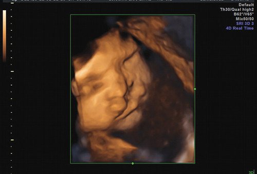
Facial cleft in fetus on ultrasound
The study of facial structures is also important. Visualization of the face is possible as early as 14-15 weeks of pregnancy, but this is very difficult and a detailed assessment of the structures is problematic . The introduction of three-dimensional echography into widespread practice has simplified the task and made it possible to diagnose pathology earlier. At 35 weeks, it is possible not only to visualize the baby’s face, but also to see his facial expressions and emotions. A separate line indicates the visualization of the nasolabial triangle and its condition.
The formation of the lungs is a very important stage in the development of a child. For diagnosing pulmonary pathology at 35 weeks, informative indicators are the echogenicity of the tissue and the ratio of the chest circumference to the abdominal circumference. By this time, the echogenicity of the lungs becomes higher than the echogenicity of the liver, which corresponds to the second degree of maturity of the lung tissue. The chest to abdominal circumference ratio is 0.89 . The introduction of three-dimensional echography has not found wide application due to the great complexity and low information content of the study.
Cardiac examination is important in prenatal diagnosis due to the fact that heart defects are the most common defects and the high mortality rate from this pathology. The main one in ultrasound is the four-chamber projection of the heart. When this section is obtained correctly, the position of the heart, the condition of its chambers and partitions are assessed, and the valve apparatus is also assessed. It is possible to diagnose heart defects starting from 20-24 weeks of pregnancy. Based on this, the study at 35 weeks is of an auxiliary nature to determine the tactics of postpartum monitoring of the child.
Projection through three vessels also refers to the mandatory stages of diagnosing fetal pathology. A section through the aorta, pulmonary artery and superior vena cava helps diagnose heart defects and vascular anomalies, which are difficult to identify in the four-chamber view.
Visualization and assessment of the condition of the stomach and intestines at 35 weeks is not difficult. Due to good visualization in the early stages, almost all developmental anomalies are detected during the second screening study.
Examination of the urinary system is also a relatively easy task. In the third trimester, the kidneys occupy a typical position and have a length of 35-45 mm. It is important in the study to assess the size of the pelvis, since pyeloectasia is a fairly common pathology. By this time, the bladder already has a volume of about 25 ml, however, due to periodic emptying, its visualization is not always possible. To exclude developmental defects, in such situations, an additional study is carried out after 40-45 minutes.
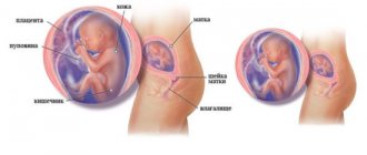
Provisional structures
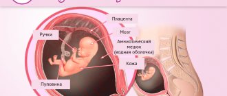
During ultrasound, the placenta is assessed according to the following criteria: location, thickness, internal structure, level of maturity, presence of pathology. The type of placenta previa can only be accurately determined in the third trimester. These data are important for determining the method of labor management.
By the 35th week of pregnancy, its thickness ranges from 27 to 44.8 mm. It is important to take the measurement at the level of the umbilical vessels, since the thickness of the placenta may vary in different places. On ultrasound of the 35th week, the placenta should correspond to the second degree of maturity: have a linear basal layer, depressions of the chorionic membrane to the basal layer, linear echogenic density.
The assessment of amniotic fluid is mainly based on its quantity, since its echogenicity is very variable and does not have reliable clinical significance. An amniotic fluid index has been developed to assess volume. It is the sum of the vertical dimensions of the four pockets of amniotic fluid. This indicator has a value from 79 to 249 mm. For values less than 79 mm, oligohydramnios is indicated, and for values exceeding 250 mm, polyhydramnios is indicated.
The process of ultrasound examination at 35 weeks is quite complex, routine and lengthy. To simplify the task, the specialist conducting the ultrasound must understand the purpose of the additional study and have data from previous screening ultrasounds.
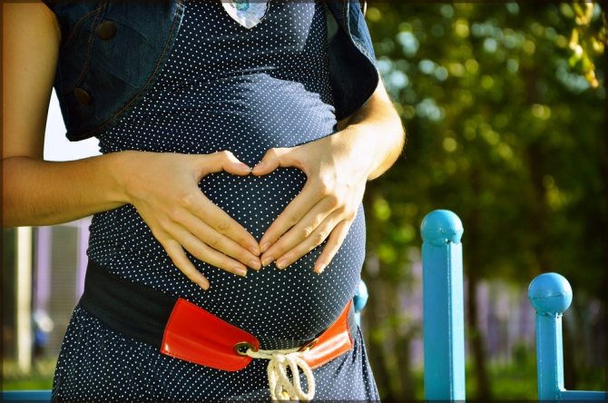
Preparing for childbirth
After just 2 weeks, your baby will be considered fully term (that is, from a full 36 weeks), and therefore labor can begin any day. All you have to do is make the final preparations.
We advise you to complete the purchase of the children's dowry and a set of things for the maternity hospital this week. The longer you are pregnant, the more difficult it will be for you to do this. And besides, you may need all these things a little earlier than you expect. We remind you: a pregnancy is considered full-term at 36 full weeks. However, birth occurs between 37 and 43 weeks after the start of the countdown (day 1 of your last menstrual period). And since it is impossible to predict the exact date of birth of your baby, it is better to prepare for everything in advance. Wash and iron everything that is necessary. Pack your things in bags or special plastic bags (most maternity hospitals do not allow fabric travel bags, as they are difficult to wash and disinfect, unlike plastic bags). Place the collected bag in a visible place in the maternity hospital, preferably in the hallway of your apartment. Draw your husband's attention to where it is, so that when you get ready for the maternity hospital, in a hurry, he does not forget to take it.
And of course, at such a late date, you are prohibited from long voyages and travel. For example, you simply won’t be allowed on the plane for completely legal reasons (many air carriers have restrictions in their instructions on the permissible period of pregnancy for passengers). Traveling by train or car, firstly, will be very difficult for you, and secondly, it can threaten early delivery.
It’s better not to travel even outside the city to visit your parents or to the country (if such a trip cannot be avoided, ask your husband to take his maternity hospital bag with him in the car).
Nutrition

Nutrition of a pregnant woman in the 35th week of gestation
At the 35th week, some pregnant women will notice the beginning of the process of drooping of the abdomen, as a result of the body preparing for childbirth. For others, this change will begin a little later. As a result, women feel relief from digestion, but this is not a reason to break the nutritional rules. The uterus still puts pressure on the intestines, and a violation of the diet can cause constipation. It is worth adhering to moderation and some dietary features during pregnancy:
- split meals: eat 5-6 times a day, every 2.5-3 hours;
- finish your meal before you feel full;
- do not mix different dishes at one meal;
- limit food intake 2-3 hours before bedtime;
- drink about 1.5 liters of fluid per day, unless otherwise prescribed by the gynecologist;
- exclude all harmful products: smoked meats, pickles, semi-finished products, fast food, etc.;
- consume a minimum amount of salt;
- Do not drink strong tea or coffee unless otherwise prescribed by your gynecologist.
Vitamins
At the 35th week, most gynecologists suggest that pregnant women stop taking all multivitamins and dietary supplements, especially those containing calcium. It is recommended to strengthen control over the intake of essential nutrients from food, and to refuse those created chemically.
This is done so that in the last weeks, when the main development processes are completed, the fetus does not direct all unused resources into weight, thereby growing greatly. A large fetus is more difficult to deliver. If a gynecologist advises you to stop taking dietary supplements, it is better to listen to him.
Sex at 35 weeks pregnant
If all the inevitable symptoms of pregnancy still do not bother the expectant mother, and also if there is no risk of early birth and a doctor’s ban on sexual activity (in the case of multiple pregnancies, placental abruption or previa, leakage of amniotic fluid or other threats of early birth), you should not refuse from pleasures.
Modern obstetricians and gynecologists even consider sex in later stages to be an excellent preparation for childbirth for absolutely healthy women. Of course, the husband should also be confident in his health. In this case, unprotected sex will bring the couple not only joy and satisfaction, but also undoubted benefits, since sperm softens the cervix and makes it more elastic. If for some reason unprotected sex is contraindicated for you, it is better to abstain altogether, since using a condom can cause allergic reactions in a woman in late pregnancy.

35th week of pregnancy: you can have sex, provided that both partners are healthy
Otherwise, if both partners are healthy, there is no need to worry that your love will harm the baby - he is sufficiently protected in the amniotic sac, and neither the penetration of the father’s penis nor the mother’s orgasm can disturb him.
Sexual intercourse is unacceptable only after depressurization of the amniotic sac - if the mucus plug and, of course, the water have come out. After these events, any microbes can easily get to the child, so sex is excluded.
In the meantime, given the size of your belly, as well as the need to avoid too deep penetration, of course, many of the usual positions will have to be left for later, when the baby is born. In the meantime, our Kama Sutra for pregnant women will help you choose comfortable positions, where we have carefully divided the poses into groups suitable for each trimester of pregnancy.
Good to know
Childbirth with a scar on the uterus
What awaits a premature baby immediately after birth in the hospital?
How to choose a nursing bra
Is a pregnant woman beautiful?
What worries a woman during pregnancy?
What are the methods of preparing for childbirth?
All texts for pages about mother and baby were kindly provided by RAMA Publishing - these are chapters from the book by Svetlana Klaas “Your Favorite Little Man from Conception to Birth”, reviewer Irina Nikolaevna Kononova, Candidate of Medical Sciences, Associate Professor of the Department of Obstetrics and Gynecology of the Ural State Medical Academy (Ekaterinburg).
It is possible and it is not possible
How to behave as a pregnant woman at 35 weeks.
What is possible, what is not. By the 35th week of pregnancy, many rules of behavior are followed by a woman not so much out of necessity, but because there is no other option. Increased weight, increased fatigue, and difficulties in moving force the expectant mother to move smoothly, not make sudden movements, and think about her actions.
Pregnancy is approaching the end, but this is not a reason to relax. The goal of every expectant mother is to allow the baby to achieve maximum intrauterine development in order to be born fully prepared. To do this, you should continue to adhere to the basic principles of correct behavior:
- listen carefully and follow the gynecologist’s instructions;
- take tests and undergo prescribed studies in a timely manner;
- complete abstinence from alcohol, nicotine, and drugs;
- minimize contact with household and construction chemicals;
- proper nutrition and drinking regime;
- daily routine: sufficient sleep, daily walks, periodic warm-ups;
- careful body hygiene;
- order in the house;
- daily ventilation of the room in which the pregnant woman is located;
- minimal contact with possible carriers of viruses and bacteria;
- psychological calm.
Sex
Most gynecologists agree that at the 35th week it is worth limiting your intimate life. This is primarily due to the risk of premature birth. Unexpected movement, stress, or emotional upsurge can trigger the onset of labor, which cannot always be stopped within a medical facility.
Weight
At the 35th week, the weight gained from the moment of registration of a pregnant woman can range from 8 to 14.5 kg. In some cases, deviations from these standards may be greater or lesser. Deciding how much the gained kilograms correspond to the term, whether there is a potentially dangerous underweight or overweight is the task of the gynecologist monitoring the development of pregnancy.
If a deviation from the calculated norm for a particular woman is detected, the doctor may recommend changes to the diet or suggest hospitalization.