The primary methods for diagnosing diseases of the scrotal organs include examination, palpation and ultrasound. However, examination and palpation are not as informative as ultrasound diagnostics, which can be used to determine structural and functional changes in the organ.
- What it is?
- Indications for ultrasound of the scrotum
- What are the normal indicators for an ultrasound of the scrotum?
- What pathologies can be found on ultrasound of the scrotum?
- Preparing for an ultrasound of the scrotum
- How does the procedure work?
- Advantages and disadvantages of scrotal ultrasound
What is ultrasound of the scrotum?
Ultrasound (sonography) is a method of examining internal organs using ultrasound. For ultrasound of the scrotal organs, high-frequency waves are used, which allows one to visualize the internal structure, determine the size, shape, contours and structure of the tissues of the organ being examined. Diagnostics is quick and painless for the patient and does not require special preparation. The procedure is safe for the body, so it can be performed at any age.
To conduct ultrasound of the scrotum, linear sensors with a frequency of 5-10 MHz are used. This equipment has a small scanning depth - up to 10 cm, but the resulting image has a high resolution. Such a sensor is used to study superficially located organs: the thyroid and mammary glands, scrotum, small muscles and joints. Using ultrasound, it is possible to determine the size of the organ under study with an accuracy of up to a millimeter, which makes it possible to detect even minor changes and diagnose pathology at an early stage of its development.
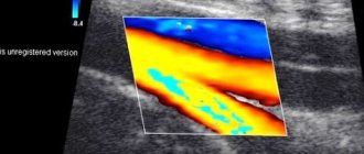
A separate type of ultrasound examination is Doppler ultrasound, or Doppler ultrasound of blood vessels. It is prescribed for a detailed study of the blood circulation of the organ. It allows you to determine the speed and volume of blood flow in the arteries and veins, see blood clots and plaques in them, and detect vasoconstriction. Doppler ultrasound is prescribed to patients to diagnose varicose veins of the scrotum (varicocele). To detect other pathologies, a standard ultrasound is sufficient.
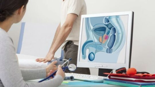
What will a transrectal pelvic ultrasound show in men?
Transrectal ultrasound scanning is prescribed for urinary incontinence, cystitis and when the patient is unable to fill the bladder for medical reasons. TRUS allows you to assess the condition of the prostate gland in more detail and see formations of various types. In addition, doctors use this method of pelvic ultrasound for puncture biopsy of the prostate, with an increased PSA value in the blood. TRUS will also be prescribed for complicated prostatitis and gland abscess.
| Ultrasound service of pelvic organs | Price, rub | Promotion Price |
| Ultrasound of the bladder with determination of residual urine | 800 rub. | |
| Ultrasound of the pelvis in women and men with an abdominal sensor | 1200 rub. | |
| Ultrasound of the pelvis in women with a vaginal probe | 1300 rub. | |
| Comprehensive pelvic ultrasound with abdominal and vaginal probe | 1400 rub. | |
| Ultrasound of folliculogenesis | 750 rub. | |
| Ultrasound cervicometry | 1100 rub. | |
| Ultrasound of the prostate gland with a rectal probe | 1500 rub. | |
| Ultrasound of the scrotum (testicles, appendages) | 1000 rub. | |
| Ultrasound of the prostate and bladder with an abdominal probe | 1900 rub. | |
| Comprehensive ultrasound (ultrasound of the abdominal organs + ultrasound of the kidneys + ultrasound of the thyroid gland + ultrasound of the pelvis with an abdominal probe + ultrasound of the mammary glands) | 4200 rub. | 2999 rub. |
| Comprehensive ultrasound (ultrasound of the abdominal organs + ultrasound of the kidneys + ultrasound of the thyroid gland + ultrasound of the prostate gland with an abdominal probe) | 3300 rub. | 2499 rub. |
Indications for the procedure
Patients are prescribed an ultrasound for the following symptoms:
- pain in the groin,
- the appearance of tumor tumors in the scrotum,
- enlargement of the testicle or epididymis,
- swelling of the scrotum,
- enlargement of the femoral or inguinal lymph nodes,
- erectile disfunction,
- infertility.
Ultrasound of the scrotum is performed to prevent various diseases and to monitor the rate of recovery of the operated organ. The procedure is also prescribed after injuries to assess the nature of the damage and prescribe surgical treatment.
For children, the procedure is prescribed for cryptorchidism, which is the absence of a testicle in the scrotum. The pathology manifests itself as a result of delayed descent of the testicle along the inguinal canal. To determine its location, children undergo a series of diagnostic tests, including ultrasound.
In childhood and adolescence, the procedure is prescribed for:
- dwarfism or gigantism,
- premature puberty, as well as its delay,
- underweight or overweight.
When these symptoms appear, the child’s entire endocrine system, which includes the gonads, is examined.
Indications
Specialists send patients for an ultrasound test of the testicles if there is:
- suspicion of a neoplastic process, signs of inflammation;
- absence of one or both testicles;
- enlargement of regional lymph nodes;
- for infertility;
- to monitor treatment and perform biopsies;
- when the testicles increase or decrease and their contours change;
- if the patient complains of painful manifestations in the testicular area;
- if there is a suspicion of torsion of the spermatic cord and with injuries in the scrotum area.
What are the normal indicators for an ultrasound of the scrotum?
The ultrasound protocol of the scrotal organs includes the following points:
- testicular parameters,
- the sizes and contours of their appendages,
- presence of free fluid in the scrotum,
- thickness of the organ walls.
The testicles are oval in shape, with one of them usually slightly larger than the other. As a rule, the left one is lowered slightly lower than the right one. For men, it is considered normal for testicle sizes not to exceed 4-5 cm in length, 3 cm in width, and 2 cm in thickness. A slight deviation of one of the parameters is considered acceptable. In a normal state, the contours of the testicles should be smooth and clear, and their echo density should be average.
The epididymis are ducts necessary for the maturation, accumulation and movement of sperm. The ultrasound image shows only the head of the appendage, the dimensions of which range from 1-1.5 cm. The tail and body of the appendage are not visible, since their sizes do not exceed a few mm. The appendages have a homogeneous echostructure, and they should not have any formations.
The walls of the scrotum are 0.7-0.8 cm thick. Between the walls and the testicles there is a small amount of free fluid within 1-2 ml. The above indicators are typical for an adult healthy man; for boys the indicators will be different. The norm is determined based on their age.
Survey results
During diagnosis, all parameters of the organs being examined are displayed on the monitor screen, so the doctor can immediately establish a preliminary diagnosis. Ultrasound data is recorded in a protocol, which is immediately given to the patient. With the conclusion, you need to go to the doctor who gave the referral for examination.
The main parameters assessed by the diagnostician first of all:
- shape and size of the pelvic organs;
- location relative to neighboring organs and the spinal column;
- echogenicity of tissues.
Normal indicators:
- The size of the seminal vesicles is no more than 10 mm.
- Prostate volume – up to 30 cm3;
- The thickness of the bladder walls is no more than 5 mm.
What pathologies can be found on ultrasound of the scrotum?
Ultrasound can detect:
- cryptorchidism (absence of testicle),
- inflammation of the testicle and its epididymis (orchitis and epididymitis),
- dropsy of the testicle (hydrocele),
- seminal cyst (spermatocele),
- varicose veins of the testicular vessels,
- tumors and cysts of the scrotal organs,
- testicular torsion,
- abscesses,
- organ ruptures after injuries.
Ultrasound of the scrotum helps to identify obstructive forms of infertility, in which there is an obstacle to the path of sperm from the testicle to the urethra. This is prevented by scars from operations, adhesions after infections, and various neoplasms in the scrotum that compress the vas deferens.
Preparation
- A man needs to prepare for a pelvic ultrasound in advance. Preparation consists, first of all, of bowel movements naturally several hours before the procedure. In a situation of constipation, you can free the rectum from excess toxins using an enema.
- A bladder with moderate filling will improve the results of ultrasound diagnostics.
- Before an ultrasound with a puncture biopsy, the attending physician prescribes a laboratory blood test for coagulation, a general blood test and a urinalysis. This is done in order to protect the subject from complications after the puncture, since there is a category of patients for whom this type of manipulation will be contraindicated. As an alternative, they will be offered an MRI of the prostate with contrast.
Author: Telegina Natalya Dmitrievna
Therapist with 25 years of experience
How should you prepare for an ultrasound of the scrotum?
No preparation is required from the patient other than maintaining genital hygiene. If you have been prescribed a Doppler ultrasound of the scrotal organs, you should refrain from cigarettes an hour before the procedure. Nicotine causes vasoconstriction, which can affect the accuracy of the test.
To avoid discomfort after an ultrasound of the scrotum, men are recommended to shave the hair in the groin area. The gel that is used for ultrasound then remains on the hair. It is not easy to remove it with a napkin or paper towel. Remains of the gel after the procedure will cause discomfort.
If ultrasound of the scrotum is performed on children or adolescents, then parents need to psychologically prepare the child for the examination. In young children, fear can cause the testicles to retract into the inguinal canals, making examination difficult. Therefore, talk to your child before the procedure so that he is not afraid.
Is Doppler ultrasound of the testicle safe?
To perform an ultrasound scan, it is not necessary to introduce additional stimulating or contrasting drugs by puncturing the skin of the scrotum. During the examination, the genitals are not exposed to harmful ionizing radiation. It follows that the procedure is safe and harmless. It can be prescribed to young children in the first year of life.
The frequency of ultrasound examination of the testicles can be any, because this examination is safe. This is important when it is necessary to periodically evaluate the condition of the scrotum during treatment, because, for example, such types of diagnostics as CT or MRI are not allowed to be used frequently.
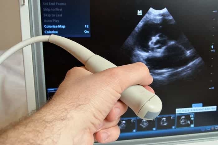
How is an ultrasound of the scrotum performed?
The ultrasound lasts 10-20 minutes. The patient takes off his underwear and lies down on the couch. The doctor applies the gel to the skin of the scrotum. The gel is used to ensure good contact between the ultrasound probe and the patient's skin. The gel must be warm. If it is cold, it can cause the testicles to retract into the inguinal canals. If the patient experiences pain from touching the sensor, which happens with epididymitis or orchitis, he is given superficial anesthesia.
The scrotum is examined in three projections: transverse, longitudinal and oblique. The ultrasound doctor records the data obtained in the study protocol and hands it to the patient. Your attending physician will decipher this data and make a diagnosis.
Decoding the research results
An ultrasound diagnostic specialist has extensive experience and extensive medical knowledge to correctly interpret what a scrotal ultrasound shows.
A healthy testicle is characterized by a dense, homogeneous, fine-grained structure. On the outside there is a protein shell in the form of a thin white stripe. If a dark stripe is visible between the protein membranes, a hydrocele is diagnosed. When fluid spills into the inguinal canal, they speak of hydrocele of the testicles. Excess fluid may indicate a hematoma. Testicular cysts are observed as round black spots with clear contours. Epididymitis is indicated by an increased testicular size and a loose, heterogeneous structure. In acute orchitis, there is a cellular structure and an uneven outer contour of the testicle. Varicocele looks like a tense cord with a cellular heterogeneous structure.
The cause of pain may be calcifications, which are small dense areas that usually do not pose a danger, but if there are a large number of them, it is recommended to undergo regular examination. A malignant neoplasm is recognized as an irregularly shaped spot with a heterogeneous structure. With the help of Doppler ultrasound, increased blood flow can be detected in this place.
A transcript of the research results is issued in the form of a medical report.
Advantages and disadvantages of scrotal ultrasound
Among the advantages of the ultrasound method are:
- Non-invasive examination. Does not require surgery for diagnosis.
- Rapidity. You receive data and a primary diagnosis within 20 minutes.
- Availability. Ultrasound examination is an inexpensive procedure that can be performed in every clinic in Moscow.
- Safety. Ultrasound is harmless to our body, so the procedure has no restrictions.
- Dynamic image. Ultrasound provides a picture in real time, which allows it to be used during surgery or biopsy.
The only drawback of the ultrasound method is that it cannot accurately determine whether a malignant or benign formation has appeared in the scrotum. In this case, the patient is prescribed additional diagnostics. The method has no other disadvantages.
Preparing for an ultrasound
You can do an ultrasound of the scrotum with Doppler ultrasound in a clinic or medical center without any special preparatory measures. It is recommended to reduce the hair growth if it is too abundant, but this is not necessary. It is advisable to carry out the necessary hygiene procedures before the examination.
On the eve of the examination, it is necessary to limit the consumption of alcohol and drugs that can affect blood pressure. If you are taking vasodilators, tell your doctor. If possible, it is recommended to perform an ultrasound scan of the scrotum with Doppler Doppler Doppler after completing the course of taking these medications.
If the examination is to be carried out on a child, it is necessary to discuss the upcoming procedure with him in advance and it is advisable not to give him anything to drink for 1–1.5 hours, so as not to have to urgently interrupt the procedure.