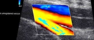
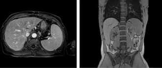
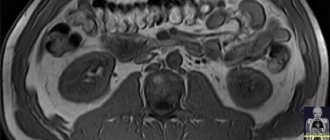
MRI of a woman's pelvic organs
MRI of the pelvis with contrast is a diagnostic procedure that uses a magnetic field, radiofrequency pulses and computer processing to obtain a detailed picture of the structure of the organs being examined, nearby tissues and bones. The introduction of a paramagnetic provides better visualization of pathological processes at an early stage of the disease and can resolve the ambiguity of previous results of ultrasound, radiography, hysteroscopy, etc. There is no radiation exposure, without which CT is impossible, when performing magnetic resonance imaging, so the study is available without harm to health and any - long-term consequences for most people, including children and women during gestation. If there are metal implants in the body, other diagnostic methods are considered.
- Prices for MRI of internal organs
MRI of the pelvis with contrast: what does it show?
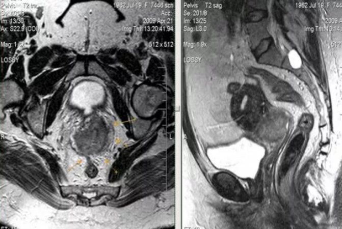
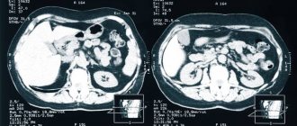
MRI of the pelvic organs in a woman: a malignant neoplasm of the cervix extends beyond its boundaries, but does not spread to the lower third of the vagina or the pelvic wall
MRI of the pelvis reliably shows a number of pathological processes, the visualization of which is not available with other methods of examination: ultrasound of the genitals, CT, cystoscopy, hysteroscopy, radiography, etc. Magnetic resonance scanning has found application in urology, gynecology, proctology, surgery and today is considered one of the most of highly informative methods for determining the cause of the disease, stages, effectiveness of treatment: conservative or surgical. MRI of the pelvic organs with contrast has no quantitative restrictions and can be performed as long as needed. The study is prescribed as a preoperative diagnosis to obtain an idea of the features of the anatomy of the pelvis, which allows you to plan the access and scope of surgical intervention, for example, before myomectomy, hysterectomy, or uterine artery embolization.
MRI of the pelvis with contrast will help evaluate the following conditions:
- inflammation: tubo-ovarian abscess, endometritis, etc.;
- benign tumors (endometriomas, fibroids), ovarian cysts, adenomyosis;
- consequences of injuries;
- obstruction of the fallopian tubes;
- pelvic floor pathologies associated with fecal and urinary incontinence;
- congenital anomalies of the pelvic organs;
- suspicion of malignant processes in the endometrium, ovaries, cervix, rectum, peritoneum, bladder, prostate, testicles;
- for the diagnosis of volumetric processes in the retroperitoneal space, for example, Ormond's disease, in which multiple tumor formations exert a compressive effect on organs and vessels. When the ureter is compressed, renal colic develops;
- neoplasms (for example, sarcomas) of soft tissues of the area under study, etc.
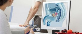
For what symptoms is pelvic tomography prescribed for men?
The attending physician may prescribe a man an MRI of the pelvis when he suspects the presence of pathologies in the relevant organs. Let us indicate what is included in an MRI of the pelvis in men: bladder, prostate gland, penis, scrotum, lymph nodes, pelvic bones, rectum. Let's look at some of the most common reasons for prescribing a pelvic MRI for a man.
After a bruise or injury to the scrotum
The MRI method in this case will be especially relevant in case of closed injury and will allow timely detection of subcutaneous testicular ruptures.
Pain in the sacrolumbar region
Tomography for such pain is necessary, as it will reveal diseases of the internal organs, including prostatitis.
Suspicion of abnormalities of the kidneys, urinary tract and scrotum
An MRI examination can confirm or refute the suspicions of the attending physician.
Preparation for MRI of the pelvis with contrast in women

It is better to resolve all questions before undergoing an MRI of the pelvic organs with contrast in advance.
MRI of the pelvic organs with contrast is primarily a routine diagnostic procedure. Preparation ensures the quality of the tomograms obtained, a more comfortable examination, safety, and minimization of side effects due to the introduction of a paramagnetic substance. To get the maximum benefit from pelvic MRI (PMR), it is necessary to take into account a number of nuances:
- choose loose clothing made from natural fabrics (if desired, you can change into a disposable shirt);
- remove all metal objects: jewelry, glasses, hairpins, removable dentures, etc. Fundamentally installed dental structures (bridges, implants, metal-ceramics) are not an obstacle to undergoing diagnostics;
- deposit keys, cell phones, coins, magnetic cards;
- take with you a referral from your attending physician indicating the type of study and the expected diagnosis, compulsory medical insurance or voluntary health insurance policy (if insurance covers the cost of MRI); medical extracts and reports.
To undergo an MRI scan at a lower cost, sign up for the study at night, find out about promotions and discounts on pelvic MRI with contrast. Information can be obtained on the clinics’ websites or by calling the indicated numbers.
On what day of the cycle is a pelvic MRI performed?
In some cases, the study is carried out on an emergency basis. Then an MRI of the pelvis is performed on any day of the cycle. For the greatest reliability of the results, it is better to plan the diagnosis in advance. MRI is recommended to be performed on days 7-12 of the cycle.
You can do a magnetic resonance imaging scan of the pelvic organs in the diagnostic department. The clinic has all the conditions for conducting studies in native mode and with contrast. When making an appointment by phone, our staff will tell you about the preparation rules and select the most convenient time.
Diet before MRI of the pelvic organs with intravenous contrast

To prevent flatulence, it is better to abstain from raw vegetables and fruits for several days, but they can be consumed in processed form.
3-4 days before the study, it is necessary to follow a special diet aimed at preventing flatulence and constipation. Is it necessary to do an enema before an MRI of the pelvic organs with contrast, take antispasmodics or sorbents (activated carbon, polysorb, etc.), it is better to check with your doctor in advance. It is preferable to steam, boil or bake in the oven. The diet is frequent (4-5 times a day), in small portions. Foods that cause fermentation in the intestines and bloating are excluded from the menu:
- sauerkraut and raw cabbage;
- legumes (beans, peas, lentils);
- corn;
- black bread;
- yeast-containing baked goods;
- raw vegetables, herbs and fruits;
- mushrooms and nuts;
- spices, marinades, freshly squeezed juices;
- carbonated drinks, alcohol, kvass;
- high fat milk.
Vegetables can be consumed in processed form. Non-rich light soups, stewed, baked, boiled lean meat, poultry or fish are suitable.
MRI OMT in women is performed with a moderately full bladder, which provides greater clarity and visibility of pathological changes on the films. The diagnostic procedure is performed on an empty stomach: the last meal is appropriate 4-6 hours before reporting to the clinic.
How to prepare for the procedure?
How can a woman prepare for an MRI of the pelvis? – One of the main questions. It arises because it is necessary to achieve the utmost accuracy of diagnosis. Preparation of MRI of the pelvic organs
in women: main stages and methods
Diet before the procedure
Diet during pelvic MRI in women is not complicated. It is not advisable to undergo the procedure with a full stomach, so immediately before the study it is better not to eat or limit yourself to a very light meal. Two days before the test, it is necessary to exclude foods that stimulate the formation of gases from the diet.
Drinking regime
Preparing for a pelvic MRI in women also involves preparing the bladder. You should not drink liquid 5-6 hours before the procedure.
MRI OMT with contrast: indications and contraindications

There are many indications for MRI; this type of diagnosis has improved the detection of pathology of the pelvic organs at an early stage
Indications for pelvic MRI with contrast include the following:
- the need to clarify the diagnosis in case of ambiguous data from previous diagnostic methods;
- tracking changes in dynamics;
- preoperative planning;
- suspicion of a pathological process in the pelvic organs.
Complaints that are considered as indications for MRI of the pelvic organs with contrast in a woman, taking into account the general clinical situation:
- absence of a desired pregnancy within 12 months, subject to regular sexual contact without contraception;
- dyspareunia (pain during intimacy);
- repeated self-termination of pregnancy with normal tests;
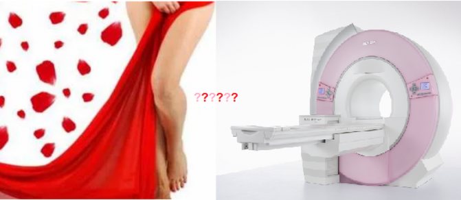
- the appearance of bloody discharge in the interval between regular menstruation, a change in its character: more abundant, scanty, with large blood clots, the appearance of an unpleasant odor;
- pain in the lower abdomen of unknown etiology;
- dysfunction of urination: the need to strain, a feeling of incomplete emptying, urine mixed with blood, frequent urges, etc.;
- irregular bowel movements: diarrhea, constipation, bloating, bloody stool.
Common symptoms that are suspicious of a neoplastic process in the pelvic organs: weakness, fatigue, unexplained fever, sweating, weight loss, anemia. MRI of the pelvic organs with contrast is performed as part of the general diagnosis if changes in the hormonal background are detected: increase/decrease in FSH, LH, prolactin. According to indications, the search can be expanded: the brain (pituitary gland), which is responsible for the production of biologically active substances that regulate the functioning of the female reproductive system, is additionally examined.
Contraindications for MRI

The risk of MRI in some cases outweighs the benefits; before going to the clinic, read the contraindications
There are a number of contraindications to MRI of the pelvic organs with contrast:
- Metal objects in the body. Any implants with electrical, magnetic or mechanical activation (cardiac and neurostimulators, insulin pumps), cochlear devices, ferromagnetic clamps, brackets, orthopedic structures will cause artifacts to appear on the images during the procedure. There is a risk of failure of equipment providing vital functions. Metal shrapnel, shavings or a bullet under the influence of a magnetic field can begin to move, as a result of which neighboring tissues are injured. The intrauterine device, as a means of contraception, can also lead to image defects.
- Inability to remain still during pelvic MRI with contrast. Some neurological pathology (Parkinson's disease, epilepsy, etc.), psychiatric illnesses in the acute stage, severe curvature of the spine, severe pain are an obstacle to magnetic scanning. For tremors and obsessive movements, the examination can be performed after the administration of sedatives, which is also important for patients suffering from claustrophobia. Immersion in medicinal sleep is one of the ways out in this situation, but the issue is always resolved on an individual basis.
- Obesity. If you have a high body mass index, it will not be possible to do an MRI of the pelvic organs for technical reasons: too much weight (120 kg or more) will not allow the patient to move inside the device. For this category of people, scanning is performed on open-type tomographs, which significantly reduces the quality of diagnosis due to low power, or they resort to other testing methods. Sometimes it is recommended to follow a diet for 1.5-2 months in order to lose weight and undergo a full examination.
An MRI of the pelvic organs with contrast in a woman has an additional obstacle to performing: pregnancy in the first trimester. In world practice, there is no data on the teratogenic effect of a magnetic field on a developing embryo, but it is more rational to conduct research after the 12th week of gestation, when the main formation of organs and systems of the fetus is completed. According to vital indications, magnetic resonance scanning can be performed at any time. MRI of the pelvic organs helps in diagnosing intrauterine malformations of the fetus, but there must be compelling reasons for carrying out the procedure, for example, if ultrasound detects abnormalities that are suspicious of a serious pathology in both the mother and the unborn child. Compared to other research methods - cordocentesis, amniocentesis - magnetic resonance scanning has no side effects.
MRI of the pelvis with contrast

Contrast based on gadolinium chelates has passed all clinical trials and is approved for use in magnetic resonance diagnostics
Contrast provides clearer visualization: on tomograms, the contours and size of the tumor, its interaction with nearby structures, the condition of blood vessels, and potential metastasis are seen much better. In modern diagnostics, a paramagnetic agent is used based on gadolinium chelates, a rare earth metal that in its native state is dangerous to the body, but when used in the form of dissolved salts does not lead to any serious consequences in 98% of patients. The drug is excreted by the kidneys. Contrasting is not applicable if:
- a generalized allergic reaction such as angioedema or anaphylaxis to the administration of the drug has previously been observed;
- the patient is diagnosed with end-stage chronic renal failure (glomerular filtration rate less than 30 ml/minute);
- the woman does not rule out pregnancy in the early stages, which she plans to prolong.
Paramagnetic agents, which provide higher-quality images during MRI of the pelvic organs, are much better tolerated and have a smaller list of adverse reactions than iodine-containing substances for CT. Contrast-induced nephropathy/systemic fibrosis is incidentally rare. Minor allergic reactions - redness of the injection site, urticaria-type rash were reported in 10% of patients.
When a paramagnetic is administered, unpleasant symptoms from the autonomic nervous system may appear: a metallic taste in the mouth, a diffuse feeling of heat, salivation, nausea, headache, which does not require cancellation of the procedure. After 2-5 minutes the phenomena stop on their own. To minimize the likelihood of their development, you need to eat a light breakfast 40-50 minutes before, for example, a sandwich with sweet tea or a few spoons of porridge boiled in water.
The contrast is delivered into the vein using an injector during certain phases of the study or administered by a nurse.
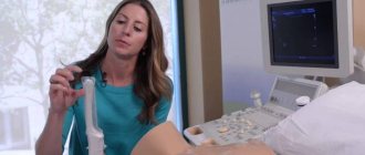
MRI of the pelvis with contrast: how is it done?
Maximum immobility during the procedure is a prerequisite for obtaining high-quality images
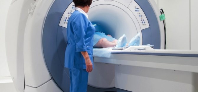
MRI OMT with contrast is done by appointment; an unscheduled study on the day of treatment may cost more; the prospects for its implementation must be checked with the operators. Before the procedure, the doctor talks with the patient to identify contraindications. Medical documentation is filled out; without signing voluntary informed consent for an MRI, the procedure cannot be performed. At the appointed hour, the subject is placed on the tomograph table, lying on his back. The patient is automatically transported deep into the drum ring; sensors rotate in the body of the device around the area under study, capturing the response of organs and tissues to the influence of the magnetic field. There are no unpleasant sensations; to reduce mechanical noise from operating equipment, you can use headphones or earplugs.
Duration of MRI OMT with contrast is 45-60 minutes. During certain phases of the examination, you need to hold your breath for a few seconds; the technician will tell you this over the speakerphone.
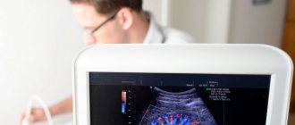
The resulting tomograms are interpreted by a radiologist, the conclusion will be ready in an hour, unless otherwise agreed. The results can be printed on film, copied to electronic media, or sent by e-mail. There are no contraindications to normal activities after MRI with contrast.
Is there a relationship between the menstrual cycle and pelvic MRI?
Physiological processes do not interfere with MRI
During menstruation, under the influence of hormones, a woman’s psycho-emotional state is unstable, excessive nervousness can lead to headaches and increased bleeding, so it is better to plan diagnostics for another time. An MRI of a woman’s pelvic organs can be performed at any phase of the menstrual cycle without harm to health or loss of information, but it would be more comfortable for the patient not to be distracted by cramps in the lower abdomen caused by immobility, and not to think about urgently replacing a pad or tampon. There are situations when uterine bleeding occurs for a long time (for example, with uterine fibroids or a tumor), and waiting weeks for it to end in order to do an MRI and find out the cause is far from the most correct decision. Experts believe that in this case, the sooner the diagnosis is made and therapy is started, the better the life prognosis.