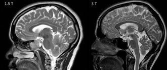
What is transcranial Doppler ultrasound (TCD)?
Transcranial Dopplerography is nothing more than an ultrasound scan of the brain
. Since the vessels of the brain are securely “hidden” under the skull, which does not transmit ultrasound waves well, ultrasound of the brain is done using a special technique in open areas of the skull, and this examination method is called TCD (transcranial Doppler).
When is TKDG prescribed?
TCD, like ultrasound, is indicated for various symptoms: fatigue, dizziness and headaches, attention disorders, frequent fainting, numbness of the extremities. All this may indicate brain damage, including after trauma or surgery. TCD is also prescribed for diagnosed vascular diseases to control their course. TCD is indicated for infants after neurosonography, which gave alarming results.
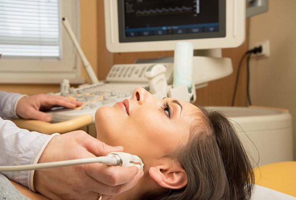
How is the TCD procedure performed?
To assess the condition of the vessels located in the cranium, the sonologist uses three “windows”: temporal (in the temporal part of the head), suboccipital (in the occipital part) and orbital (in the eye area). Through these areas (the doctor briefly places the probe on them), ultrasound waves can easily reach most of the intracranial vessels. The TCD procedure itself takes about twenty minutes, but it is usually complemented by an ultrasound scan of the neck
to obtain more complete information about the condition of the patient’s blood vessels, therefore it requires more time.
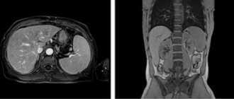
Carrying out a scan
To undergo duplex scanning
the patient lies down on the couch, his skin above the waist is treated with gel for attaching the sensors.
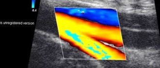
Ultrasound of cerebral vessels
is performed with data transmitted to the screen. This allows you to evaluate the activity of blood flow and the location of blood vessels. If the doctor asks you to hold your breath or inhale or exhale, this must be done. The patient receives the result immediately after completing the examination. Total time is about half an hour.
Triplex scanning
combines the technique described above and Doppler. The movement of blood through the vessels can be viewed in color in more detail. This is necessary for the convenience of visual assessment of the image.
What is Doppler ultrasound (USD)?

Doppler ultrasound is based on the Doppler effect and is aimed at determining the speed and trajectory of red blood cells in the bloodstream, from which ultrasound is reflected. The method allows you to identify the patency of blood vessels and, based on the data obtained, draw conclusions about possible pathologies and vascular diseases.
When is ultrasound ultrasound prescribed?
Most often, ultrasound examination is prescribed for the initial examination if vascular diseases are suspected, the signs of which may be: heaviness in the limbs, headaches and impaired concentration, dizziness and tinnitus. Mainly the upper and lower extremities and the neck are examined with TCD, which is often accompanied by ultrasound.
How is the ultrasound procedure performed?
The ultrasound examination procedure is performed with the patient in a sitting or lying position, depending on the area of the body being examined. The doctor places the sensor on certain areas of the body and guides it in the direction of blood flow. Unlike TCD, ultrasound of the lower extremities
and neck does not encounter obstacles in the form of the skull, so it takes less time and is more informative. If necessary, as with TCD, before the ultrasound procedure, functional tests can be performed, during which the patient can raise his arms and legs, as well as follow other doctor’s instructions.
Indications for ultrasound of cerebral vessels
Indications for ultrasound examination of the extracranial carotid and vertebral arteries include:
- Evaluation of patients with hemispheric neurologic symptoms including stroke, transient ischemic attack, and amaurosis
- Assessment of the condition of patients with cervical osteochondrosis
- Evaluation of pulsating massive neck masses
- Preoperative evaluation of patients scheduled for major cardiovascular surgical procedures
- Evaluation of non-empirical or unexplained neurological symptoms
- Dynamic examination of patients with proven carotid artery disease.
- Assessment of the condition of patients after surgery or after intervention after cerebrovascular revascularization, including carotid endarterectomy, stenting
- Intraoperative monitoring of vascular surgery
- Evaluation of suspected subclavian artery steal syndrome
- Examination for suspected carotid artery dissection, arteriovenous fistula, or pseudoaneurysm.
- Evaluation of patients with carotid artery reconstruction after ECMO (extracorporeal membrane oxygenation) bypass
- Assessing patients with fainting, seizures, or dizziness
- Screen for high-risk patients: atherosclerosis elsewhere, history of head and neck radiation, fibromuscular dysplasia, Takayasu arteritis, or other vasculopathies.
- Neck injury
Indications for transcranial ultrasound examination:
- Assessing sickle cell disease to determine stroke risk.
- Detection and follow-up of stenosis or occlusion in the main intracranial artery or vertebrobasilar system, including monitoring and rationale for thrombolytic therapy in patients with acute stroke.
- Detection of cerebral vasculopathy.
- Detection and monitoring of vasospasm in patients with spontaneous or traumatic subarachnoid hemorrhage.
- Assessment of collateral pathways of intracranial blood flow, including after intervention.
- Detection of circulating cerebral microembolisms
- Detection of shunts from right to left.
- Assessment of cerebral vasomotor reactivity.
- As an adjunct to the clinical diagnosis of brain death.
- Intraoperative and postoperative monitoring to detect cerebral thrombosis, embolization, hypoperfusion and hyperperfusion.
- Assessment of arteriovenous malformations before and after treatment.
- Detection and follow-up of intracranial aneurysms.
- Evaluation of positional vertigo.
- Assessment of intracranial pressure and hydrocephalus
TCD and USDG - what is the difference?
The main difference between TCD and ultrasound is their purpose. Ultrasound Dopplerography is not only a common name for all methods of sound Dopplerography, but also a way to assess the condition of the vessels of the extremities and other accessible parts of the body using indirect signs of blood flow. Due to the complexity of its implementation, TCD is a separate examination technique, but there is no other difference between it and ultrasound, since the same problems are solved, and the principle of both diagnostics is based on the Doppler effect.
About transcranial Dopplerography
An ultrasound research method that is used to study blood flow and detect its disorders is called transcranial Dopplerography (TCDG, ultrasound of the brain, Dopplerography of cerebral vessels, etc.). This type of diagnosis is based on the Doppler effect. Unlike conventional ultrasound, Doppler ultrasound is capable of recording reflected ultrasound waves from moving blood cells. Thus, receiving data, the device processes the information and displays it on the monitor.
During the examination, the sonologist examines the important blood vessels supplying the brain. And blood supply to the brain is a very important element of the health of any person. Not only does the brain absorb more than any other organ, it is also extremely sensitive to hypoxia.
Hypoxia is a lack of oxygen in tissues. Thus, blood supply to the brain is one of the most important functions of the circulatory system in the body of every person.
TCD or ultrasound ultrasound - which is better?
If you understand the difference between TCD and ultrasound, the question of which is better will not arise in the future. For pathologies of the lower extremities, TCD will not be prescribed, while for problems with brachycephalic vessels, an additional ultrasound of the neck vessels
. Thus, these two methods are complementary and cannot replace each other. The importance of ultrasound research methods in modern medicine is very great, since they are completely safe even for pregnant women and newborns.
Types of Doppler ultrasound for vascular pathology of the brain
Depending on what the disease is associated with, there are two main types of research:
- Transcranial Dopplerography. This diagnosis is performed if the disease is caused directly by pathology of the vessels supplying the brain, that is, those located under the bone.
- Doppler ultrasound of extracranial vessels. In this diagnosis, large vessels located in the neck are studied. Although they are not located inside the skull, it is through them that blood flows to the brain.
Ultrasound safety question
The vast majority of scientists and practicing doctors say that ultrasound examination methods are absolutely safe. In simple terms, ultrasonic waves are extremely high-frequency sounds that the human ear cannot detect. Bats and dolphins use such waves in nature.
These waves are safe not only for adults, but also for children. Ultrasound is performed even on pregnant women. In the history of medicine, there are no cases in which pathology arose due to exposure of humans to ultrasonic waves for medical purposes.
About the results
After the examination, which lasts from 15 to 20 minutes, you need to wait a little time to receive the results. Then you should take them to the doctor, since you won’t be able to evaluate them yourself. Also, you should not seek help with decoding and prescriptions from a sonologist. Part of his job is to initially evaluate the results according to standards and conduct the actual research. It is also worth understanding that each artery has its own standards.
The parameters assessed during transcranial Doppler ultrasound are as follows:
- lumen diameter;
- wall thickness;
- description of the nature of blood flow;
- when studying symmetrical sections, synchrony is assessed;
- speed in systole and diastole;
- in the presence of stenosis (narrowing), its degree is assessed, as well as the condition of the vessel after the detected stenosis;
- resistive index;
- pulsation index;
- systole-diastolic ratio.
The above parameters are typical for arteries. For veins, the list of measured characteristics is smaller. It includes:
- lumen diameter;
- nature of blood flow;
- condition of the venous wall.
When assessing the condition of a vein, more attention is paid to a comprehensive assessment rather than to a specific parameter. Although it cannot be said that when studying arteries, any parameter can be studied in isolation.
When going to see a doctor, the best option would be a situation in which there would be the same specialist who knows all the features of the person being examined and has an idea of where the pathology could be “hiding”.
Neurosonography and TCD
Some patients may confuse these tests. In order to understand their differences, it is necessary to understand the goals of these types of diagnostics.
Neurosonography (NSG) is an ultrasound examination of the brain of infants, which studies possible pathologies associated with intrauterine development of the fetus. This is necessary if complications such as toxicosis, colds, infectious diseases, risk of miscarriage, etc. were present during pregnancy. NSG is performed for up to 3 months. During this period, the fontanelles are still quite open, and research can be done through them.
Transcranial Doppler sonography is performed after three months and is a diagnostic test that studies blood flow. Based on this, it should be understood that transcranial Doppler ultrasound, including neurosonography, are very important methods, but they are used in people of different age groups and study different structures, but they have the same goal - to identify brain pathologies as early as possible and prevent them.